This article contains AI generated content.
· UpdatedGlomus Tumor Genes Biomarkers - 6 Genes and 7 Biomarkers to Track
Introduction
A glomus tumor diagnosis tends to arrive with very little context. Whether the tumor is a small, painful nodule beneath a fingernail or a larger mass discovered in the neck, ear, or skull base — a condition clinicians may call a paraganglioma or glomus jugulare — most people leave their first appointment knowing very little beyond "we found something." The anatomy is unfamiliar, the genetic terminology is dense, and the standard clinical roadmap rarely addresses the questions that matter most to the person sitting in the chair: Why did this happen? What is driving it? And what can I actually track?
What makes glomus tumors and their close relatives, head-and-neck paragangliomas, especially worth examining at the molecular level is that a significant proportion of them are directly caused by inherited gene mutations that science has now characterized in detail. This is not speculative biology. These are clinically actionable findings that change surveillance frequency, surgical decisions, and the recommendation to test first-degree relatives. Knowing your genetic landscape is not academic — it genuinely changes management.
The biochemical picture adds another layer of clarity. Functional glomus tumors and paragangliomas produce measurable signals in blood and urine: catecholamines, proliferation markers, metabolic byproducts. Tracking these over time tells a different story than imaging alone. It reveals tumor activity, treatment response, and early signs of recurrence long before a scan changes.
This article takes both approaches seriously. The primary focus is on the seven most clinically meaningful biomarkers — what they measure, how to get tested, and what abnormal results mean in practice. The second approach covers the six genes most associated with hereditary glomus tumor risk, with practical steps for each. Further sections cover relevant research on catecholamine biology, complementary strategies with real supporting evidence, and a clear path forward for anyone trying to make sense of a complicated diagnosis.
Summary
This article breaks down the molecular and biochemical landscape of glomus tumors and paragangliomas — the genes that create vulnerability and the blood or tissue markers that reveal what is happening right now.
The primary section covers 7 biomarkers worth tracking: plasma fractionated metanephrines, chromogranin A, SDHB immunohistochemistry, dopamine and 3-methoxytyramine, the Ki-67 proliferation index, plasma succinate, and neuron-specific enolase. Each one includes what it measures, realistic testing options with cost ranges, what an abnormal result reveals, and evidence-based steps — both with and without supplements — to address it.
The genetics section breaks down 6 key genes — SDHD, SDHB, SDHC, VHL, NF1, and MAX — showing how each one disrupts normal cellular function and what targeted steps exist when a mutation is confirmed.
Beyond lab work, the article also covers what catecholamine research tells us about tumor-related autonomic biology, four complementary approaches with meaningful clinical evidence, and a clear framework for the next smart steps.
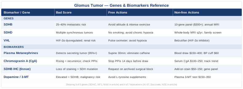
7 Key Biomarkers for Monitoring Glomus Tumors
Biomarkers are the translation layer between complex tumor biology and something you can actually measure, track, and respond to. For glomus tumors and paragangliomas, this is especially valuable because many of these tumors grow slowly, cause subtle symptoms for years, and are difficult to monitor with imaging alone between follow-up scans. The seven markers below span tumor function, proliferative risk, genetic surrogate signals, and metabolic disruption — giving a much more complete picture than any single test.
Biomarker 1: Plasma Fractionated Metanephrines
Why it matters: Metanephrines — metanephrine and normetanephrine — are the stable, inactive metabolites of epinephrine and norepinephrine. Functional paragangliomas and glomus tumors that secrete catecholamines continuously release these metabolites into the bloodstream, even during periods when the tumor is not actively firing. This continuous leakage makes plasma fractionated metanephrines the most sensitive biochemical test available for detecting a secreting glomus tumor or paraganglioma, with sensitivity often cited above 95 percent in large studies.
What it reveals: Elevated plasma normetanephrine with a relatively low metanephrine suggests a norepinephrine-predominant tumor — typical of sympathetic paragangliomas. A pattern showing predominantly elevated methoxytyramine (see Biomarker 4) shifts suspicion toward SDHB-mutated or dopaminergic tumors. The degree of elevation helps estimate secretory volume and treatment urgency.
How to measure it
Testing is done via a single blood draw, ideally after 20 to 30 minutes of supine rest. Some labs also offer 24-hour urine fractionated metanephrines as an alternative. Plasma testing is preferred for sensitivity. Cost ranges from approximately $150 to $400 USD at most reference labs in the US, often covered by insurance with appropriate clinical indication. The Mayo Clinic Laboratories and Quest Diagnostics both offer reliable fractionated plasma panels.
Key interferences to rule out before testing: tricyclic antidepressants, sympathomimetic medications, caffeine excess, and recent intense physical exercise can all raise results. Your clinician should review your medication list carefully before attributing an elevated result to a tumor.
If the score is elevated: the plan without supplements
The first step is eliminating medication and lifestyle confounders. Discontinue any interfering drugs under medical supervision at least two weeks before retesting. Reduce or eliminate caffeine, avoid intense aerobic exercise 48 hours before draws, and ensure the blood draw is done after proper supine rest. If results remain elevated on repeat testing, referral to an endocrinologist experienced in neuroendocrine tumors is essential. Imaging (MRI, CT with contrast, or DOTATATE PET-CT) is typically the next step. Medical management of catecholamine excess with alpha-blockade — using phenoxybenzamine or doxazosin — is standard before any surgical intervention and may be initiated even before surgery is definitive.
If the score is elevated: the plan with supplements or equipment
While no supplement replaces medical management of a truly secreting tumor, several adjunctive strategies may support the cardiovascular burden and oxidative stress that catecholamine excess creates. Magnesium glycinate (300 to 400 mg/day) supports vascular smooth muscle relaxation and is well-tolerated long-term. CoQ10 (100 to 300 mg/day with meals) addresses mitochondrial oxidative stress linked to catecholamine metabolism. For home monitoring, a validated cuff sphygmomanometer (devices like the Omron Platinum meet AHA standards at around $60 to $80) with daily blood pressure logging is highly practical. Target resting blood pressure below 130/80. Avoid ephedrine, pseudoephedrine, or high-dose caffeine supplements entirely. Cycling CoQ10 is generally not necessary — it can be used continuously without established harm.
Biomarker 2: Chromogranin A (CgA)
Why it matters: Chromogranin A is a glycoprotein released by neuroendocrine cells alongside secretory granule contents. Most paragangliomas and glomus tumors, whether functional or not, express and secrete CgA. It serves as a general neuroendocrine tumor marker that correlates with tumor mass and secretory activity. While it is less specific than metanephrines, its advantage is that it can be elevated even in non-secreting tumors where metanephrines are normal — making it particularly useful as a follow-up marker after treatment.
What it reveals: Rising CgA after surgical resection or during watchful waiting may signal recurrence or growth before imaging shows it. Very high CgA values (above 300 to 400 ng/mL) are associated with larger tumor burden and, in some series, with metastatic disease.
How to measure it
Serum or plasma CgA is measured by a standard blood draw. Cost ranges from $100 to $250 USD. Normal reference ranges vary by lab — most use an upper limit of around 100 to 120 ng/mL for the standard immunoradiometric assay. Importantly, proton pump inhibitors (PPIs) substantially elevate CgA — sometimes doubling or tripling levels — making any result meaningless if the patient is on omeprazole, lansoprazole, or similar drugs. PPIs must be stopped for at least two weeks before testing.
Renal insufficiency also raises CgA artificially. Creatinine should be measured simultaneously to interpret CgA results correctly.
If the score is elevated: the plan without supplements
First confirm there are no confounders: discontinue PPIs under medical guidance at least 14 days before retesting, and rule out chronic kidney disease or inflammatory bowel disease (both of which elevate CgA). If CgA remains elevated on clean testing, it should be tracked at consistent intervals — typically every 3 to 6 months — alongside imaging. Any sustained upward trend warrants imaging even before the scheduled surveillance scan. Work with your specialist to define a personal threshold at which to escalate.
If the score is elevated: the plan with supplements or equipment
To support overall neuroendocrine health and reduce cellular stress that may contribute to elevated secretion, berberine (500 mg twice daily with meals, cycled 8 weeks on, 2 weeks off to preserve gut microbiome diversity) has demonstrated anti-proliferative effects in neuroendocrine tumor models. N-acetylcysteine (600 mg twice daily) supports glutathione pathways that buffer oxidative damage from catecholamine excess. Neither replaces surveillance, and neither has been tested specifically in glomus tumor patients in large trials — evidence is primarily from in vitro and early-phase neuroendocrine tumor studies.
Biomarker 3: SDHB Immunohistochemistry (IHC)
Why it matters: SDHB immunohistochemistry is not a blood test — it is a staining pattern applied to tumor tissue after biopsy or resection. When succinate dehydrogenase function is lost due to a mutation in any of the four SDH subunits (SDHB, SDHC, SDHD, or SDHAF2), the entire SDH complex becomes unstable and SDHB protein disappears from the cell. A pathologist can detect this loss of staining, and its presence is a powerful surrogate marker for an underlying SDH gene mutation — even before genetic testing confirms which specific subunit is affected.
What it reveals: Loss of SDHB staining in a glomus tumor or paraganglioma immediately triggers the recommendation for germline genetic testing. It identifies the subset of patients who are most likely to carry a hereditary mutation, have a risk of additional tumors, and need first-degree relatives screened.
How to measure it
SDHB IHC is performed on formalin-fixed paraffin-embedded tumor tissue by a pathologist. It adds minimal cost to routine tumor pathology ($50 to $150 additional), and should now be considered standard of care for all paragangliomas and glomus tumors at major centers. Patients who had surgery without this staining performed should ask whether residual paraffin blocks exist and whether retrospective staining is possible.
If the score is abnormal (loss of staining): the plan without supplements
Loss of SDHB staining directly triggers a management cascade: germline genetic testing for the full SDH panel (SDHB, SDHC, SDHD, SDHAF2), imaging to assess for other tumors (whole-body MRI or DOTATATE PET-CT), and cascade testing for first-degree relatives. Surveillance intervals are tightened — typically annual biochemical testing and imaging every 12 to 24 months. This is a non-negotiable step that cannot be addressed by lifestyle alone. Engage a genetic counselor alongside your oncologist or endocrinologist.
If the score is abnormal: the plan with supplements or equipment
SDH-deficient tumors accumulate succinate, which acts as an oncometabolite by inhibiting key epigenetic enzymes. Strategies to reduce the cellular burden of succinate accumulation and support mitochondrial health include alpha-ketoglutarate (AKG) supplementation (1 to 2 grams daily) — which can partially compete with succinate's epigenetic inhibition — and vitamin C (1 gram daily), which supports TET enzyme activity that succinate suppresses. These are early-stage strategies supported by mechanistic reasoning and in vitro data, not large human trials. Disclose all supplements to your oncologist, particularly if systemic therapies are being considered.
Biomarker 4: Dopamine and 3-Methoxytyramine (3-MT)
Why it matters: While epinephrine and norepinephrine are the classic catecholamines tested in suspected pheochromocytoma, many paragangliomas — especially those driven by SDHB mutations — preferentially secrete dopamine rather than norepinephrine. Dopamine is often invisible on standard metanephrine panels because its metabolite, 3-methoxytyramine (3-MT), is not always included. Patients with SDHB-related paragangliomas who present with apparently normal metanephrines may have markedly elevated plasma 3-MT — and without testing for it specifically, the biochemical signature of their tumor goes undetected.
What it reveals: Elevated 3-MT in the context of a known or suspected glomus tumor is a red flag for SDHB mutation and is associated with a higher risk of metastatic disease. It can also explain cardiovascular symptoms in patients whose standard catecholamine panel was reported as normal.
How to measure it
Plasma 3-MT is available through specialized reference labs (Esoterix/LabCorp, Mayo Medical Laboratories, and Vanderbilt's reference laboratory offer validated assays). Cost ranges from $150 to $350 USD. It must be specifically ordered — it is not automatically included in most standard catecholamine panels. The reference range cutoff for plasma 3-MT is typically around 0.10 nmol/L, with values above 0.20 nmol/L considered clearly elevated. A 24-hour urine methoxytyramine measurement is an alternative if plasma is unavailable locally.
If the score is elevated: the plan without supplements
Elevated 3-MT substantially raises the probability of an SDHB-mutated, dopaminergic paraganglioma. The clinical response is the same as for elevated metanephrines — imaging, genetic referral, and specialist co-management — but the urgency is higher because SDHB tumors carry the greatest malignancy risk of any SDH variant. Dopamine excess does not respond as well to alpha-blockade as norepinephrine excess does; the blood pressure pattern is also different (often normotensive at rest with episodic symptoms). Make sure your endocrinologist is aware of the dopamine-dominant pattern so preoperative preparation is adjusted accordingly.
If the score is elevated: the plan with supplements or equipment
Dopamine excess creates a distinct oxidative footprint — dopamine auto-oxidizes to reactive quinones that damage mitochondria and cell membranes. Vitamin E (mixed tocopherols, 400 IU/day) and NAC (600 mg twice daily) can buffer quinone-mediated oxidative stress. Avoid high-dose L-tyrosine or L-DOPA supplements, as these are dopamine precursors that could increase tumor substrate. Similarly, avoid combining MAOI supplements (such as high-dose quercetin or grapefruit extract) with any catecholamine-active medications without specialist clearance.
Biomarker 5: Ki-67 Proliferation Index
Why it matters: Ki-67 is a nuclear protein expressed only in actively dividing cells. Its percentage within a tumor sample — the Ki-67 index — is a direct readout of how aggressively the tumor is proliferating. For glomus tumors and paragangliomas, where there is no universally agreed histological definition of malignancy, Ki-67 provides one of the most useful risk-stratification signals available from tissue. A Ki-67 below 3 percent is generally associated with slow-growing, favorable-prognosis tumors. Values above 5 to 10 percent, especially in the context of vascular invasion, raise concern for aggressive behavior.
What it reveals: In the context of the paraganglioma-pheochromocytoma pathology scoring system (PASS or GAPP scoring), Ki-67 contributes to predicting recurrence and metastatic potential. A high Ki-67 in combination with SDHB mutation is a particularly adverse pairing.
How to measure it
Ki-67 is measured by IHC on resected tumor tissue, performed as part of standard pathology. It is typically reported as a percentage of positively-staining cells per high-power field. Cost is included within routine surgical pathology in most cases. If not reported on your pathology report, request that it be added retrospectively on archived tissue — most major pathology labs can perform this.
If the index is elevated: the plan without supplements
A high Ki-67 (above 5 percent) in a glomus tumor or paraganglioma should prompt discussion of more aggressive surveillance — shortening the imaging interval to 6 to 12 months — and consideration of adjuvant therapies such as PRRT (peptide receptor radionuclide therapy) if the tumor is somatostatin receptor-positive on DOTATATE imaging. High-Ki-67 tumors that are metastatic may qualify for CAPTEM (capecitabine plus temozolomide) chemotherapy or sunitinib trials. This decision belongs firmly in specialist hands.
If the index is elevated: the plan with supplements or equipment
Anti-proliferative nutritional strategies with supporting (though often indirect) evidence include melatonin (3 to 10 mg at night), which has shown anti-proliferative effects in neuroendocrine cell lines via multiple mechanisms. Curcumin with piperine (500 mg curcumin, 5 mg piperine, twice daily with food) has demonstrated NF-kB and mTOR inhibition in neuroendocrine models. Neither should be used as monotherapy for a high-Ki-67 tumor, and curcumin may interact with certain chemotherapy agents — always disclose to your oncologist before starting.
Biomarker 6: Plasma Succinate
Why it matters: Succinate is a Krebs cycle intermediate that accumulates when succinate dehydrogenase is not functioning — which is precisely what happens in SDH-mutated glomus tumors. This accumulated succinate acts as an oncometabolite: it leaves the mitochondria, enters the cytoplasm and nucleus, and inhibits a family of alpha-ketoglutarate-dependent dioxygenases including the TET methylcytosine dioxygenases and histone demethylases. The result is a globally hypermethylated epigenetic state that silences tumor suppressor genes and promotes tumor progression. Measuring plasma succinate provides a window into this process.
What it reveals: Elevated plasma succinate in a patient with a known SDH mutation confirms that the metabolic consequence of the mutation is active. It can also serve as a treatment response marker — falling succinate after resection suggests that the main tumor source has been removed. Research from the Wellcome Sanger Institute and others has used succinate-to-fumarate ratios as diagnostic adjuncts for SDH-deficient tumors.
How to measure it
Plasma succinate measurement is not yet a mainstream clinical test, but it is available through metabolomics platforms at academic medical centers (including some Mayo Clinic and Cleveland Clinic programs). Research panels from companies like Metabolon or Genoptix measure succinate alongside a broad organic acid panel. Cost ranges from $200 to $600 out of pocket. In clinical practice, urine organic acids (available at most large reference labs for $100 to $300) can provide a proxy measure — elevated urinary succinate is seen in SDH-deficient states.
If the level is elevated: the plan without supplements
Elevated succinate signals active SDH dysfunction. The primary intervention is addressing the underlying cause — confirming the gene mutation, pursuing resection if the tumor is localized, and ensuring the genetic management plan is in place. Serial measurement can help confirm whether a partial resection removed the dominant source.
If the level is elevated: the plan with supplements or equipment
Alpha-ketoglutarate (AKG) is the direct competitive substrate against succinate for the dioxygenase enzymes that succinate inhibits. At 1 to 3 grams daily, AKG may partially buffer epigenetic silencing caused by succinate accumulation. Longevity researchers including David Sinclair have studied AKG for its epigenetic properties, though no clinical trial in SDH-mutated tumors has been completed. Vitamin C (1 gram daily) supports TET enzyme activity — the same family inhibited by succinate. Combined use of AKG and vitamin C is theoretically synergistic in this context and has a favorable safety profile. Avoid very high supplemental iron alongside these, as iron dyshomeostasis may worsen hypoxia-inducible factor dysregulation in SDH-mutated tumors.
Biomarker 7: Neuron-Specific Enolase (NSE)
Why it matters: Neuron-specific enolase is a glycolytic enzyme expressed in neurons and neuroendocrine cells. While it is less specific than metanephrines or chromogranin A, it adds value as a secondary neuroendocrine marker — particularly useful when CgA is confounded by PPI use or when a patient cannot stop PPIs. Rising NSE in the context of known glomus tumor or paraganglioma may signal increased tumor activity or dedifferentiation.
What it reveals: NSE above 16.3 ng/mL (a common upper reference limit) in this context warrants repeat testing and correlation with other markers. Very high NSE (above 50 ng/mL) is associated with large tumor burden and, in small cell and neuroendocrine carcinoma literature, with poor prognosis. In well-differentiated paragangliomas, NSE is usually only modestly elevated — so dramatic rises are clinically meaningful.
How to measure it
Serum NSE is a standard clinical lab test available at most major labs. Cost ranges from $50 to $150 USD. Importantly, hemolysis falsely elevates NSE because red blood cells also contain enolase — the blood draw must be processed quickly and without mechanical trauma to the sample. Confirm with your lab that the result was obtained from a non-hemolyzed specimen before acting on an elevated value.
If the score is elevated: the plan without supplements
Rule out hemolysis first (your lab report should flag this). If confirmed elevated on clean specimens, add NSE to the quarterly or semi-annual monitoring panel alongside CgA. A sustained rise in NSE — even while CgA remains stable — warrants imaging reassessment. In discussion with your oncologist, rising NSE may influence the decision about treatment timing.
If the score is elevated: the plan with supplements or equipment
The lifestyle interventions most directly applicable to NSE reduction are those that reduce systemic inflammation and support neuronal health: omega-3 fatty acids (2 to 3 grams EPA/DHA daily) have shown neuroprotective and mild anti-inflammatory effects in neuroendocrine contexts. Lion's mane mushroom extract (500 to 1000 mg daily standardized to hericenones) supports nerve growth factor — though evidence in neuroendocrine tumor contexts is theoretical. Consistent aerobic exercise at moderate intensity (zone 2, 150 minutes per week) reduces systemic inflammation and supports mitochondrial health without excessively elevating catecholamines — an important balance to strike in patients with secreting tumors.
Now that the biomarker picture is mapped, it is worth understanding the genetic architecture that often drives these abnormalities in the first place. Knowing your gene status does not just explain the past — it shapes every future decision.
The 6 Genes Behind Glomus Tumor Risk and Progression
Hereditary paraganglioma-pheochromocytoma syndromes represent one of the most genetically heterogeneous tumor syndromes in medicine. Roughly 30 to 40 percent of all paragangliomas carry a germline mutation, a proportion far higher than most common cancers. For clinicians following the work of researchers like Ali Torkamani at Scripps Research — whose work on population-scale genomics and variant interpretation has shaped precision medicine — these hereditary mutations represent exactly the kind of high-penetrance, actionable variants that genetic testing was designed to detect.
Gene 1: SDHD — The Most Common Head and Neck Culprit
SDHD encodes subunit D of the succinate dehydrogenase complex (also called mitochondrial complex II). Mutations in SDHD are the most common cause of hereditary head and neck paragangliomas and are strongly associated with multiple synchronous tumors — meaning patients can present with bilateral carotid body tumors, jugulotympanic paragangliomas, and vagal paragangliomas simultaneously.
A critical nuance: SDHD follows maternal imprinting. Only mutations inherited from the father lead to tumor development. If you inherited your SDHD mutation from your mother, you are a carrier but are very unlikely to develop tumors yourself — though your children who inherit it from you as a father remain at risk. This makes family tree analysis essential.
The original 2000 paper by Baysal et al. in Science established SDHD as the first hereditary paraganglioma gene, fundamentally changing how these tumors are managed.
If the gene variant is present: the plan without supplements
Annual biochemical screening (plasma fractionated metanephrines plus dopamine/3-MT) from age 6 to 10 onward. Whole-body MRI every 2 years from puberty. Avoid smoking and chronic hypoxia exposure (including high-altitude living), as hypoxia is a known tumor growth stimulus in SDH-deficient cells. Surgical referral when tumors are found — watchful waiting is sometimes appropriate for small, asymptomatic tumors in older patients, but younger patients generally benefit from earlier resection to avoid cranial nerve damage from growing tumors.
If the gene variant is present: the plan with supplements or equipment
Since SDHD loss leads to succinate accumulation and pseudohypoxia, strategies targeting the HIF pathway and epigenetic machinery are theoretically relevant. AKG (1 to 2 grams daily) and vitamin C (500 to 1000 mg daily) are the most mechanistically justified adjuncts. A personal pulse oximeter ($25 to $40 for a reliable device) is practical for monitoring resting oxygen saturation — maintain above 95 percent. Avoid repeated extreme altitude exposure. Home blood pressure monitoring is standard.
Gene 2: SDHB — The Highest Malignancy Risk
Of all the SDH genes, SDHB mutations carry the worst prognosis. Approximately 25 to 40 percent of patients with SDHB germline mutations develop metastatic disease over their lifetime — a proportion dramatically higher than SDHD or SDHC. SDHB-mutated tumors also tend to be extra-adrenal, dopaminergic (secreting dopamine rather than norepinephrine), and biochemically subtle, making them easier to miss with standard catecholamine panels.
Astuti et al. (2001) in the American Journal of Human Genetics first identified SDHB germline mutations as the cause of familial paraganglioma type 4 and pheochromocytoma, establishing the mutation-malignancy link that now guides surveillance intensity.
SDHB loss also creates a specific epigenetic signature — the CpG island methylator phenotype (CIMP) — that silences tumor suppressor genes broadly. This is why SDHB tumors can behave aggressively even when Ki-67 appears relatively low.
If the gene variant is present: the plan without supplements
Annual plasma metanephrines plus 3-MT (dopamine metabolite) — do not rely on standard norepinephrine-focused panels. Annual whole-body MRI (skull base to pelvis) or DOTATATE PET-CT every 2 years. Prompt surgical resection is strongly recommended when tumors are identified, given malignancy risk. Referral to a high-volume center with experience in hereditary paraganglioma syndromes is important. Genetic counseling for first-degree relatives is mandatory.
If the gene variant is present: the plan with supplements or equipment
The SDHB-CIMP link means epigenetic strategies are particularly relevant here. EGCG from green tea extract (400 to 600 mg decaffeinated extract daily) has demonstrated DNA methylation-modifying effects in cancer cell lines. Sulforaphane (30 to 50 mg daily from broccoli sprout extract) activates Nrf2 and has shown epigenetic benefits in neuroendocrine tumor models. Cycle sulforaphane 8 weeks on, 2 to 4 weeks off. Avoid high-dose isolated DIM or indole-3-carbinol without specialist clearance in patients on targeted therapies.
Gene 3: SDHC — Rarer, But Worth Knowing
SDHC mutations are less common than SDHB or SDHD variants but follow a similar biochemical mechanism — loss of SDH complex function, succinate accumulation, and pseudohypoxia. SDHC-related tumors tend to be predominantly head and neck (similar to SDHD) with a lower malignancy risk than SDHB. They are more often solitary and are less likely to be associated with multiple synchronous tumors than SDHD variants.
Surveillance protocols for confirmed SDHC mutation carriers are similar to SDHD, but with somewhat less urgency: annual biochemical testing from the second decade of life, imaging every 2 to 3 years. Surgical decisions are made on a case-by-case basis given the lower malignancy risk, and watchful waiting is more commonly appropriate for small, asymptomatic SDHC-related tumors.
If the gene variant is present: plan without and with supplements
Without supplements: annual biochemical testing, imaging per specialist protocol, cascade family testing. Avoid lifestyle factors that drive hypoxia-inducible factor signaling — smoking, chronic sleep apnea, sustained altitude exposure.
With supplements: the same AKG and vitamin C approach described for SDHD applies here. Riboflavin (vitamin B2, 100 mg daily) is a cofactor for SDHB protein stability and may partially support residual SDH complex function in some heterozygous mutation contexts — though evidence is primarily theoretical. Well-tolerated long-term.
Gene 4: VHL — The Hypoxia Gene
Von Hippel-Lindau disease, caused by germline VHL mutations, is associated with a syndrome that includes clear cell renal cell carcinoma, hemangioblastomas of the brain and spine, retinal angiomas — and paragangliomas. VHL protein is the critical regulator of HIF-1α and HIF-2α. When VHL is lost, HIF proteins accumulate even under normoxic conditions, driving a pseudohypoxia state that upregulates angiogenesis and cell survival pathways.
VHL-related paragangliomas tend to be noradrenergic (secreting norepinephrine), and bilateral adrenal pheochromocytomas are seen in roughly 10 to 20 percent of VHL patients. Genetic testing for VHL is also relevant because belzutifan — an HIF-2α inhibitor now FDA-approved for VHL disease — represents a targeted molecular therapy that specifically addresses VHL tumor biology.
If the gene variant is present: plan without and with supplements
Without supplements: annual plasma metanephrines, ophthalmic evaluation, renal imaging, MRI brain and spine per VHL Alliance guidelines. Belzutifan should be discussed with a VHL-specialist oncologist for patients with growing renal or CNS lesions.
With supplements: HIF pathway support includes antioxidant strategies that reduce ROS-driven HIF stabilization: vitamin C (500 to 1000 mg daily), quercetin (500 mg daily with food — note this has HIF-inhibitory properties in some cell models), and adequate selenium (100 to 200 mcg daily as selenomethionine). A portable continuous pulse oximeter during sleep is worth using if sleep apnea is suspected — hypoxia-driven HIF activation is additive to VHL loss.
Gene 5: NF1 — The Neurofibromatosis Connection
Neurofibromatosis type 1 (NF1), caused by mutations in the neurofibromin gene, is primarily known for its characteristic skin lesions and neurofibromas. However, NF1 also carries a 2 to 5 percent lifetime risk of pheochromocytoma and paraganglioma — lower than the SDH genes but significant given the prevalence of NF1 (1 in 3000 people). NF1 protein normally acts as a RAS-GAP (GTPase-activating protein), suppressing the RAS/MAPK pathway. Loss of NF1 function leads to constitutively active RAS signaling, driving cell proliferation.
NF1-related pheochromocytomas tend to be epinephrine-secreting and bilateral adrenal. They are less commonly head-and-neck paragangliomas. Any NF1 patient with hypertension, palpitations, or episodic sweating should have plasma metanephrines measured promptly.
If the gene variant is present: plan without and with supplements
Without supplements: the NF1-specific clinical guidelines (from the Children's Tumor Foundation and Legius syndrome community) do not mandate routine catecholamine screening in asymptomatic adults — but any cardiovascular symptoms should trigger testing. Annual blood pressure monitoring at minimum. Avoid MEK inhibitor supplements without medical guidance, as MEK is downstream of the pathway NF1 normally suppresses.
With supplements: Lovastatin (prescription) has been studied in NF1 for its RAS-modifying effects. Supplement-level strategies targeting RAS/MAPK: berberine (500 mg twice daily) has MEK-inhibitory properties at pharmacological doses in some models. Omega-3 fatty acids (2 to 3 grams EPA/DHA) modulate RAS membrane dynamics. Evidence is primarily preclinical.
Gene 6: MAX — The Underrecognized Risk
MAX (MYC-associated factor X) gene mutations are among the most recently characterized hereditary pheochromocytoma/paraganglioma genes. MAX protein normally dimerizes with MYC to regulate transcription of growth-promoting genes. When MAX is lost, MYC activity becomes unregulated, driving proliferation. MAX-mutated tumors are predominantly bilateral adrenal pheochromocytomas with a strong family history pattern, particularly in males (though female carriers are also affected). Malignant transformation occurs in some MAX cases, and the tumors are often catecholamine-secreting.
MAX testing is not yet included in all hereditary paraganglioma panels — you may need to specifically request a comprehensive 10+ gene panel (including SDHA, SDHAF2, FH, TMEM127, and MAX) rather than a limited 4-gene SDH panel to capture this.
If the gene variant is present: plan without and with supplements
Without supplements: annual plasma metanephrines, bilateral adrenal imaging (MRI preferred over CT to avoid contrast-related catecholamine release risk), and cascade testing for all first-degree relatives. Given bilateral adrenal risk, patients need counseling about adrenal insufficiency risk if both adrenals require surgery — cortisol-sparing approaches should be discussed with a surgeon experienced in adrenal-preserving techniques.
With supplements: MYC pathway is notoriously difficult to drug, but nutritional strategies that reduce MYC transcriptional activity include bromodomain inhibitor-adjacent nutrients: EGCG from green tea (400 mg standardized extract daily) and resveratrol (500 mg daily with meals) have demonstrated MYC downregulation in cell culture models. These are adjunctive only — not substitutes for surveillance or surgical management.
Understanding these six genes transforms the glomus tumor narrative from a mysterious local finding to a system-level picture with clear management implications. The research on catecholamine biology adds yet another dimension — one that connects tumor chemistry to everyday physiology in ways that matter for monitoring and symptom management.
What Catecholamine Research Reveals About Your Tumor's Biochemistry
The Huberman Lab podcast episode on dopamine — hosted by Andrew Huberman, professor of neurobiology and ophthalmology at Stanford — is not specifically about paragangliomas, but it contains some of the most accessible explanations of catecholamine biology available to a general audience. For patients with catecholamine-secreting glomus tumors or paragangliomas, this episode (available as Episode 39 on the Huberman Lab feed, titled "Controlling Your Dopamine for Motivation, Focus and Satisfaction") reframes the biology of dopamine in ways that directly apply to understanding their condition.
The following are the ten most practically relevant insights from this body of catecholamine research, contextualized for glomus tumor and paraganglioma patients:
1. Dopamine Is Released Continuously, Not Just in Spikes
Huberman explains that dopamine has two release modes: tonic (baseline continuous release) and phasic (surge-based release tied to reward or novelty). Paragangliomas can disrupt both by continuously secreting dopamine or its precursors into circulation, elevating baseline levels and distorting normal reward and motivation circuits. This biochemical interference partly explains why some patients feel persistent restlessness, anxiety, or mood instability before diagnosis.
2. Epinephrine vs. Norepinephrine vs. Dopamine Have Distinct Effects
The three main catecholamines drive different symptoms: epinephrine raises heart rate and blood sugar, norepinephrine raises blood pressure predominantly, and dopamine at high levels can cause a surprising range of cardiovascular effects including orthostatic hypotension. Understanding which catecholamine your tumor predominantly secretes (revealed by the biomarker pattern) directly predicts your symptom profile.
3. The Gut-Brain Axis Produces Catecholamines Too
Roughly 50 percent of the body's dopamine is produced in the gut, not the brain. Gut microbiome health influences catecholamine turnover, which means gut-directed strategies — prebiotic fiber, fermented foods, minimizing gut dysbiosis — can modestly influence the background biochemical environment even in patients with secreting tumors.
4. Stress Layering Amplifies Catecholamine Dysregulation
Huberman discusses how psychological stressors compound catecholamine release — a finding directly relevant to secreting paraganglioma patients, where stress-triggered catecholamine surges can cause hypertensive crises. Stress management is not a soft recommendation here — it is a biochemically rational intervention.
5. Cold Exposure Drives Sustained Norepinephrine Release
Cold showers or cold water immersion significantly elevate norepinephrine (reported 2 to 3-fold increases in studies Huberman cites). For patients with norepinephrine-secreting glomus tumors, cold water immersion is contraindicated and should be explicitly avoided until the tumor is resected and biochemical normalization is confirmed.
6. Caffeine Elevates Catecholamines Through Adenosine Blockade
Caffeine does not directly release catecholamines but prevents the adenosine-driven suppression of adrenergic signaling — the net effect is amplified catecholamine action. In secreting tumor patients, even moderate caffeine intake can meaningfully worsen blood pressure, palpitations, and anxiety. Caffeine reduction or elimination before testing and ideally throughout the pre-surgical period is rational.
7. Intermittent Fasting Elevates Epinephrine
Huberman has discussed how extended fasting or very low calorie intake raises epinephrine as part of the counter-regulatory response. For patients with epinephrine-secreting tumors, prolonged fasting protocols should be used cautiously. Smaller, more frequent meals may be preferable for metabolic stability.
8. Vigorous Exercise Acutely Raises Catecholamines
High-intensity interval training (HIIT) and anaerobic exercise drive large catecholamine surges. For patients with secreting paragangliomas or glomus tumors awaiting resection, vigorous exercise is a trigger for hypertensive episodes. Zone 2 aerobic exercise (conversational pace, sustained for 30 to 60 minutes) is much safer because catecholamine elevation is modest and sustained rather than large and acute.
9. Bright Light in the Morning Modulates Dopamine Circuitry
Morning sunlight exposure regulates dopamine and serotonin receptor expression in the brain, partly through melanopsin activation in the retina. While not directly impacting tumor catecholamine output, maintaining circadian consistency reduces stress-related catecholamine variability and supports sleep — itself important for tumor surveillance compliance and overall autonomic tone.
10. Tyrosine Is the Upstream Catecholamine Precursor — Supplementation Has Risks
L-tyrosine is the dietary amino acid from which all catecholamines are synthesized. High-dose L-tyrosine supplements (sometimes marketed for focus or adrenal support) are a direct substrate for catecholamine synthesis. In patients with secreting glomus tumors, supplemental L-tyrosine could theoretically increase tumor catecholamine output. Avoid L-tyrosine, L-DOPA, Mucuna pruriens, and phenylalanine supplements in any patient with a known or suspected catecholamine-secreting tumor.
Complementary Approaches That May Support Conventional Treatment
Given that glomus tumors and paragangliomas are primarily surgical conditions, complementary approaches play a supporting role — managing symptoms, reducing autonomic burden, and supporting recovery — rather than a primary role. The following four modalities have meaningful clinical evidence in related conditions and can be applied thoughtfully alongside conventional treatment.
Mindfulness Meditation and MBSR
Mindfulness-Based Stress Reduction (MBSR) is an 8-week structured program developed by Jon Kabat-Zinn at the University of Massachusetts that has been studied in dozens of clinical trials for anxiety, blood pressure, and catecholamine regulation. Its relevance to glomus tumor patients lies in its demonstrated ability to reduce sympathetic nervous system tone — directly opposing the autonomic excess driven by catecholamine-secreting tumors.
A randomized controlled trial published in Hypertension (Blom et al., 2012) showed that mindfulness practices reduced 24-hour urinary catecholamine excretion in patients with elevated cardiovascular risk — a finding that directly maps to the biochemical concerns of paraganglioma patients. Meta-analyses of MBSR in hypertension consistently show modest but statistically significant reductions in systolic blood pressure (3 to 5 mmHg on average), which is clinically meaningful in patients whose blood pressure is already driven upward by tumor catecholamine output.
Practically, MBSR can be accessed through the Mindfulness-Based Stress Reduction program at local hospitals, certified instructors, or online programs. Daily 10 to 20 minute sitting practice is sufficient for initial benefit. For glomus tumor patients specifically, body scan practices that increase cardiovascular awareness may help patients recognize early catecholamine surges and respond with relaxation techniques rather than anxiety amplification. Introduce gradually and do not practice during active catecholamine crises.
Breathing-Based Therapies
Controlled slow breathing — specifically at a rate of 5 to 6 breaths per minute (0.1 Hz breathing, sometimes called resonance frequency breathing) — activates the baroreceptor reflex and increases heart rate variability (HRV), a direct measure of vagal tone and autonomic flexibility. For patients with catecholamine excess, improving vagal tone via breathing is one of the few evidence-based non-pharmacological interventions that directly opposes sympathetic overdrive.
A 2018 meta-analysis in Frontiers in Human Neuroscience found that slow-paced breathing significantly increased HRV and reduced blood pressure across multiple populations. While no study has specifically tested slow breathing in paraganglioma patients, the autonomic mechanism is directly relevant. Slow breathing at 5 to 6 breaths per minute (approximately 5 seconds in, 5 seconds out) is the target.
For glomus tumor patients, 10 to 15 minutes of resonance frequency breathing daily — ideally in the morning before the sympathetic system ramps up — is a practical starting protocol. A biofeedback device such as the Muse headband or HeartMath Inner Balance sensor ($100 to $250 range) can guide and measure HRV during practice, providing objective feedback on whether the technique is achieving physiological benefit. Avoid forced Wim Hof hyperventilation-style breathing, which acutely alters blood gases and may trigger catecholamine surges.
Biofeedback
Biofeedback — using real-time physiological monitoring to train voluntary regulation of normally involuntary functions like blood pressure, heart rate, or skin conductance — is directly applicable to the blood pressure management challenges faced by patients with functioning glomus tumors. Several clinical studies have demonstrated clinically meaningful blood pressure reductions with HRV biofeedback in hypertensive patients (5 to 10 mmHg systolic reductions in some trials).
For glomus tumor patients on alpha-blockade awaiting surgery, biofeedback can serve as an adjunct to medication by training relaxation responses that blunt adrenergic surges. HRV biofeedback devices (HeartMath, Garmin HRV monitoring, Polar chest straps paired with apps) are accessible without prescription. Session frequency of 3 to 5 times per week, 10 to 20 minutes per session, is supported by the literature for blood pressure effects.
Practically, identify a biofeedback practitioner with experience in cardiovascular applications if possible — many hospital cardiac rehabilitation programs offer this service. Home devices are a reasonable starting point for motivated patients. Biofeedback should be suspended during active hypertensive episodes and resumed after stabilization. Do not reduce medications based on biofeedback results alone without specialist oversight.
Music Therapy
Music therapy — specifically slow-tempo, live or recorded music at 60 to 80 beats per minute — has documented anxiolytic and blood-pressure-lowering effects in perioperative and intensive care settings. For glomus tumor patients who face substantial surgical procedures (sometimes involving the skull base, cranial nerves, or major neck vessels), preoperative anxiety is a significant contributor to catecholamine surge risk around the time of surgery.
A Cochrane systematic review of music interventions in perioperative anxiety found significant reductions in anxiety and blood pressure compared to control. The effect size is modest but consistent across multiple randomized trials. For a patient whose catecholamine levels are already elevated by tumor output, even modest anxiety-driven amplification is worth reducing.
Practically, 20 to 30 minutes of slow, familiar, or nature-based music daily — particularly in the weeks before major surgery — is a simple, free, and safe adjunct. Playlists specifically designed for relaxation (60 to 72 BPM, major key, familiar or instrumental) are widely available on streaming platforms. Music therapy is fully compatible with all other treatments and carries no meaningful risk in this population.
Conclusion
Glomus tumors and paragangliomas sit at a genuinely fascinating intersection of genetics, metabolic biology, and clinical endocrinology. The science has moved far enough forward that a patient who understands the six key genes and seven tracked biomarkers covered in this article is meaningfully better equipped than one who does not — not to replace specialist care, but to participate in it far more effectively.
The most important takeaway is that this is not a condition to navigate passively. Whether you are a confirmed SDH mutation carrier, someone post-surgical monitoring for recurrence, or a patient still in the diagnostic process, the tools described here — plasma metanephrines, chromogranin A, dopamine and 3-MT, genetic panel testing, SDHB immunohistochemistry, Ki-67, and succinate — give you a concrete biomarker framework to track. The complementary strategies add meaningful support without replacing surgery or medical management.
The next smart step is to review your most recent lab results with the biomarker framework in mind, ask your endocrinologist whether your panel includes dopamine and 3-methoxytyramine, and — if genetic testing has not been done — request a comprehensive hereditary paraganglioma gene panel. These are simple, concrete questions that a single specialist conversation can answer. Better information, as always, leads to better decisions.
Ear, Nose & Throat Cancer & Oncology Endocrine & Metabolic
Neurological: Nerve Conditions
Cardiovascular: Blood Pressure Conditions
Endocrine & Metabolic: Adrenal Conditions