This article contains AI generated content.
Glomuvenous Malformation Genes And Biomarkers: 3 Genes And 5 Biomarkers To Track
If you or your child have been told this is "just a genetic vascular thing" and left with little else, that reaction is fair. Glomuvenous malformation (GVM) usually shows up as small, firm, bluish-purple nodules that are tender to the touch, sometimes clustered, sometimes scattered across a limb, and the standard advice tends to stop at "avoid bumping it" and "come back if it grows." That advice isn't wrong, but it's thin. It doesn't explain why the lesions appeared where they did, why a parent with the same mutation might have almost nothing visible while a child has a dozen patches, or what a doctor is actually checking for when they order bloodwork or imaging.
Generic dermatology guidance is built for the average case, and GVM doesn't have an average case. It's driven by a specific gene, a specific inheritance quirk, and a small set of lab and imaging findings that genuinely change what a specialist decides to do next. Knowing that a single mutation sits quietly in every cell of your body until a second, local event switches it on in one specific spot is the kind of detail that reframes the whole condition — from "random bad luck" to "a two-step process with a name and a mechanism."
This article goes past the surface-level advice. It walks through the three genes most relevant to GVM and the overlapping conditions it's often confused with, what each one actually does, and what a realistic management plan looks like — with and without medical intervention. It then covers the handful of blood and imaging biomarkers that specialists actually track, what a good or concerning result means, and how to act on it. A section on preventive-health thinking and a short review of complementary approaches for pain and flare management round things out.
None of this reverses a mutation or dissolves a malformation on its own. But understanding the mechanism changes real decisions: whether to test other family members, when imaging is worth the cost, when a lesion needs a procedure versus watchful waiting, and what "getting worse" actually looks like on a lab report before it looks like anything on the skin.
Summary
Here's the short version of what's ahead. Glomuvenous malformation isn't caused by one bad gene switch that's always "on" — it's caused by a mutation that sits silently in every cell of the body, doing nothing, until a second and completely separate genetic event happens in one specific patch of tissue. That second event is what actually turns a normal-looking piece of skin into a painful nodule, and it explains why the same mutation can produce one lesion in one family member and thirty in another. The article breaks down GLMN, the gene responsible for the great majority of cases, along with two related genes, TEK and PIK3CA, that cause look-alike conditions doctors need to rule out because the treatment path is genuinely different.
Beyond the genetics, there's a practical side most people never hear about: five specific lab and imaging markers — including a coagulation marker most patients have never had checked — that tell a specialist how active or extensive a malformation is, and when it's time to move from watching to treating. The article also pulls out ten ideas from a widely read longevity book that map surprisingly well onto living with a lifelong vascular condition, and closes with a short, honest look at which complementary approaches (like biofeedback and slow breathing) have real evidence for managing the pain that comes with these lesions, rather than pretending any of them treat the malformation itself.
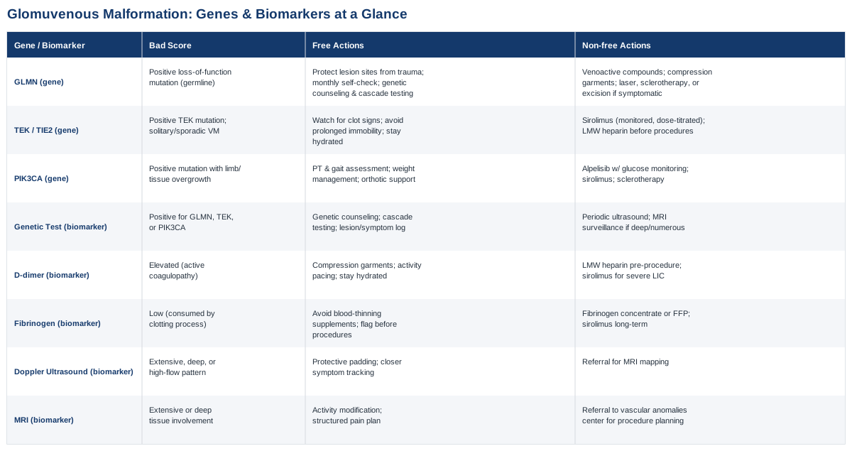
The Three Genes Behind Glomuvenous Malformation
Glomuvenous malformation is one of the more genetically well-understood vascular skin conditions, which is actually good news: unlike many chronic conditions where genetics only nudges probability, here the biology is close to deterministic and has been mapped in real family studies going back over two decades. Three genes matter for anyone trying to understand this condition properly — one that causes it directly, and two others that cause conditions doctors must distinguish it from, because mixing them up changes treatment.
GLMN: The Gene Where It Usually Starts
GLMN sits on chromosome 1p21-22 and was first linked to inherited "glomangiomas" in 1999, with the causative gene — glomulin — identified in 2002 (Boon et al., 1999; Brouillard et al., 2002). Glomulin is a small protein that interacts with cullin-RING ubiquitin ligase complexes and helps regulate signaling pathways involved in how vein walls are built — specifically, how smooth muscle-like glomus cells are laid down around the vessel. Nearly all disease-causing GLMN mutations are loss-of-function: frameshift or premature stop-codon changes that produce little or no working protein.
Four specific mutations account for roughly 70% of familial cases, with one deletion (often written as 157delAAGAA) responsible for close to half of them on its own (Brouillard et al., 2013). That same study of 162 families found penetrance around 90% by adulthood — meaning most people who inherit the mutation eventually develop at least one visible lesion, but not everyone, and not on the same timeline.
Why One Mutation Isn't Enough: The Two-Hit Mechanism
This is the part that explains almost everything confusing about GVM. Having a GLMN mutation in your germline (inherited in every cell) is not, by itself, sufficient to produce a lesion. A 2013 study found that visible malformations only form where a second, separate mutation knocks out the one remaining healthy copy of GLMN in a localized cluster of cells — most often through a process called somatic uniparental isodisomy, where a small patch of tissue ends up with two copies of the mutated chromosome instead of one mutated and one normal (Amyere et al., 2013).
This is why GVM behaves so unevenly within the same family: the germline mutation sets the stage everywhere, but a lesion only appears wherever that second, random, local event happens to occur. It also explains multifocality — new lesions over the years usually represent new independent second hits, not spread of an existing one. This mechanism is sometimes loosely called "paradominant" inheritance, and while it isn't classic epigenetics (it's a somatic genetic event, not a change in gene expression via methylation or histone modification), it functions similarly in practice: an inherited predisposition that stays silent until a local, largely unpredictable trigger flips it on. Human evidence for this mechanism is strong for GLMN specifically — it's been demonstrated directly in lesion tissue, not inferred from population statistics — but evidence on what environmental or physical factors increase the odds of a second hit (trauma, hormonal shifts, pressure) remains thin and mostly anecdotal from case series rather than controlled studies.
If the Gene Is Bad: The Plan Without Supplements or Procedures
A GLMN mutation is not something lifestyle changes reverse, and it's worth being direct about that rather than implying otherwise. What actually helps is reducing the odds that existing lesions become symptomatic and catching new ones early. That means: physically protecting known lesion sites from repeated pressure or trauma (padding over a lesion on a weight-bearing area, avoiding tight straps or repetitive rubbing gear at that spot); doing a monthly self-check of known lesion areas plus a visual skin sweep for new nodules, logged with a date and rough size so genuine growth is easy to spot; and pursuing genetic counseling and cascade testing for first-degree relatives, since a confirmed familial mutation lets relatives skip years of "is this something?" uncertainty. None of this changes the biology, but it changes how early problems get caught, which is where the real leverage is with a two-hit condition.
If the Gene Is Bad: The Plan With Supplements or Medical Equipment
For lesions causing ongoing discomfort, a few options have actual use, each with real trade-offs. Venoactive compounds — most commonly micronized purified flavonoid fractions (diosmin-hesperidin blends), typically dosed around 500 mg twice daily — are used off-label for the heaviness and tenderness associated with venous-type malformations; evidence is drawn mainly from broader venous insufficiency research rather than GVM-specific trials, side effects are generally mild (occasional GI upset), and there's no established cycling protocol, so it's typically taken continuously and reassessed every few months with a clinician. Medical-grade compression garments (20–30 mmHg) can meaningfully reduce discomfort for larger or deeper lesions on limbs and are worn daily during waking hours, with the main downside being consistency and skin irritation in hot weather.
For lesions that are persistently painful or cosmetically significant, procedural equipment options include pulsed-dye or Nd:YAG laser sessions (typically spaced four to six weeks apart for a course of three to six sessions, with risks of temporary pigment change, blistering, or scarring), sclerotherapy for larger venous components, and surgical excision for well-localized, symptomatic lesions. These are decisions made with a dermatologist or vascular anomalies specialist, not self-directed — and it's worth knowing upfront that recurrence is possible with all of them, since the underlying genetic tissue predisposition in the surrounding area isn't removed along with the treated nodule.
TEK (TIE2): The Look-Alike Gene That Changes Management
TEK encodes Tie2, a receptor on the surface of blood vessel lining cells that responds to angiopoietin signals and helps coordinate the relationship between the vessel lining and the smooth muscle wrapped around it. Germline TEK mutations cause inherited cutaneomucosal venous malformations, and somatic (localized) TEK mutations are found in roughly half of sporadic, solitary venous malformations that are not GVM at all (Limaye et al., 2009).
Why this matters practically: GVM and ordinary venous malformations can look similar to an untrained eye, but they are managed differently and have different complication profiles. Boon and colleagues laid out the clinical distinctions clearly — GVM lesions tend to be firmer, more clearly tender on direct pressure, and often multiple and familial, while TEK-related venous malformations are typically softer, more compressible, and carry a higher likelihood of localized intravascular coagulopathy (Boon et al., 2004). Getting the gene right on a test isn't academic — it determines whether coagulation monitoring and certain medications become relevant.
TEK: The Plan Without Supplements or Procedures
If genetic testing points to TEK rather than GLMN, the non-medical plan shifts toward coagulation awareness: knowing the signs of a clot forming within the malformation (sudden localized pain, firmness, or swelling), avoiding prolonged immobility on long flights or car rides if the malformation is in a limb, and staying appropriately hydrated. This isn't a treatment for the malformation — it's risk-awareness for its most common complication.
TEK: The Plan With Supplements or Equipment
For extensive or symptomatic TEK-related venous malformations, sirolimus (an mTOR inhibitor) has emerging use under specialist supervision, typically dosed daily with periodic blood-level monitoring to keep it in a safe range; side effects include mouth ulcers, elevated cholesterol, and increased infection risk, which is why it's cycled and monitored rather than started casually. Before any planned procedure on a TEK-related lesion, clinicians often check coagulation markers first (covered in the biomarker section below) and may use low molecular weight heparin around the procedure if localized coagulopathy is present.
PIK3CA: The Gene That Matters When Malformations Overlap With Overgrowth
PIK3CA is the third gene worth knowing, not because it causes GVM, but because roughly 30% of venous malformations that test negative for TEK carry a somatic PIK3CA mutation instead, often as part of a broader PIK3CA-related overgrowth spectrum where the vascular lesion appears alongside asymmetric limb or tissue overgrowth (Luks et al., 2015). This activates the PI3K-AKT-mTOR growth pathway in the affected tissue.
PIK3CA: The Plan Without Medication or Equipment
Where overgrowth is present alongside a vascular lesion, the realistic non-drug plan centers on physical therapy and gait assessment (since limb-length or girth asymmetry changes mechanics over years), weight management to reduce mechanical strain on the affected limb, and properly fitted compression or orthotic support.
PIK3CA: The Plan With Medication or Equipment
Targeted PI3K-pathway inhibitors such as alpelisib are being used in specialist and trial settings for PIK3CA-related overgrowth with vascular involvement, dosed daily with regular glucose monitoring because hyperglycemia is the main dose-limiting side effect; it requires active co-management with endocrinology. Sirolimus, sclerotherapy, and embolization remain options for symptomatic venous components regardless of which second-line gene is driving them.
What Consumer Genetic Tests Get Wrong About This Condition
This is worth addressing directly because it trips people up. Researchers like Ali Torkamani, who has spent years studying how genomic data translates into clinical risk prediction, and popularizers like Gary Brecka, who has brought methylation and epigenetic-style testing into mainstream wellness conversations, have done a lot to make genetic information feel accessible. But their frameworks are built around common polygenic risk (heart disease, metabolic risk, biological aging markers) using SNP microarray data — and that approach is largely the wrong tool for GVM. Consumer SNP-chip tests are not designed to reliably detect the small deletions, insertions, and structural events that cause most GLMN, TEK, and PIK3CA-related conditions, and they don't detect somatic second-hit mutations at all, since those aren't present in a blood or saliva sample drawn from the whole body. Confirming or ruling out GVM requires clinical-grade sequencing of the specific gene (or a targeted vascular-malformation gene panel) ordered through a genetics clinic, ideally with a genetic counselor involved to interpret results in family context — not a direct-to-consumer raw-data download.
Five Biomarkers Worth Tracking Alongside Your Genetic Result
A confirmed gene result tells you what's driving the condition; these five markers tell you how active it currently is. None of them are exotic — most are markers vascular medicine specialists already use routinely, but rarely explained to patients in plain language.
The Genetic Test Itself, as a Diagnostic Biomarker
How to measure it: a blood or saliva sample sent for targeted GLMN sequencing, or a broader vascular-malformation gene panel that also covers TEK, PIK3CA, and related genes. Cost typically runs from around $250 for a single-gene test to $1,000–$1,500 for a multi-gene panel, often partly covered by insurance when ordered with a clear clinical indication; turnaround is usually two to six weeks. If the result is positive: the plan without equipment is genetic counseling, family cascade testing, and a symptom/lesion log kept over time. The plan with equipment is periodic imaging surveillance (ultrasound as a first step, MRI if lesions are deep or numerous) on a schedule set by a specialist rather than a fixed annual default, since surveillance frequency depends on lesion location and symptom trend.
D-dimer
Why it matters: D-dimer rises when the body is breaking down small clots, and in vascular malformations it's the main marker of localized intravascular coagulopathy (LIC) — a low-grade clotting process that happens inside the abnormal, slow-flowing vessels themselves. Dompmartin and colleagues found D-dimer elevation correlates with lesion size and extent, particularly in larger, deeper malformations (Dompmartin et al., 2008). How to measure it: a standard blood draw, typically $10–$50, available at any lab. If the score is bad (elevated): the plan without supplements is compression garments, activity pacing, avoiding long periods of immobility, and staying hydrated. The plan with supplements or equipment involves specialist-directed use of low molecular weight heparin around planned procedures, and in more severe or extensive LIC, sirolimus has been studied as a way to reduce the coagulopathy over a period of months, with monitoring every few weeks initially and mouth ulcers or elevated lipids as the main side effects to track.
Fibrinogen
Why it matters: fibrinogen is consumed during the clotting process described above, so a low result alongside a high D-dimer indicates more severe LIC and higher bleeding risk during any procedure on the malformation. How to measure it: part of a standard coagulation panel blood draw, roughly $15–$40. If the score is bad (low): the plan without supplements is straightforward but important — avoid supplements with blood-thinning properties (high-dose fish oil, high-dose vitamin E, excess garlic extract) and flag the result before any dental or surgical procedure. The plan with equipment involves perioperative fibrinogen concentrate or fresh frozen plasma when a procedure is unavoidable, managed in a hospital setting, and sirolimus again as a longer-term option for chronically low fibrinogen tied to extensive disease.
Doppler Ultrasound Flow Pattern
Why it matters: Doppler ultrasound is usually the first imaging step because it's non-invasive, has no radiation, and quickly classifies a lesion as low-flow (typical of GVM and venous malformations) versus higher-flow, which would point toward a different diagnosis entirely and a different urgency level. How to measure it: an outpatient ultrasound, typically $150–$600 depending on region and facility, taking 20–40 minutes. If the finding is concerning (extensive, deep, or high-flow): the plan without equipment is protective padding and closer symptom tracking while awaiting specialist review. The plan with equipment is referral for MRI to map the lesion fully before deciding between sclerotherapy, laser treatment, or surgical planning.
MRI Signal and Enhancement Pattern
Why it matters: MRI is the gold-standard imaging biomarker for mapping how deep and extensive a malformation actually is — something a visual skin exam alone consistently underestimates, since GVM and venous malformations often extend below what's visible at the surface. This matters directly for procedural planning. How to measure it: an MRI with contrast, typically $1,000–$3,000 or more depending on location and insurance, no radiation involved, usually 30–60 minutes. It's not something to repeat on a routine annual basis; it's ordered at baseline diagnosis and again if symptoms change meaningfully or before a procedure. If the finding is bad (extensive or deep involvement): the plan without equipment is activity modification and a structured pain-management plan with a physician. The plan with equipment is referral to a multidisciplinary vascular anomalies center for image-guided sclerotherapy, laser treatment, or surgery, since deep or extensive lesions generally do better under a team that treats these regularly rather than as one-off cases.
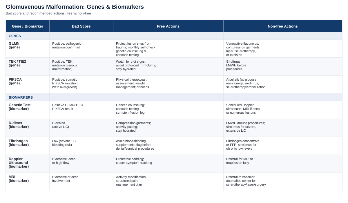
Together, these five give a fuller picture than the skin exam alone: the genetic result explains the mechanism, the coagulation markers flag active internal activity before it's visible or painful, and the imaging defines the physical extent that guides any procedure. Knowing which of these your specialist is actually checking — and why — turns a vague "let's keep an eye on it" into a concrete monitoring plan with clear next steps.
Ten Ideas From Peter Attia's Outlive That Apply Here
Outlive: The Science and Art of Longevity by Peter Attia isn't written about vascular malformations, but its central argument — that medicine works better when it acts on early markers rather than waiting for a crisis — maps unusually well onto a two-hit genetic condition where the "crisis" (a symptomatic lesion) is preceded by a silent, testable state. Here are the ten ideas from the book most worth borrowing.
1. Medicine 3.0: Act on the Marker, Not Just the Symptom
Attia's core distinction is between reactive medicine, which waits for disease to become symptomatic, and a more proactive model that intervenes on measurable risk markers earlier. For GVM, that means treating a positive GLMN result and elevated D-dimer as information worth acting on — through monitoring and planning — well before a lesion becomes painful.
2. Define Your "Centenarian Decathlon"
Attia asks readers to define the specific physical tasks they want to still do decades from now, then train backward from that goal. For someone with lesions on a load-bearing limb, this reframes protection and physical therapy not as restriction, but as preserving specific future function — walking distances, carrying grandchildren, whatever the personal list includes.
3. Zone 2 Cardio Still Matters, With One Caveat
Attia is a strong advocate for low-intensity aerobic training for metabolic and vascular health broadly. That guidance still applies here, with the practical caveat that activity involving repetitive impact or friction directly over a known lesion site should be modified — swimming or cycling instead of running when a lesion sits on a shin or foot, for instance.
4. VO2 Max as a Marker Worth Knowing
Attia treats VO2 max as one of the strongest predictors of longevity across large population studies. It's unrelated to GVM directly, but for someone managing a lifelong condition, tracking a broad marker of cardiovascular fitness alongside the condition-specific ones keeps the bigger health picture from being crowded out by the more visible issue.
5. A Genetic Diagnosis Isn't a Verdict
One of the book's steadier points is that genetic risk is probabilistic and actionable, not fatalistic. A confirmed GLMN mutation defines a mechanism and a monitoring plan — it doesn't define an outcome, especially since penetrance and lesion burden vary widely even within the same family.
6. Ask Different Questions at Checkups
Attia pushes patients to bring specific questions and specific markers to their doctor rather than a general "check me out." For GVM, that translates directly into the biomarker section above — arriving at an appointment already knowing what D-dimer and fibrinogen results might mean, rather than hearing about them for the first time.
7. Build a Care Team, Not a Single Doctor
Attia argues that complex, chronic conditions are best managed by a coordinated team rather than one generalist. Vascular anomalies centers exist specifically for this — combining dermatology, vascular surgery, hematology, and genetics — and they're worth seeking out for anything beyond a single small lesion.
8. Stability Training Reduces Injury Risk
Attia emphasizes stability and balance work as injury prevention, particularly for older adults. For someone with lesions in areas vulnerable to bumps or falls, this has a direct secondary benefit: fewer falls means fewer traumatic impacts on a malformation site.
9. Emotional Health Is Part of the Plan, Not an Add-On
Attia is candid that physical longevity work means little without emotional stability, and chronic, visible, occasionally painful conditions carry a real psychological load. Addressing that directly — rather than treating it as separate from "the real treatment" — is consistent with how the book frames whole-person health.
10. The Marginal Decade Argument
Attia's closing argument is that the effort invested in health now compounds most visibly in the decade before it would otherwise decline. Applied here: consistent monitoring and lesion protection habits built now are what prevent a manageable condition from becoming a harder one to treat later, simply through fewer untreated flare-ups accumulating over years.
Complementary Approaches That Can Help You Manage Pain and Flare-Ups
None of the following treat the malformation itself, and it's worth being upfront that direct clinical evidence in glomuvenous malformation specifically doesn't exist — these lesions are too rare for dedicated trials. What follows is evidence from closely related pain and vascular-symptom contexts, applied cautiously.
Mindfulness Meditation / MBSR
Chronic, intermittently painful conditions carry a psychological load that plain pain medication doesn't address, and mindfulness-based stress reduction is one of the more studied non-drug approaches for chronic pain generally. It's relevant here less as a treatment for the lesions and more as a way to manage the day-to-day experience of a condition with unpredictable flare-ups.
A 2017 systematic review and meta-analysis found mindfulness meditation associated with a small but consistent reduction in pain compared with controls across dozens of randomized trials, alongside improvements in depression symptoms and quality of life (Hilton et al., 2017). The effect size is modest, and the authors were clear that evidence quality was still rated low to moderate.
Realistically, this means treating a structured MBSR course (typically eight weekly sessions) as a genuine adjunct to pain management rather than a primary strategy — useful alongside monitoring and procedural care, not instead of it, and most helpful during periods when a lesion is tender but not requiring active intervention.
Biofeedback
Biofeedback trains conscious control over normally automatic functions like skin temperature or muscle tension, using real-time sensor feedback. It's included here specifically because thermal biofeedback has a genuine evidence base for a thematically related problem: vascular symptom control in Raynaud's phenomenon, a different condition but one that shares the theme of managing peripheral vascular discomfort through learned physiological control.
Evidence is mixed rather than uniformly positive: some controlled trials found biofeedback improved finger temperature and symptom scores in Raynaud's (Raynolds review), while a broader meta-analysis of complementary approaches for Raynaud's found biofeedback did not significantly change attack frequency or severity overall. That inconsistency should be stated plainly rather than glossed over.
For GVM, biofeedback has no direct evidence at all, so it's reasonable to consider only as a general relaxation and pain-coping tool — sessions with a trained biofeedback therapist, typically weekly for four to six weeks — rather than something expected to change lesion behavior.
Guided Imagery
Guided imagery uses structured mental visualization to reduce perceived pain and anxiety, and it has one of the more consistent evidence bases among the complementary options for procedural and postoperative pain specifically — relevant here because many people with GVM eventually go through a laser, sclerotherapy, or excision procedure.
A randomized trial in geriatric orthopedic postoperative patients found guided imagery significantly reduced pain and improved comfort compared with standard care (Topcu & Findik, 2023), consistent with older trials showing similar benefits around colorectal and cardiac surgery.
The realistic application is around procedures rather than daily management: a short guided imagery session or recording used in the days before and after a planned laser or sclerotherapy session for a GVM lesion, as a low-risk complement to standard pain control — not a replacement for it.
Breathing-Based Therapies
Slow, deep breathing has a plausible mechanism for pain modulation through baroreceptor and vagal activation, and it's the lowest-cost, lowest-risk option on this list, which matters for a chronic condition where sustainability counts.
An experimental study found that slow, deep breathing reduced pain perception and altered autonomic activity and mood compared with normal breathing (Busch et al., 2012), though later work has questioned whether the effect is driven specifically by breathing rate or partly by distraction and relaxation more broadly — worth knowing so the technique isn't oversold.
In practice, this looks like a five-to-ten-minute session of paced breathing (roughly six breaths per minute) during a flare or before a procedure, which has essentially no side effects or cost and can be layered on top of any of the other approaches above.
The Bottom Line
Glomuvenous malformation is unusual among genetic conditions in that the mechanism is genuinely well understood: a germline GLMN mutation sets the stage everywhere in the body, but a lesion only forms where a second, local mutation removes the last working copy of the gene — which is why the same family history can look so different from one person to the next. Knowing whether GLMN, TEK, or PIK3CA is driving a given lesion changes real decisions, not just labels, and pairing that genetic result with a small set of blood and imaging markers — D-dimer, fibrinogen, Doppler ultrasound, and MRI — gives a specialist (and you) a concrete way to track whether things are stable or moving.
None of this promises a cure, and none of the complementary approaches above change the underlying biology — they help with the pain and stress that come along with it, which is a legitimate goal on its own. The most useful next step is usually the least dramatic one: confirm the genetic diagnosis if you haven't, ask your doctor directly which of the five biomarkers they're tracking and why, and start a simple log of lesion size, tenderness, and any new spots. That log is what turns "it feels different lately" into something a specialist can actually act on.
Cardiovascular: Blood Vessel Conditions Vascular Conditions