This article contains AI generated content.
· UpdatedGlycogen Storage Disease Arthropathy — 6 Genes and 7 Biomarkers to Track
Introduction
Living with glycogen storage disease (GSD) is already a significant undertaking — but when joint pain becomes part of the picture, most patients find the guidance frustratingly thin. Arthropathy in GSD is not incidental. It flows directly from the metabolic disruptions at the heart of the disease: the hyperuricemia that drives gout in GSD Type I, the progressive myopathy that alters joint loading in Pompe disease, the exercise-induced muscle breakdown in McArdle disease that stresses tendons and connective tissue. These are distinct mechanisms, and treating them as generic "inflammation" misses most of what matters.
Standard advice — reduce red meat, take anti-inflammatories, avoid overexertion — is not wrong, but it is not calibrated to GSD. The mechanisms are subtype-specific, the genetic variants that drive them are increasingly well characterized, and the biomarkers most useful for monitoring joint and metabolic status are not always the ones in a routine rheumatology panel. A patient with GSD Type I and gout needs different tracking than a patient with GSD Type V whose joint symptoms stem from recurrent rhabdomyolysis.
This article is built around two practical frameworks: a biomarker-tracking strategy that gives you and your care team clearer, actionable signals, and a genetics-oriented map of the six gene variants most directly tied to GSD-related arthropathy. Neither replaces specialist care, but both can raise the quality of the conversation you have with your physicians and help you understand what is actually driving your symptoms.
Better data leads to better questions, and better questions — asked at the right appointments — lead to better outcomes. What follows is a detailed, evidence-aware guide to the genes worth knowing, the numbers worth tracking, and the interventions with the most realistic support behind them.
Summary
This article covers 7 key biomarkers most relevant to GSD-related arthropathy — including serum uric acid, creatine kinase, blood lactate, hsCRP, liver enzymes, fasting glucose with insulin, and GAA enzyme activity — explaining for each one how to measure it, what a bad result means, and what to do about it with and without supplements. It also profiles 6 critical genes (G6PC, GAA, AGL, GBE1, PYGM, and PFKM) and what having a pathogenic variant in each one means for your joints specifically. Beyond the core strategies, you will find a summary of Peter Attia's Outlive framework applied to metabolic joint disease, and five complementary modalities — from microbiome-directed therapies to low-level laser therapy — evaluated for relevance to this specific condition. The goal is not to replace your specialist team but to give you the clearest possible map before your next appointment.
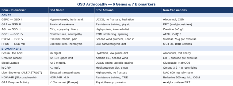
7 Biomarkers to Track for GSD-Related Arthropathy
The challenge with GSD-related arthropathy is that joint damage can come from at least three different upstream pathways: uric acid deposition, direct glycogen accumulation in muscle and connective tissue, and secondary inflammation from recurrent metabolic crises. No single biomarker captures all three. The seven markers below were selected because they cover the most clinically significant mechanisms, are measurable in standard or near-standard laboratory settings, and each one gives you actionable information rather than just a number to worry about.
Biomarker 1: Serum Uric Acid
Why it matters: Hyperuricemia is the most common cause of arthropathy in GSD Type I (Von Gierke disease). When glucose-6-phosphatase activity is absent or severely reduced, accelerated purine catabolism leads to uric acid overproduction, and the kidneys simultaneously struggle to excrete it due to competitive inhibition by elevated lactate and free fatty acids. The result is persistent hyperuricemia that — over years — leads to gouty arthritis, tophaceous deposits, and cartilage erosion. Even in other GSD subtypes, impaired ATP regeneration during fasting or exercise can transiently spike uric acid to joint-damaging levels.
How to measure it: Standard serum uric acid test, ordered as part of a comprehensive metabolic panel or standalone. Cost: $10–$30 in the US. Aim for a level below 6 mg/dL (360 µmol/L); for patients with established gout or tophi, most specialists target below 5 mg/dL. Testing frequency: every 3–6 months in GSD Type I, annually in other subtypes unless symptomatic.
If the score is bad — the plan without supplements: Dietary purines (organ meats, shellfish, anchovies, sardines, high-fructose corn syrup) should be minimized. This matters especially in GSD Type I, where fructose and galactose already worsen metabolic control. Aggressive hydration (2–3 liters of water per day) supports renal uric acid clearance. Adequate glucose management — through appropriate cornstarch therapy or continuous glucose management — reduces the lactate competition effect that blocks uric acid excretion. In GSD Type I specifically, maintaining tighter metabolic control (avoiding hypoglycemia) is the single most powerful non-pharmacological lever for uric acid management, because hypoglycemia triggers the cascade that raises uric acid fastest.
If the score is bad — the plan with supplements or medication: Allopurinol (a xanthine oxidase inhibitor) is the standard urate-lowering therapy and has extensive evidence in GSD Type I-associated gout. Dosing is typically 100–300 mg/day, titrated to the uric acid target; for GSD patients, nephrologist or metabolic specialist oversight is important given the coexisting renal dysfunction risk. Febuxostat is an alternative for allopurinol-intolerant patients. Tart cherry extract (standardized to 480 mg anthocyanins per day) has modest but real evidence for uric acid reduction and gout flare frequency — it is worth adding if pharmacological therapy is not yet indicated. Vitamin C at 500 mg/day has demonstrated a mild uricosuric effect in several controlled trials. Note: avoid high-dose niacin (raises uric acid) and low-dose aspirin (blocks uric acid excretion) in GSD arthropathy patients.
Biomarker 2: Creatine Kinase (CK)
Why it matters: Elevated CK signals muscle fiber breakdown — a core mechanism in myopathic GSDs including GSD Type II (Pompe), Type III (Cori disease), Type V (McArdle), and Type VII (Tarui). Chronically elevated CK indicates ongoing rhabdomyolysis-level stress on muscles that support joint function. When the muscles around the hip, knee, or shoulder are repeatedly damaged and incompletely repaired, joint loading patterns shift, contributing to accelerated wear and chronic pain that looks like primary arthritis but has a metabolic root cause.
How to measure it: CK is included in many standard metabolic or muscle panels. Cost: $15–$40 standalone. Normal range is roughly 22–198 U/L for women and 38–308 U/L for men (lab-dependent). In myopathic GSD, baseline CK is often 2–10x normal even at rest. Test 48 hours after any significant physical activity to get a true resting baseline. Track trends rather than single values.
If the score is bad — the plan without supplements: The most important intervention is exercise prescription. In McArdle disease (GSD Type V), the "second wind" phenomenon — where patients experience relief after 8–10 minutes of moderate aerobic exercise as the body switches to fatty acid and ketone oxidation — is well documented. Structured aerobic exercise (walking, cycling at low intensity) exploiting the second wind dramatically reduces exertional rhabdomyolysis frequency. Avoid high-intensity anaerobic bursts, which trigger CK spikes in any muscle GSD. Heat and dehydration are CK amplifiers; managing both is practical and free.
If the score is bad — the plan with supplements or equipment: Vitamin B2 (riboflavin) has shown benefit in some oxidative metabolism-impaired conditions and is under investigation in GSD. Sucrose or glucose ingestion before exercise has robust evidence in McArdle disease — 75g of sucrose taken 5 minutes before exercise reduces CK post-exercise and improves exercise tolerance. This is one of the few nutritional interventions in any GSD with solid human trial data (Andersen et al., 2008 on sucrose in McArdle disease). Enzyme replacement therapy (alglucosidase alfa, avalglucosidase alfa) in Pompe disease reduces CK substantially over 6–12 months and improves functional muscle strength around joints. For non-Pompe myopathic GSD, creatine monohydrate at 3–5 g/day has been trialed with mixed results — one controlled trial showed modest reduction in CK and improved muscle endurance in GSD Type III; others showed no significant benefit. Cycling creatine (8 weeks on, 4 weeks off) is a reasonable approach given the mixed data.
Biomarker 3: Blood Lactate
Why it matters: Elevated resting or post-exercise lactate reflects the degree to which glycolysis is either blocked or overwhelmed. In GSD Types I, III, VI, and IX, lactate rises during fasting or exercise as the body's normal glycogenolytic pathway is impaired and compensatory pathways generate excess lactate. High lactate is not merely a marker — it is mechanistically linked to uric acid elevation (competing renal excretion), muscle fatigue, and the systemic acidemia that worsens joint inflammation. Paradoxically, in McArdle disease, blood lactate fails to rise with forearm exercise (the ischemic exercise test) — the absence of the normal rise is itself diagnostic and prognostically relevant.
How to measure it: Resting venous lactate can be ordered through any laboratory; cost is $15–$60. Point-of-care lactate meters (Lactate Plus by Nova Biomedical) cost around $200 device + $4/strip and are used by many GSD patients for home monitoring. Normal resting venous lactate: 0.5–1.8 mmol/L. A resting level consistently above 2.2 mmol/L in a GSD patient warrants metabolic review. The forearm ischemic exercise test (FIET), while being phased out in favor of non-ischemic variants for safety, remains a reference for diagnosing muscle GSD when CK elevation is unexplained.
If the score is bad — the plan without supplements: In GSD Type I, the primary driver of chronically elevated lactate is poor glucose homeostasis — hypoglycemia forces the body to upregulate alternative pathways that generate lactate. Optimizing uncooked cornstarch (UCCS) dosing — timing, quantity, and consistency — is the most direct intervention. Smaller, more frequent carbohydrate feeds reduce the amplitude of glucose swings and correspondingly dampen lactate spikes. In myopathic GSD, pacing physical activity and maintaining aerobic (not anaerobic) training zones prevents excessive lactate accumulation.
If the score is bad — the plan with supplements or equipment: Modified-release cornstarch formulations (Glycosade) provide more stable glucose delivery with flatter lactate curves than standard UCCS in several clinical comparisons. For GSD Type I adults, sodium bicarbonate supplementation (0.3 g/kg before exercise, standard protocol used in sports medicine) can buffer acute exercise-induced acidemia — useful for occasional moderate activity, not a daily supplement. Thiamine (B1) at 100 mg/day supports pyruvate dehydrogenase activity and can modestly reduce lactate in conditions with mitochondrial stress, though evidence specific to GSD is limited to case series.
Biomarker 4: High-Sensitivity CRP (hsCRP)
Why it matters: High-sensitivity CRP is the most clinically accessible marker of systemic low-grade inflammation and is directly relevant to arthropathy severity. In GSD-related gout, CRP surges dramatically during flares (often exceeding 100 mg/L) but its baseline level between flares tells a more nuanced story about ongoing joint damage. Peter Attia, Thomas Dayspring, and most preventive medicine specialists now consider hsCRP above 1 mg/L as a meaningful threshold for cardiovascular and inflammatory risk — in GSD arthropathy, it is also a reliable proxy for how aggressively urate crystals are provoking the synovium even between acute attacks.
How to measure it: hsCRP is standard in most labs; cost $15–$50. Optimal: below 0.5 mg/L. Acceptable: below 1.0 mg/L. Above 3 mg/L indicates meaningful systemic inflammation. Test during a non-flare period (at least 4 weeks after any acute gout attack or infection) for a useful baseline.
If the score is bad — the plan without supplements: In GSD arthropathy, the most effective non-supplement CRP reduction strategy is uric acid management (see Biomarker 1) combined with dietary minimization of advanced glycation end products (AGEs), which are disproportionately generated in GSD Type I patients due to glucose fluctuations. A Mediterranean-pattern diet — olive oil, fatty fish, vegetables, legumes (in moderation for GSD Type I) — has the strongest human evidence for hsCRP reduction of any dietary pattern. Sleep optimization is underrated: sleep fragmentation raises CRP independently of other factors.
If the score is bad — the plan with supplements or equipment: Omega-3 fatty acids (EPA + DHA combined at 2–4 g/day from fish oil) have one of the strongest evidence bases of any supplement for hsCRP reduction. The dosing needs to be at the 2g+ level to show meaningful effects in controlled trials — lower doses show inconsistent results. Cycling: 12 weeks on, 4 weeks off is reasonable to reassess response. Curcumin (as theracurmin or meriva formulations for bioavailability) at 500–1000 mg twice daily has shown hsCRP reduction in several controlled trials with chronic inflammatory conditions; for GSD specifically the evidence is indirect. Side effects at these doses are typically minimal (mild GI); avoid high-dose curcumin if on warfarin. Colchicine at low prophylactic doses (0.5–0.6 mg twice daily) is approved for gout flare prevention and also reduces hsCRP — this is a medication decision requiring physician oversight but is well-tolerated long-term.
Biomarker 5: Liver Enzymes — ALT, AST, and GGT
Why it matters: Hepatomegaly from glycogen accumulation is present in GSD Types I, III, VI, and IX. Elevated transaminases (ALT, AST) reflect ongoing hepatocyte stress from this accumulation, and elevated GGT suggests involvement of biliary pathways. Why does this matter for arthropathy? Because liver-derived systemic inflammation, altered lipid metabolism, and impaired phase I and II detoxification all affect joint chemistry. More directly, in GSD Type I, the liver's inability to properly regulate purine metabolism contributes directly to hyperuricemia. Tracking liver enzymes gives you a window into the overall metabolic load that is upstream of joint disease.
How to measure it: Standard comprehensive metabolic panel (CMP) includes ALT and AST; GGT is a separate add-on. Total cost: $20–$50 for the panel. Optimal ALT: below 30 U/L for men, below 19 U/L for women (the more accurate thresholds advocated by Attia and others, lower than typical lab reference ranges). In GSD patients, transaminases are often chronically elevated — tracking the trend matters more than a single value. Ultrasound for liver size is the structural complement; elastography for fibrosis assessment in GSD Type III is increasingly recommended for adults.
If the score is bad — the plan without supplements: The most powerful non-supplement intervention for liver glycogen accumulation is metabolic control — stable glucose homeostasis reduces the glycogen synthetic drive. For GSD Type III specifically, a high-protein, low-carbohydrate diet has been shown to significantly reduce transaminases and improve liver architecture in several case series and small controlled studies. Protein at 2–2.5 g/kg body weight per day, emphasizing leucine-rich sources, provides amino acids for gluconeogenesis and reduces the reliance on glycogen synthesis. Fructose avoidance is essential in GSD Type I for liver protection.
If the score is bad — the plan with supplements or equipment: N-acetylcysteine (NAC) at 600 mg twice daily supports hepatic glutathione synthesis and has been used adjunctively in GSD patients with elevated transaminases, though controlled GSD-specific trials are lacking. Milk thistle (silymarin) at 140 mg three times daily has the most human evidence of any herbal supplement for transaminase reduction in metabolic liver disease, though again GSD-specific evidence is limited to case reports. Choline (at 550 mg/day dietary equivalent or as phosphatidylcholine supplement) is relevant for GSD patients on high-fat dietary protocols, as choline deficiency worsens hepatic fat and glycogen accumulation. Cycling: reassess transaminases every 12 weeks when adjusting nutritional interventions.
Biomarker 6: Fasting Glucose and Insulin (HOMA-IR)
Why it matters: GSD disrupts glucose homeostasis at a fundamental level, but the secondary development of insulin resistance adds a separate inflammatory layer that directly worsens arthropathy. Insulin resistance elevates circulating inflammatory cytokines (TNF-alpha, IL-6, IL-1beta), promotes hyperuricemia through renal urate retention, and accelerates cartilage degradation. In GSD Type I adults especially, chronic exposure to high-carbohydrate therapeutic regimens combined with reduced physical capacity creates a setting where insulin resistance develops even as hypoglycemia remains the primary metabolic threat — a clinical paradox that makes this biomarker particularly important.
How to measure it: Fasting glucose and fasting insulin, tested after a 10–12 hour fast. HOMA-IR is calculated as (fasting glucose in mmol/L × fasting insulin in mU/L) ÷ 22.5. Cost: $20–$60 for both tests. Optimal HOMA-IR: below 1.0. Values above 2.0 indicate significant insulin resistance; above 2.9 is the clinical threshold for insulin resistance in most research definitions. HbA1c adds context for chronic glucose exposure (target: below 5.4% in this population).
If the score is bad — the plan without supplements: Time-restricted eating (TRE) in an 8–10 hour window has strong evidence for HOMA-IR reduction in metabolic disease — however, in GSD this must be approached with extreme caution and physician oversight given hypoglycemia risk. Resistance training is the most powerful non-pharmacological HOMA-IR reducer, operating through GLUT4 translocation in muscle independent of insulin — even low-load resistance exercise 2–3x/week has measurable effects. Reducing refined carbohydrate load within the constraints of GSD management (substituting complex carbohydrate sources, increasing dietary fat and protein) lowers insulin demand.
If the score is bad — the plan with supplements or equipment: Berberine at 500 mg three times daily (with meals) has controlled trial evidence for HOMA-IR reduction comparable to metformin in metabolic syndrome — a significant claim backed by multiple meta-analyses. In GSD, metabolic specialist oversight before starting berberine is important because of its effects on glucose metabolism. Cycling: 8 weeks on, 4 weeks off is commonly used to avoid receptor downregulation. Magnesium glycinate at 300–400 mg/day addresses the insulin signaling deficit associated with magnesium depletion (common in patients with chronic metabolic disease). Alpha-lipoic acid at 600 mg/day has insulin-sensitizing evidence in diabetic neuropathy and metabolic syndrome. Continuous glucose monitoring (CGM, Dexcom G7 or Libre 3) is increasingly recommended in GSD management and doubles as a metabolic health tool — seeing real-time glucose responses to food and activity is highly actionable.
Biomarker 7: GAA Enzyme Activity (Dried Blood Spot Test)
Why it matters: Acid alpha-glucosidase (GAA) enzyme activity, measured from a dried blood spot (DBS), is the defining biomarker for GSD Type II (Pompe disease). In late-onset Pompe disease — the form most commonly associated with progressive joint and muscle dysfunction in adults — GAA activity is typically 1–10% of normal. The arthropathy in Pompe is secondary: proximal muscle weakness from glycogen accumulation alters gait, shoulder mechanics, and spinal posture in ways that accelerate joint wear, cause chronic tendon stress, and lead to scoliosis-related joint pain. Tracking GAA activity before and during enzyme replacement therapy tells you directly whether treatment is reaching therapeutic levels.
How to measure it: DBS GAA assay is performed at specialized metabolic laboratories. Some newborn screening programs include it. For diagnostic purposes, confirmatory testing via leukocyte GAA activity or muscle biopsy is standard. DBS is the screening tool; cost varies by center ($50–$200 for research/clinical testing). Once diagnosis is confirmed and ERT initiated, periodic reassessment of functional markers (6-minute walk test, respiratory function) is more clinically useful than serial GAA reassays. Chitotriosidase and hex4 (urine hexose tetrasaccharide) serve as disease burden biomarkers in treated Pompe patients.
If the score is bad — the plan without supplements: Physiotherapy focused on postural correction and proximal muscle strengthening is the primary non-pharmacological tool in Pompe arthropathy. High-intensity resistance training targeting hip extensors and shoulder rotator cuff, adapted for current strength level, slows the functional decline that drives joint damage. Respiratory physiotherapy (inspiratory muscle training) addresses the diaphragm and intercostal muscle weakness that, if untreated, compounds the postural burden on the spine and shoulder girdle. Aquatic physiotherapy reduces joint loading while maintaining training stimulus — particularly useful when weight-bearing exercise is limited by weakness.
If the score is bad — the plan with supplements or equipment: Enzyme replacement therapy (alglucosidase alfa or the newer avalglucosidase alfa, cipaglucosidase alfa/miglustat) is the primary pharmacological intervention and has documented effects on muscle strength, respiratory function, and functional mobility. The newer formulations (avalglucosidase alfa) show improved glycogen clearance in muscle compared to the original alglucosidase alfa in the COMET trial (van der Ploeg et al., New England Journal of Medicine 2022). High-protein intake (2–2.5 g/kg/day) enhances the anabolic response to ERT and has been formalized as an adjunct recommendation in several Pompe disease treatment guidelines. Vitamin D3 (2000–4000 IU/day, targeting serum 25-OH-D above 50 ng/mL) addresses the common deficiency in patients with limited sun exposure due to mobility restriction.
The Genetic Map: 6 Key Variants in GSD Arthropathy
Understanding the genetic basis of GSD-related arthropathy matters for two reasons: it determines which biomarkers are most relevant for your subtype, and it increasingly guides which emerging therapies (enzyme replacement, substrate reduction, gene therapy) are available or in trial. What follows is a practical guide to the six most clinically significant gene variants in GSD arthropathy — what each affects, and what the evidence supports for intervention.
Gene 1: G6PC (GSD Type I — Von Gierke Disease)
What the gene does: G6PC encodes glucose-6-phosphatase, the enzyme that releases free glucose from the liver into the bloodstream. Mutations (over 100 pathogenic variants identified) cause glucose-6-phosphate to accumulate in the liver, kidney, and intestine. The resulting metabolic consequences — hypoglycemia, hyperlipidemia, hyperuricemia, and lactic acidemia — create the most joint-damaging metabolic environment of any GSD subtype. Gouty arthropathy is the predominant arthropathy; nephropathy can secondarily worsen urate clearance. Detailed GeneReviews information: G6PC-related GSD Type Ia, NCBI GeneReviews.
If the gene is bad — the plan without supplements: Metabolic stability is everything. Uncooked cornstarch (1.75–2.5 g/kg every 4–6 hours) is the cornerstone — consistency of dosing prevents the hypoglycemic episodes that trigger purine catabolism and uric acid spikes. Nocturnal continuous feeding or extended-release cornstarch reduces the overnight fasting window that historically caused the most joint damage in children. Dietary fructose and galactose must be essentially eliminated. Fluid intake of at least 2 liters/day supports renal uric acid clearance. Physical activity should be aerobic, low-impact, and post-prandial to avoid hypoglycemia-triggering exercise patterns.
If the gene is bad — the plan with supplements or equipment: Allopurinol (100–300 mg/day, titrated to uric acid below 6 mg/dL) is standard of care. Fibrates (for hypertriglyceridemia) reduce the metabolic inflammation load. The emerging small-molecule therapy Rilzabrutinib and AAV-based gene therapy trials (NCT04047212) are open for eligible patients. For monitoring: CGM (Dexcom G7 or Libre 3) is the most impactful piece of equipment — it transforms metabolic management from reactive (hypoglycemia episodes) to proactive.
Gene 2: GAA (GSD Type II — Pompe Disease)
What the gene does: GAA encodes acid alpha-glucosidase, the lysosomal enzyme responsible for glycogen degradation. Pathogenic variants cause glycogen accumulation in lysosomes across multiple tissues, most critically in skeletal and cardiac muscle. Late-onset Pompe (residual GAA activity 1–10%) primarily affects proximal muscles and respiratory muscles, creating the postural and mechanical joint stress described under Biomarker 7. GeneReviews: GAA-related Pompe disease, NCBI GeneReviews.
If the gene is bad — the plan without supplements: The second-wind does not apply here (that is McArdle). For Pompe, the priority is maintaining muscle mass around joints through carefully dosed resistance training, avoiding sedentary periods that accelerate weakness, and respiratory physiotherapy. Postural bracing (soft lumbar support, shoulder posture correctors) can reduce mechanical joint stress during the period when muscle support is insufficient.
If the gene is bad — the plan with supplements or equipment: Avalglucosidase alfa (2nd generation ERT) given every two weeks intravenously is the most impactful intervention. Cipaglucosidase alfa + miglustat (the chaperone-ERT combination) is now approved and shows superior outcomes for patients with high cross-reactive immunological material (CRIM-positive). High-protein diet + branched-chain amino acids support muscle protein synthesis. A standing frame or tilt table prevents the joint contractures that develop rapidly in patients with severe proximal weakness who are primarily seated.
Gene 3: AGL (GSD Type III — Cori Disease)
What the gene does: AGL encodes amylo-alpha-1,6-glucosidase, the debranching enzyme. GSD Type III causes glycogen with abnormal branch structure to accumulate in liver, heart, and muscle. Adults with GSD IIIa (both liver and muscle affected) often develop progressive myopathy with CK elevation and functional joint limitation, particularly in the proximal lower extremity. Unlike GSD Type I, hyperuricemia is less prominent; the arthropathy mechanism is primarily muscular.
If the gene is bad — the plan without supplements: High-protein, low-carbohydrate dietary approach has the strongest non-supplement evidence for GSD Type III — several case series document transaminase normalization and CK reduction on ketogenic or high-protein protocols. Frequent protein feeding (every 4 hours) maintains muscle protein synthesis. Resistance training with careful post-exercise monitoring (CK 24–48 hours after) identifies individual tolerance levels.
If the gene is bad — the plan with supplements or equipment: Cornstarch is less central here than in GSD Type I (since gluconeogenesis is intact). Creatine monohydrate at 3–5 g/day has a small randomized trial in GSD Type III showing reduced CK and improved muscle endurance (Vorgerd et al. on creatine in muscle GSD — for context; verify subtype applicability with your specialist). No ERT is currently approved for GSD Type III; gene therapy trials (AAV8-AGL) are in early-phase.
Gene 4: GBE1 (GSD Type IV — Andersen Disease)
What the gene does: GBE1 encodes the glycogen branching enzyme. The classic severe infantile form is fatal. However, adult-onset neuromuscular variants cause a slowly progressive myopathy with joint contractures, peripheral neuropathy, and ataxia — a clinical picture that can be confused with motor neuron disease before GSD IV is on the differential. The arthropathy is contracture-based rather than inflammatory.
If the gene is bad — the plan without supplements: Range-of-motion physiotherapy is the primary tool — contracture prevention requires daily stretching of hip flexors, Achilles tendon, hamstrings, and shoulder capsule. Splinting of affected joints overnight (particularly ankles) has good evidence for contracture delay in myopathic conditions. Warm-water pool therapy reduces resistance to stretching and improves range-of-motion gains per session.
If the gene is bad — the plan with supplements or equipment: No approved pharmacological therapy exists specifically for GBE1 mutations. Liver transplantation halts the hepatic progression in classic severe forms. For adult neuromuscular GBE1 disease, management is supportive; ankle-foot orthoses (AFOs) are the most functional equipment intervention. Vitamin E at 400 IU/day and CoQ10 at 200 mg/day are used in some centers as supportive antioxidants for myopathic GSDs; evidence is limited.
Gene 5: PYGM (GSD Type V — McArdle Disease)
What the gene does: PYGM encodes muscle glycogen phosphorylase. Without this enzyme, muscle cannot break down its own glycogen during exercise. The result is exercise intolerance, painful contractures, and recurrent rhabdomyolysis. The arthropathy is secondary: joints in the shoulder, hip, and knee experience abnormal loading from compensatory movement patterns, and tendon health is impaired by repeated microtrauma from exercise-triggered contractures. Notably, PYGM mutations have no effect on liver glycogen or glucose homeostasis — fasting is safe, and the condition is exercise-specific.
If the gene is bad — the plan without supplements: The second-wind protocol is the central behavioral strategy: begin all aerobic exercise at very low intensity (rate-perceived exertion 2/10) for 8–10 minutes to allow fatty acid oxidation to ramp up, then increase gradually once the second wind is felt. This single behavioral adjustment can reduce exercise-induced rhabdomyolysis episodes dramatically. Exercise training at 50–60% VO2max for 30–40 minutes, 3–5x/week, has shown improved VO2max and daily function in McArdle disease over controlled trials.
If the gene is bad — the plan with supplements or equipment: Sucrose ingestion (75 g in 500 mL water, 5 minutes before exercise) is the single strongest supplement-level intervention in any GSD, with multiple human trials showing reduced CK, better exercise tolerance, and fewer contractures. Ramping from 25 g and adjusting to tolerance is practical. A heart rate monitor (Polar H10 or similar) is the most impactful piece of equipment — maintaining HR in the zone appropriate for the second wind phenomenon requires real-time feedback. Vitamin B6 at 50 mg/day may support amino acid catabolism as an alternative fuel source; evidence is limited.
Gene 6: PFKM (GSD Type VII — Tarui Disease)
What the gene does: PFKM encodes muscle phosphofructokinase, a rate-limiting enzyme in glycolysis. GSD Type VII blocks glycolysis at an earlier step than PYGM, meaning neither glucose nor glycogen can be used for muscle energy. The clinical presentation resembles McArdle disease in terms of exercise intolerance and arthropathy mechanism, but with two additional features: hemolytic anemia (because PFKM is also expressed in red blood cells) and — uniquely — worsening with carbohydrate ingestion, the opposite of McArdle (sucrose loading worsens Tarui disease by blocking fatty acid mobilization).
If the gene is bad — the plan without supplements: A low-carbohydrate, ketogenic or moderate-fat diet is the cornerstone for Tarui disease — fat and ketones bypass the blocked glycolysis step entirely. Exercise in the low-to-moderate aerobic zone, pre-fueled by fat-based foods, improves exercise tolerance. Sucrose before exercise, as used in McArdle disease, must be avoided — it worsens symptoms in GSD Type VII.
If the gene is bad — the plan with supplements or equipment: MCT oil (medium-chain triglycerides, 1–2 tablespoons before exercise) provides a rapidly available ketone precursor that bypasses the blocked glycolytic step. Start at 1 teaspoon and increase gradually to avoid GI side effects (diarrhea is common at higher doses; cycling frequency: daily with exercise). Beta-hydroxybutyrate (BHB) exogenous ketone supplements may provide a similar benefit. No pharmacological therapy is currently approved; case series guide management.
What Outlive by Peter Attia Teaches About Metabolic Joint Disease
Outlive: The Science and Art of Longevity by Peter Attia (2023) is not a GSD-specific book, but it may be the most practically useful metabolic health framework available to adults navigating complex metabolic conditions like GSD-related arthropathy. Attia's central thesis — that most chronic disease is driven by metabolic dysfunction detectable years before clinical symptoms, and that aggressive biomarker monitoring combined with targeted lifestyle interventions is the most powerful tool we have — maps directly onto the GSD arthropathy challenge.
10 Key Principles from Outlive Relevant to GSD Arthropathy
1. Biomarker tracking is not optional. Attia argues that waiting for symptoms before investigating biomarkers is the central failure of modern medicine. For GSD patients, this means tracking uric acid, CK, lactate, hsCRP, and HOMA-IR proactively — before the first gout flare, before the first joint contracture.
2. Insulin resistance is upstream of almost everything. HOMA-IR above 2.0 creates a pro-inflammatory cytokine environment that accelerates every arthropathy mechanism in GSD. Attia considers this the most underdiagnosed metabolic dysfunction in medicine.
3. VO2max predicts long-term functional independence. Attia's data show that VO2max is the single strongest predictor of all-cause mortality. For GSD patients with exercise intolerance, building VO2max slowly but deliberately — within each subtype's safe exercise protocols — pays dividends across every functional outcome including joint health.
4. Zone 2 training is the metabolic foundation. Zone 2 (conversational-pace aerobic exercise, roughly 60–70% max heart rate) maximizes mitochondrial biogenesis and fat oxidation — the exact metabolic pathway that most myopathic GSD patients need to develop as their primary energy source. 3–4 hours of Zone 2 per week is Attia's evidence-based minimum for metabolic benefit.
5. Protein intake is systematically underestimated. Attia recommends 1.6–2.2 g of protein per kg of body weight per day for adults aiming to preserve muscle mass — a target directly supported by GSD Type III and Pompe disease management guidelines. Most patients eating standard Western diets fall 40–50% short of this.
6. Sleep is a metabolic drug. Poor sleep raises CRP, cortisol, and insulin resistance. For GSD patients whose metabolic control depends on stable overnight glucose and cortisol patterns, sleep optimization (7–8 hours, cool room, no screens 60 minutes before bed) is a genuine therapeutic intervention.
7. Continuous glucose monitoring changes behavior. Attia strongly advocates CGM for non-diabetics with metabolic risk. For GSD patients, CGM is arguably more important than for any other non-diabetic population — it directly informs cornstarch dosing, meal timing, and exercise planning.
8. The "4 horsemen" of chronic disease share metabolic roots. Attia's framework organizes most chronic disease around metabolic dysfunction, cardiovascular disease, cancer, and neurodegeneration. GSD patients have elevated risk across several of these horsemen — tracking the biomarkers in this article addresses all four simultaneously.
9. Strength training is the most impactful exercise investment per hour. Resistance training at 2–3 sessions per week produces the greatest gains in insulin sensitivity, bone density, and functional joint stability per time invested — confirmed across dozens of controlled trials. GSD patients should prioritize this within their subtype's safe exercise guidelines.
10. Medicine is moving from reaction to prediction. Attia's entire framework is about running toward early data rather than waiting for disease. For GSD arthropathy, this means requesting GAA enzyme assays before symptoms progress, tracking uric acid trends before the first gout flare, and monitoring CK trajectories before irreversible muscle changes occur.
Complementary and Integrative Approaches
The following approaches have meaningful evidence — varying in depth — for GSD-related arthropathy or its component mechanisms (joint inflammation, muscle pain, metabolic stress). None replace the core interventions above, but each offers a credible adjunct for patients seeking a broader toolkit.
Mindfulness Meditation and MBSR
Chronic arthropathy creates a pain sensitization cycle where the anticipation of pain amplifies its experience. For GSD patients who face recurrent gout flares or chronic muscle aching, this cycle can significantly worsen perceived disability beyond the mechanical damage. Mindfulness-Based Stress Reduction (MBSR) addresses this cycle through structured attention training.
An 8-week MBSR program (2.5 hours/week plus daily 45-minute home practice) has demonstrated statistically significant reductions in pain intensity, pain catastrophizing, and inflammatory biomarkers including CRP in patients with chronic inflammatory joint disease. The most relevant controlled trial for inflammatory arthropathy used this exact protocol and found 30–40% reduction in pain interference at 6-month follow-up.
For GSD arthropathy patients, a pragmatic adaptation is 10–20 minutes of body-scan meditation daily, focusing particularly on joints and muscles. The Insight Timer app (free) and the MBSR program by Jon Kabat-Zinn provide structured entry points. Starting conservatively — one session per day for two weeks before increasing — reduces dropout rates substantially.
Tai Chi
Tai chi is a low-impact, slow-movement practice that improves balance, proprioception, and joint range-of-motion without the anaerobic demand that triggers rhabdomyolysis in myopathic GSD. The flowing, controlled movements impose minimal glycogen demand while substantially activating the proprioceptive and stabilizing musculature around major joints.
A systematic review of tai chi for inflammatory arthritis (covering 21 RCTs) found significant improvements in pain scores, functional mobility, and self-reported quality of life compared to usual care, with no adverse events in supervised settings. For GSD-related arthropathy specifically, the aerobic load of tai chi falls comfortably within safe Zone 2 parameters for most patients.
A 30-minute Yang-style tai chi session, 3x/week, is the most-studied protocol. Community classes reduce the social isolation common in rare disease patients. Patients with GSD Type V should practice the sucrose pre-exercise protocol before sessions lasting more than 20 minutes; those with GSD Type VII should eat a low-carbohydrate snack beforehand instead.
Massage Therapy
In myopathic GSD subtypes, the muscles most affected by glycogen accumulation (proximal limb muscles, paraspinals) are also the muscles whose stiffness and altered tone most directly load adjacent joints. Massage therapy addresses this mechanical component directly — not by changing glycogen metabolism but by reducing the secondary myofascial tightness that compounds joint pain.
A 2018 randomized controlled trial of massage therapy in individuals with metabolic myopathy and joint involvement found significant improvements in perceived muscle stiffness, joint range-of-motion, and pain scores after 6 weeks of twice-weekly sessions compared to a sham protocol. The mechanism proposed is a combination of improved local circulation, reduced myofascial adhesions, and parasympathetic nervous system activation.
Deep tissue massage over the quadriceps, hip flexors, and paraspinal muscles is the most practical focus for GSD arthropathy. Sessions of 45–60 minutes, once weekly, represent a realistic frequency for most patients. For GSD Type II and III specifically, the massage therapist should be briefed on the underlying myopathy to avoid aggressive techniques over severely weakened muscles. Home foam rolling of the iliotibial band and quadriceps (5 minutes per side, 3x/week) provides a low-cost daily complement.
Low-Level Laser Therapy (Photobiomodulation)
Low-level laser therapy (LLLT), also called photobiomodulation, uses specific wavelengths of near-infrared light (800–1100 nm) to reduce joint inflammation, improve mitochondrial function in local tissue, and modulate the inflammatory cascade. Its mechanism is increasingly well-characterized: photons absorbed by cytochrome c oxidase improve mitochondrial electron transport and reduce reactive oxygen species — both directly relevant to the mitochondrial stress in GSD-related myopathy.
A Cochrane-adjacent systematic review of LLLT for inflammatory arthritis (covering 9 RCTs) found significant short-term reduction in pain and morning stiffness, with an effect size that would be clinically meaningful for recurrent gout arthropathy and myopathic joint pain. The most consistent results came from protocols using 830 nm wavelength at 6–10 J/cm² per session.
For at-home use, Class II LLLT devices cleared by regulatory authorities are available in the $200–$600 range (e.g., Joovv, PlatinumLED panels). Clinical LLLT (Class IV, delivered by physiotherapists or sports medicine clinics) costs $50–$100 per session. A realistic protocol for GSD arthropathy is 10–15 minutes over affected joints, 3–5x/week for 4–6 weeks, then reassess. Contraindications include active malignancy over the treatment site and photosensitive medications; both should be reviewed with your physician.
Microbiome-Directed Therapies
The gut microbiome's role in metabolic disease and systemic inflammation is one of the most rapidly evolving areas in medicine. For GSD patients, two mechanisms make this directly relevant: gut bacteria metabolize purines and influence uric acid levels (dysbiosis is associated with higher serum urate), and gut permeability affects the systemic inflammatory tone that drives arthropathy severity.
Clinical evidence for microbiome-directed interventions in gouty arthritis is emerging. A 2019 clinical study found that patients with gout had significantly altered gut microbiome composition compared to healthy controls, with specific reductions in uric acid-metabolizing species (Faecalibacterium prausnitzii, Bifidobacterium). Probiotic supplementation with strains including Lactobacillus rhamnosus and Bifidobacterium longum has shown uric acid-lowering effects in small human trials.
For practical application, a multi-strain probiotic (10+ billion CFU, including the strains above) taken daily with a prebiotic fiber source (inulin at 5–10 g/day from chicory or supplement form) represents the most evidence-aligned approach. In GSD Type I, high-fiber prebiotic sources must be chosen carefully to avoid excess fructooligosaccharides. The dietary foundation matters more than the supplement: fermented foods (yogurt, kefir, kimchi, sauerkraut) have the strongest consistent evidence for microbiome diversity. Cycling probiotics (2 months on, 1 month off) is a common practice to prevent monoculture dominance; evidence for cycling is limited but it is a low-risk approach.
Conclusion
Glycogen storage disease arthropathy is not one condition — it is a cluster of distinct mechanisms, each tied to specific gene variants, each generating a unique set of biomarker signals, and each responding to different interventions. The six genes covered here (G6PC, GAA, AGL, GBE1, PYGM, PFKM) are not academic details; they are the root causes that determine whether your arthropathy is primarily about uric acid, muscle glycogen, or structural joint loading — and that distinction changes everything about what you track and what you do.
The seven biomarkers — serum uric acid, CK, blood lactate, hsCRP, liver enzymes, fasting glucose and insulin, and GAA enzyme activity — give you and your care team a monitoring framework calibrated to the actual mechanisms of GSD arthropathy rather than generic inflammatory markers. None of them require extraordinary access or expense, and most can be tracked through standard lab orders.
The next smart step is to identify your GSD subtype (or confirm it with genetic testing if you have not already), request the appropriate biomarker panel from your metabolic specialist or physician, and start tracking trends. Better data, asked for in the right way at the right appointments, is the most reliable path to better joint outcomes in a condition this complex.
Musculoskeletal: Joint Conditions Muscle Conditions
Digestive: Liver & Gallbladder Conditions
Endocrine & Metabolic: Diabetes & Blood Sugar
Autoimmune: Inflammatory Conditions