This article contains AI generated content.
GM1 Gangliosidosis Genes And Biomarkers: 1 Gene And 6 Biomarkers To Track
When One Diagnosis Raises A Hundred Questions
If you are reading this, you have probably already heard the phrase "acid beta-galactosidase deficiency" from a geneticist, or you are a parent trying to understand what a positive newborn screen or an enzyme assay result actually means for your child. Maybe you are an adult with new, unexplained gait or speech changes whose doctor has just mentioned a rare storage disorder you had never heard of before that appointment. The vocabulary is dense, the disease is rare enough that most clinicians have seen only a handful of cases, and the internet tends to offer either outdated textbook summaries or forum posts written in fear.
Generic advice does not help much here. "Eat well, exercise, manage stress" is reasonable for most chronic conditions, but GM1 gangliosidosis is not a lifestyle-influenced condition in the way high cholesterol or insulin resistance can be. It is caused by a specific, identifiable defect in a single gene, and the biology that follows from that defect is precise: an enzyme fails to do its job, a lipid accumulates where it should not, and cells - especially neurons - are damaged by the buildup. Understanding the disease well enough to make good decisions means understanding that specific chain of events, not applying broad wellness advice to a narrow biochemical problem.
That precision is actually useful. Because the mechanism is well characterized, there is a defined set of biomarkers that can tell you, with real biochemical evidence, how active the disease process is, how it is likely to behave, and whether a given intervention (including experimental ones in clinical trials) is doing anything measurable. That is a very different, and more actionable, situation than many rare diseases are in.
This article walks through those biomarkers in detail, then looks at the genetics behind them, summarizes what recent research is converging on, and reviews which supportive, non-curative therapies have any real evidence behind them. None of this replaces a metabolic geneticist or a genetic counselor. But knowing which numbers matter, why they move, and what can - and cannot - be done about them tends to turn a frightening diagnosis into a manageable set of decisions.
Summary
GM1 gangliosidosis comes down to one gene, GLB1, and a small number of biomarkers that actually matter for tracking it. This article explains what happens when GLB1 fails, why enzyme activity, GM1 ganglioside, urinary oligosaccharides, neurofilament light chain, GFAP, and brain imaging are the measurements clinicians and researchers rely on, what each one can realistically reveal, and - honestly - what can and cannot be done when a result comes back abnormal. It also covers the genetic details worth understanding (including a common testing trap called pseudodeficiency), ten research developments changing how specialists think about this disease, and which supportive therapies have any real evidence behind them for symptom management. The goal is not a miracle fix - there isn't one yet - but a clear, accurate map of what the science actually supports.
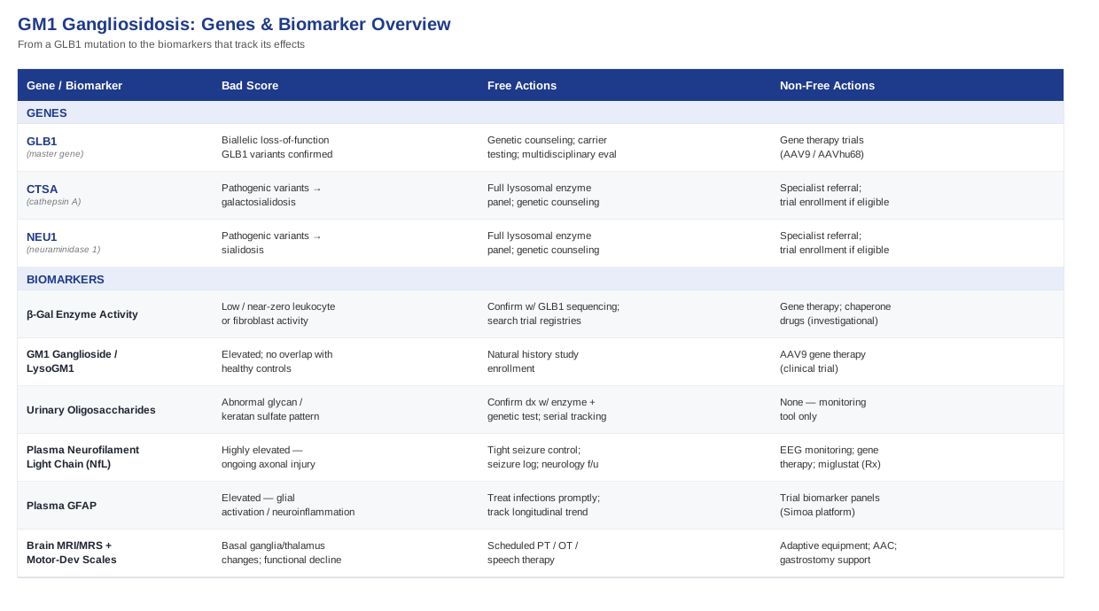
The Six Biomarkers That Matter Most In GM1 Gangliosidosis
GM1 gangliosidosis is diagnosed and monitored through a specific set of biochemical, molecular, and imaging measurements. Unlike biomarkers for common metabolic conditions, most of these are not things you can order casually through a consumer lab. They are run through biochemical genetics laboratories, often in coordination with a metabolic specialist, and increasingly through the natural history studies and clinical trials now open for this disease. Below are the six that carry the most clinical and research weight, in roughly the order a family or clinician encounters them.
1. Acid Beta-Galactosidase Enzyme Activity
This is the foundational biomarker: the direct measurement of the enzyme that GLB1 encodes. Reduced activity of acid beta-galactosidase in leukocytes or cultured fibroblasts is what confirms the biochemical diagnosis, and the degree of residual activity correlates loosely with disease severity - patients with a small amount of residual function tend to fall into the later-onset, milder end of the spectrum, while near-zero activity is typical of the infantile form. This assay is also the basis for dried blood spot newborn screening pilot programs now running in several countries, as described in a tandem mass spectrometry pilot study of six lysosomal storage diseases (NIH-indexed pilot screening study).
How To Measure It
Enzyme activity is measured from a blood draw (leukocytes) or a skin biopsy (cultured fibroblasts), processed at a specialized biochemical genetics laboratory. In the United States, a leukocyte enzyme panel typically costs somewhere between 200 and 600 dollars when billed outside full insurance coverage, though it is frequently covered when ordered by a genetics or metabolic specialist as part of a diagnostic workup. Dried blood spot testing, used in newborn screening pilots, is considerably cheaper per sample but is not yet part of routine standard-of-care screening in most regions.If Activity Is Low: The Approach Without Supplements
No supplement, diet, or lifestyle change restores acid beta-galactosidase activity - this is a structural enzyme deficiency caused by a misfolded or non-functional protein, not a nutrient deficiency or a modifiable metabolic state. The non-supplement plan is entirely about accurate characterization and next steps: confirmatory GLB1 sequencing to identify the specific variants, genetic counseling for the family (this is autosomal recessive, so both parents are typically carriers), testing of siblings where relevant, and connecting with a center that runs natural history studies or clinical trials, such as those tracked through the CureGM1 foundation's trial registry and clinicaltrials.gov. This costs no money beyond the clinical visits themselves and carries no side effects, but it does require time and persistence, since the number of centers with deep experience in this disease is small.If Activity Is Low: What Equipment Or Medical Therapies Can Help
There is no supplement or over-the-counter product that meaningfully raises enzyme activity in a person with two loss-of-function GLB1 variants. The only interventions that have shown any biochemical effect on enzyme function are investigational: gene transfer therapies that deliver a working copy of GLB1 (discussed below) and pharmacological chaperones still in preclinical development, such as N-octyl-4-epi-beta-valienamine, which has enhanced mutant enzyme activity in animal models but has not yet been established in human trials (preclinical chaperone research). These are not accessible outside of research studies, have real risks (including immune reactions to viral vectors), and should only ever be pursued through a formal trial with informed consent, not purchased independently.2. Plasma And CSF GM1 Ganglioside And LysoGM1
If enzyme activity tells you the machinery is broken, GM1 ganglioside and its related lysosphingolipid, lysoGM1, tell you how much material is actually piling up as a result. These substrate-accumulation markers are detectable in blood and cerebrospinal fluid and have been shown to be elevated in GM1 patients with essentially no overlap with unaffected controls, making them a strong pharmacodynamic biomarker - meaning they are a good way to check whether an experimental treatment is doing what it is supposed to do at the biochemical level (landscape of pharmacodynamic biomarkers in GM1 and GM2 gangliosidosis).
How To Measure It
This requires mass spectrometry-based lipidomic analysis, run almost exclusively at specialized research or reference laboratories rather than standard commercial labs. It is not yet a routine clinical order in most health systems; it is most commonly obtained through participation in a natural history study or clinical trial, where the cost is typically absorbed by the research protocol rather than billed to the family. Where available outside of research, costs can run from several hundred to over a thousand dollars per sample given the specialized methodology involved.If Levels Are Elevated: The Approach Without Supplements
There is no dietary or supplement strategy that reduces ganglioside accumulation, because the substrate builds up as a direct consequence of the missing enzyme activity, not from dietary intake of the lipid itself. The realistic non-supplement step is enrollment in a monitored research protocol where these levels are tracked over time as part of natural history data, which helps characterize the individual's disease trajectory and clarifies eligibility for interventional trials.If Levels Are Elevated: What Equipment Or Medical Therapies Can Help
The only interventions currently showing a measurable effect on ganglioside burden are gene therapies delivered under clinical trial protocols. A phase 1/2 trial of an intravenous AAV9 vector encoding beta-galactosidase in Type II GM1 gangliosidosis reported improvements in biochemical markers alongside stabilized or reduced rates of developmental deterioration in early participants (AAV9 gene therapy phase 1/2 trial). These are single-dose, one-time interventions administered in a hospital setting under a research protocol, not repeatable "cycles," and they carry real risks including infusion reactions and immune responses to the viral vector, which is why they are only available through formal trial enrollment with close medical supervision.3. Urinary Oligosaccharides And Glycan Biomarkers
Because acid beta-galactosidase is also needed to break down certain glycans and the glycosaminoglycan keratan sulfate, GM1 gangliosidosis produces a distinctive pattern of abnormal oligosaccharides in urine. This is one of the more accessible, non-invasive biomarkers, and it has become a useful part of both diagnostic confirmation and newborn screening biomarker panels being piloted for lysosomal storage diseases.
How To Measure It
A spot urine sample is analyzed by tandem mass spectrometry or thin-layer chromatography at a metabolic or biochemical genetics laboratory. This is generally one of the more affordable specialized tests in this group, often in the range of 100 to 300 dollars, and is non-invasive, requiring no blood draw or biopsy.If Results Are Abnormal: The Approach Without Supplements
As with the other substrate-level markers, no supplement or dietary change normalizes this pattern, since it reflects impaired glycan degradation rather than something correctable through intake changes. The useful non-supplement step is using an abnormal result to prompt (or confirm) definitive enzyme and genetic testing, and using serial measurements, where a center offers them, as one data point in tracking disease activity alongside the other biomarkers here.If Results Are Abnormal: What Equipment Or Medical Therapies Can Help
No equipment or device changes this biomarker directly. Its main practical value is as a monitoring and diagnostic tool rather than a treatment target - it is a way of watching the disease, not a lever for changing it.4. Plasma Neurofilament Light Chain (NfL)
Neurofilament light chain is a structural protein released into blood and spinal fluid when axons are damaged, and it has emerged as one of the more promising fluid biomarkers across many neurodegenerative diseases, GM1 gangliosidosis included. Studies have found NfL highly elevated in GM1 and GM2 gangliosidosis patients with no overlap with healthy controls, making it a sensitive - if not anatomically specific - signal of ongoing neuronal injury (neuronopathic biomarkers and clinical relevance in GM1).
How To Measure It
NfL is measured from a plasma sample using ultra-sensitive single-molecule array (Simoa) technology, increasingly available through specialty reference laboratories and academic research biobanks rather than routine hospital labs. Costs typically fall in the 200 to 400 dollar range per sample where offered outside of a research study, though it is frequently bundled into natural history or trial protocols at no direct cost to the family.If NfL Is Elevated: The Approach Without Supplements
Elevated NfL reflects real, ongoing axonal injury; no supplement lowers it in this context. What genuinely matters without any pill or product is aggressive, prompt management of anything that adds additional neuronal stress - most importantly, tight seizure control, since uncontrolled seizures are a well-recognized driver of additional neurological injury in progressive disorders. This means working closely with a pediatric or adult neurologist, keeping a seizure log, and not delaying escalation of anti-seizure medication when breakthrough seizures occur.If NfL Is Elevated: What Equipment Or Medical Therapies Can Help
The relevant "equipment" here is diagnostic and protective rather than curative: ambulatory or continuous EEG monitoring to catch subclinical seizures, and seizure-alert devices for home use where clinically appropriate. On the therapeutic side, the interventions that have shown any capacity to alter the broader trajectory that NfL reflects are the gene therapies and, historically, substrate reduction therapy with miglustat, which in a multicenter study of pediatric Type II GM1 patients was associated with delayed neurological involvement compared to untreated natural history (miglustat multicenter experience in GM1 Type II). Miglustat is an oral prescription medication, not a supplement, and its recognized side effects include diarrhea, weight loss, and tremor - it requires ongoing monitoring by a metabolic specialist and is not something to self-administer or cycle on and off without medical supervision.5. Plasma GFAP (Glial Fibrillary Acidic Protein)
GFAP is released by activated astrocytes, the brain's glial support cells, and rises in response to neuroinflammation and glial activation. Alongside NfL, it is being evaluated as a complementary biomarker that captures a different piece of the neurodegenerative process - inflammation and glial response rather than axonal loss itself - and the two together are increasingly used as a paired readout in gangliosidosis natural history and trial work.
How To Measure It
GFAP is measured from the same plasma sample and Simoa platform used for NfL, and the two are often run together as a panel. Cost and availability track closely with NfL testing, typically through the same specialty or research laboratories.If GFAP Is Elevated: The Approach Without Supplements
There is no supplement with credible evidence for lowering GFAP in this disease context. The non-supplement approach is making sure intercurrent infections, which can transiently worsen neuroinflammatory markers, are treated promptly, and keeping GFAP as part of a longitudinal trend rather than reacting to any single value, since some fluctuation between draws is expected.If GFAP Is Elevated: What Equipment Or Medical Therapies Can Help
As with NfL, no device or over-the-counter product directly modifies GFAP. Its main current role is as a trial-readiness and monitoring tool, helping researchers and clinicians understand whether an investigational therapy is calming glial activation over time - it is not, at this stage, an independent treatment target.6. Brain MRI/MRS And Standardized Motor-Developmental Scales
Structural and spectroscopic brain imaging rounds out the biomarker picture by showing the anatomical and metabolic consequences of substrate accumulation - signal changes in the basal ganglia and thalamus, white matter changes, and, on magnetic resonance spectroscopy, altered metabolite ratios that track with disease stage. Imaging findings have been described in detail in infantile-onset disease case series (imaging manifestations in infantile GM1 gangliosidosis), and a ten-year prospective study of Type II patients paired imaging with standardized motor and developmental scales to map disease trajectory in detail (ten-year prospective study of GM1 Type II).
How To Measure It
This is a standard MRI, often with spectroscopy sequences added, performed at a hospital or imaging center with pediatric or neurogenetic experience where possible. Cost varies widely by country and insurance status, roughly 500 to 3,000 dollars when self-paid, though it is usually considered medically necessary and covered once ordered by a treating specialist. GeneReviews' clinical surveillance recommendations for GM1 gangliosidosis outline suggested monitoring intervals for imaging and other systems (GeneReviews GLB1-related disorders overview).If Imaging Or Scores Show Progression: The Approach Without Supplements
No supplement reverses the structural or metabolic changes seen on imaging. What has genuine evidence and no cost barrier beyond access to services is consistent, scheduled physical, occupational, and speech therapy, generally recommended two to five times per week depending on functional status, which supports functional plateaus and quality of life even when imaging continues to show disease progression. This is not a treatment for the underlying biomarker, but it is the single most evidence-backed, zero-side-effect intervention families can implement immediately.If Imaging Or Scores Show Progression: What Equipment Or Medical Therapies Can Help
Adaptive equipment carries real, practical value even though it does not change imaging findings: standing frames and orthotics to manage contractures, wheelchairs or gait trainers matched to functional level, augmentative and alternative communication devices where speech is affected, and feeding equipment or gastrostomy support where swallowing safety becomes a concern. These should be introduced and adjusted through a multidisciplinary rehabilitation team, reassessed roughly every three to six months as function changes, and are not associated with medication-type side effects, though fitting and adjustment take clinical expertise to get right.Beyond GLB1: The Genetic Context Worth Understanding
The biomarkers above only make sense in light of the genetics driving them, so it is worth spending time on the gene itself and its immediate neighbors in the pathway - not because there is a supplement stack that fixes any of this, but because understanding the genetic architecture changes how test results should be interpreted and what questions to ask a genetics team.
GLB1 - The Master Gene
GM1 gangliosidosis is caused by biallelic (two-copy) pathogenic variants in GLB1, located on chromosome 3, which encodes acid beta-galactosidase. More than 200 disease-causing variants have been identified, clustering particularly in certain exons, and the disease is inherited in an autosomal recessive pattern - meaning both parents of an affected child are typically unaffected carriers, each with one normal and one altered copy. The GeneReviews chapter on GLB1-related disorders remains the most thorough, regularly updated clinical reference on this gene and its associated conditions (GLB1-related disorders, GeneReviews).
If Genetic Testing Confirms Two GLB1 Pathogenic Variants: The Plan Without Supplements
A confirmed biallelic GLB1 diagnosis calls for a specific, non-supplement sequence of steps rather than a treatment decision made in isolation. This includes referral to a biochemical genetics or metabolic disease center experienced with lysosomal storage disorders, formal genetic counseling for the parents (including reproductive options for future pregnancies, such as carrier screening or prenatal testing), enrollment in a natural history study if one is accessible, and a baseline multidisciplinary evaluation covering neurology, cardiology, ophthalmology, and orthopedics, since GM1 gangliosidosis affects multiple organ systems beyond the brain. None of this carries a monetary cost beyond the clinical visits themselves, and there are no side effects to worry about - the "risk" here is delay, not action.If Genetic Testing Confirms Two GLB1 Pathogenic Variants: What Medical-Grade Options Exist
To be direct: there is no supplement, herbal product, or nutraceutical with credible evidence of correcting a GLB1 loss-of-function genotype. The legitimate medical-grade options are all investigational and trial-based - the AAV9 and AAVhu68 gene therapy programs referenced earlier, and historically, substrate reduction therapy with miglustat in juvenile and adult-onset patients, which one study associated with possible reversal of some disease progression markers in that specific, later-onset population (miglustat in juvenile/adult GM1 gangliosidosis). Notably, an earlier miglustat trial in infantile-onset patients was far less encouraging, underscoring that these interventions are not interchangeable across the disease spectrum and must be discussed with a specialist familiar with a given patient's specific subtype.Pseudodeficiency Alleles - A Genetics Trap Worth Knowing
Not every GLB1 variant that lowers measured enzyme activity causes disease. Some common variants reduce activity on laboratory assays without causing clinical GM1 gangliosidosis - so-called pseudodeficiency alleles. This matters enormously in newborn screening and incidental testing contexts, where a low enzyme reading alone, without confirmatory sequencing and correlation with clinical findings, can cause unnecessary alarm. Any low enzyme result in an asymptomatic person should be interpreted by a genetics specialist alongside full GLB1 sequencing before it is treated as diagnostic.CTSA And NEU1 - The Enzyme's Partners And Lookalike Diseases
Acid beta-galactosidase does not work alone; in cells, it exists in a multi-enzyme complex with protective protein/cathepsin A (encoded by CTSA) and neuraminidase 1 (encoded by NEU1). Defects in CTSA cause a related but distinct disorder, galactosialidosis, while NEU1 defects cause sialidosis - both of which can resemble GM1 gangliosidosis clinically and biochemically enough to matter during diagnostic workup. Knowing this complex helps explain why a full lysosomal enzyme panel, not a single isolated test, is usually the right diagnostic approach when a storage disorder is suspected.Genotype-Phenotype Correlation And Carrier Testing
Because residual enzyme activity strongly (though imperfectly) predicts phenotype, the specific pair of GLB1 variants identified in a patient carries prognostic information: two null (fully inactivating) variants generally predict the severe infantile form, while at least one variant retaining partial function is more often associated with the later-onset, more slowly progressive juvenile or adult forms. This is why full sequencing, not just an enzyme number, is worth pursuing - it shapes realistic expectations and trial eligibility. For families, carrier testing of siblings and extended family members, combined with genetic counseling, remains the most concrete, low-cost step available for future family planning.Ten Research Insights Reshaping How We Think About GM1 Gangliosidosis
The research landscape for GM1 gangliosidosis has shifted meaningfully in the past few years, moving from a field with almost no biomarkers or trials to one with active gene therapy programs and a growing biomarker toolkit. Below are ten specific findings from that recent literature worth understanding, drawn from a comprehensive review of therapeutic developments in the field (therapeutic developments for neurodegenerative GM1 gangliosidosis) and the studies referenced throughout this article.
1. GM1 Gangliosidosis Is A Spectrum, Not A Single Disease
Clinicians increasingly emphasize that infantile, juvenile, and adult/chronic GM1 gangliosidosis are points on one continuum defined by residual enzyme activity, not separate diseases - which changes how trial eligibility and treatment expectations should be set for any individual patient.2. Biomarkers Are Finally Catching Up To The Underlying Biology
Until recently, disease monitoring relied almost entirely on clinical exams and imaging. The emergence of validated fluid biomarkers - NfL, GFAP, lysoGM1 - gives researchers and clinicians objective, repeatable numbers to track instead of relying solely on subjective developmental milestones.3. Axonal Injury Markers Reveal Damage Before It Is Fully Apparent Clinically
Elevated NfL in patients who still appear relatively stable clinically suggests neuronal injury can be detectable biochemically ahead of obvious functional decline, which is exactly the kind of early signal trial designers need to intervene sooner.4. A Single Gene Therapy Infusion May Reshape The Disease Trajectory
Early results from a Phase 1/2 trial of intravenous AAV9 gene therapy in Type II GM1 gangliosidosis reported stabilized or reduced rates of developmental deterioration in treated participants - a meaningfully different picture than the steady decline typically seen in natural history data (AAV9 gene therapy trial results).5. Delivery Route Is Turning Out To Be As Important As The Vector Itself
Parallel programs are testing intravenous delivery (AAV9) against intrathecal, cisterna magna-injected delivery (AAVhu68/PBGM01) - a direct comparison of how getting a gene therapy past the blood-brain barrier efficiently may matter as much as the therapeutic cargo it carries.6. Substrate Reduction Therapy Taught The Field A Hard Lesson About Timing
A U.S. trial of miglustat in infantile-onset patients was discontinued early after a high proportion of participants died before completion, while separate studies in juvenile and adult-onset patients using the same drug reported gradual neurological improvement - a stark illustration that the same molecule can help or fail to help depending entirely on how advanced and how fast-moving the underlying disease already is.7. Juvenile And Adult-Onset Patients Are Not Just "Milder Infantile" Cases
Because they retain more residual enzyme activity, later-onset patients appear to respond to certain interventions, like miglustat, in ways that infantile patients with near-zero enzyme activity simply do not - reinforcing that treatment strategy has to be matched to genotype and residual function, not applied uniformly.8. Newborn Screening Is Arriving Ahead Of Approved Treatment
Pilot newborn screening programs using dried blood spots can now identify GM1 gangliosidosis at birth, before symptoms appear - but with no approved disease-modifying therapy yet available outside of trials, this raises real, actively debated questions about what a family does with that information beyond early trial enrollment and closer surveillance (newborn screening pilot study).9. Imaging Is Becoming A Disease-Stage Tool, Not Just A Diagnostic One
Detailed case series correlating specific MRI findings with clinical stage suggest brain imaging is moving from a one-time diagnostic confirmation tool toward a longitudinal way of tracking how the disease is progressing over time.10. The Next Phase Will Likely Be Combination Strategies, Not One Silver Bullet
Given the multi-organ nature of the disease and the partial, subtype-specific success of individual approaches so far, researchers increasingly frame the future as combining gene therapy for the underlying enzyme deficiency with biomarker-guided monitoring and aggressive supportive care - rather than expecting any single therapy to address every aspect of the condition.Supportive Therapies Worth Considering Alongside Medical Care
None of the following approaches treat GM1 gangliosidosis itself, and it is worth being upfront that condition-specific clinical trial evidence for these modalities in GM1 gangliosidosis does not exist - the disease is too rare for dedicated trials of supportive therapies to have been run. What follows is evidence extrapolated from closely related pediatric neurodisability and chronic neurological populations, offered as reasonable, low-risk additions to a care plan rather than treatments for the underlying disease.
Massage Therapy
Massage therapy is a hands-on technique aimed at reducing muscle tension and spasticity, and it is relevant here because spasticity and abnormal muscle tone are common features of GM1 gangliosidosis, particularly in the infantile and juvenile forms. A randomized controlled trial in children with spastic cerebral palsy found that traditional massage, added to routine physical therapy, produced a significant reduction in spasticity compared with physical therapy alone (massage RCT in pediatric spastic cerebral palsy). In practice, this looks like a trained pediatric massage therapist working alongside, not instead of, the child's physical therapist, typically two to three sessions per week, with no significant side effects beyond occasional temporary soreness - though any new bodywork should be cleared with the treating neurologist first, especially where bone fragility or contractures are present.Music Therapy
Music-based interventions delivered by a trained music therapist are relevant for GM1 gangliosidosis because they have shown benefits for motor function, communication, and engagement in children with a range of neurological deficits, and separately, strong evidence in pediatric palliative and supportive care more broadly for reducing anxiety and improving quality of life (music therapy in pediatric neurorehabilitation). A realistic approach is weekly or biweekly sessions integrated into an existing therapy schedule, focused on functional goals like communication attempts or motor engagement rather than passive listening; there are no meaningful side effects, making this one of the lowest-risk additions on this list, though it works best when the therapist coordinates directly with the child's broader care team.Progressive Muscle Relaxation And Relaxation Training
For juvenile and adult-onset patients who remain cognitively able to participate actively, progressive muscle relaxation is a structured technique of tensing and releasing muscle groups to reduce physiological and psychological tension. A systematic review found consistent evidence for its effectiveness in reducing anxiety and depression across various patient populations coping with chronic illness (systematic review of progressive muscle relaxation for anxiety and depression), which is directly relevant given the real psychological burden of living with a progressive neurological condition. This can be practiced independently once taught, generally in 15 to 20 minute sessions three to five times a week, guided initially by a psychologist or trained therapist; it carries essentially no physical side effects, though it is not a substitute for professional mental health support when anxiety or depression become significant.Moving Forward With Better Information
GM1 gangliosidosis is driven by a single, well-characterized gene, GLB1, and its effects can now be tracked through a genuinely useful set of biomarkers - enzyme activity, GM1 ganglioside and lysoGM1, urinary oligosaccharides, neurofilament light chain, GFAP, and brain imaging - each of which reveals a different piece of how active the disease is and how it is likely to behave. None of these biomarkers can be corrected with a supplement, and being honest about that matters more than offering false reassurance: the real, evidence-backed options right now are genetic counseling, careful multidisciplinary supportive care, adaptive equipment matched to function, and, where eligible, enrollment in one of the active gene therapy or research programs described above.
The most useful next step is rarely dramatic. It is usually connecting with a metabolic genetics center experienced in lysosomal storage disorders, getting a full GLB1 sequencing result and residual-activity picture rather than relying on a single enzyme number, and asking directly whether a natural history study or trial is currently open for your specific subtype. Track the biomarkers that apply to your situation, keep a record of functional changes over time, and bring both to every specialist visit - that combination of accurate information and consistent follow-up is, at present, the most concrete way to make good decisions about this disease.