This article contains AI generated content.
Gracilis Tendinopathy: 6 Genes and 6 Biomarkers to Track
Introduction
Groin pain that lingers along the inner thigh, flares with cutting movements or hip abduction, and never quite resolves with the standard "rest and stretch" advice is a familiar and frustrating pattern. Gracilis tendinopathy sits in an awkward diagnostic corner: it overlaps with adductor-related groin pain, it rarely shows up cleanly on a single scan, and most general guidance for it is really guidance for tendinopathy in general, not for the specific tissue, biomechanics, or biology involved at the pes anserinus or pubic insertion.
Generic advice - stretch more, ice it, take some anti-inflammatories, give it time - is not wrong, but it is incomplete. It treats every tendon and every person as interchangeable, when in reality collagen structure, remodeling capacity, hormonal environment, and metabolic health all vary from one person to the next, and some of that variation is written into your genes and reflected in your bloodwork. A tendon that struggles to remodel efficiently needs a different plan than one that is simply overloaded.
This article goes past the generic script. It looks at the specific collagen and matrix-remodeling genes that human studies have repeatedly linked to tendon injury risk, the epigenetic layer that can shift how those genes behave, and the blood and imaging markers that let you track tendon-relevant biology in real time rather than guess at it. None of this is deterministic or a substitute for a proper diagnosis, but understanding your own biological starting point tends to produce better, more targeted decisions than following advice built for an average person who does not exist.
The goal is grounded, practical hope: not a promise that any gene test or blood panel will "fix" a tendon, but a clearer map of where your risk actually comes from, so that training load, recovery habits, and any supplement or equipment choices can be matched to your biology instead of applied blindly.
Summary
This article walks through six collagen and matrix-remodeling genes - COL5A1, COL1A1, TNC, MMP3, GDF5, and ESR1 - that human genetic-association studies have tied to tendon injury susceptibility, plus the epigenetic switch that can turn one of them up or down regardless of which version you inherited. For each gene, you'll find a practical two-track plan: what to change through training and movement alone, and what a supplement or equipment-based add-on could look like, including realistic dosing, cycling, and side effects. From there, the article turns to six bloodwork and imaging markers worth tracking alongside your genetics, a research-backed protocol from tendon scientist Keith Baar's work that challenges the "just rest it" instinct, and a short review of the complementary approaches - laser therapy, deep friction massage, and biofeedback-guided movement retraining - that actually have condition-relevant human evidence behind them.
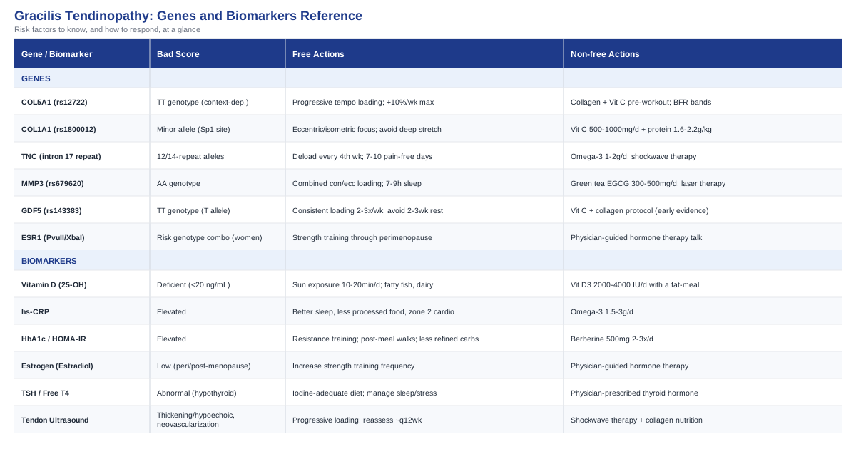
The Genetic and Epigenetic Blueprint Behind Gracilis Tendinopathy
Tendon injury genetics is one of the more mature areas of musculoskeletal genomics, built largely on decades of case-control studies out of South Africa, Australia, and the UK comparing athletes with chronic tendon pathology to injury-free controls. Researchers like Malcolm Collins, Stuart Raleigh, and Alison September have driven much of this work on the Achilles tendon specifically, and because the gracilis tendon shares the same basic collagen biology, the same variants are reasonable candidates for risk in gracilis and adductor-region tendinopathy, even though it has not been studied in isolation as often as the Achilles. If you want a general entry point into how genetics gets used in personalized health decisions, geneticist Ali Torkamani's work on polygenic risk and biohacker Gary Brecka's popularization of actionable gene panels are both reasonable starting points for understanding how a raw genotype gets turned into a practical plan - though the tendon-specific variants below come from sports-medicine genetics research, not from either of their specific panels.
Before getting into individual genes, it's worth being honest about what this evidence actually shows. These are association studies: they show a gene variant is more common in people with tendon pathology than in matched controls, not that the variant causes the injury on its own. Effect sizes are modest, results do not always replicate across populations, and a "risk" genotype simply shifts probability - it does not guarantee a problem, and a "protective" genotype does not guarantee safety. Used correctly, this information adjusts how cautious you are with load progression, not whether you can play sport at all.
COL5A1 - the collagen V fibril regulator
COL5A1 encodes one of the chains of type V collagen, a minor but critical collagen that sits at the core of type I collagen fibrils and controls how thick those fibrils grow. Thinner, more numerous fibrils generally make for a more compliant, energy-absorbing tendon, while thicker fibrils make for a stiffer one. The most studied variant is the Sp1-binding-site polymorphism rs12722 (sometimes reported via the related BstUI RFLP). Human evidence here is genuinely mixed rather than one-directional: earlier South African and Australian case-control work linked the TT genotype to increased Achilles tendon pathology risk, while a later meta-analysis pooling Caucasian cohorts found the opposite pattern, with rs12722 variants associated with a reduced injury risk overall (COL5A1 meta-analysis, Caucasian cohorts). That inconsistency is a useful lesson: population background, sport type, and tendon site all modify how a single variant behaves, so treat any single COL5A1 result as a data point, not a verdict.
If your COL5A1 result flags increased risk: the plan without supplements
Since COL5A1 affects fibril architecture rather than something you can directly correct, the highest-leverage lever is progressive tendon loading that trains stiffness gradually instead of forcing it. That means a structured strength program for the adductors and hip flexors with slow, controlled tempo (3-4 seconds down, 2-3 seconds up), performed 3 times per week with at least 48 hours between sessions for the same muscle group, and a load progression that increases no faster than about 10% per week. Avoid abrupt spikes in sprint volume, direction-change drills, or kicking repetitions, since these are exactly the loading patterns that stress an under-adapted fibril structure. There are no direct side effects to this approach beyond the normal risk of doing too much too soon, which is precisely what the gradual progression is designed to prevent.
If your COL5A1 result flags increased risk: the plan with supplements or equipment
Collagen-supportive nutrition timed around training is the most evidence-aligned add-on. Fifteen grams of hydrolyzed collagen or gelatin combined with roughly 50 mg of vitamin C, taken about 60 minutes before the loading session, is the protocol used in controlled human research on collagen synthesis (more detail on this study appears later in this article). A practical cycle is 3-5 sessions per week for 8-12 weeks, followed by a 2-4 week break to reassess symptoms and reintroduce if useful; there is no strong evidence that continuous year-round use is necessary or superior to cycling. Side effects are minimal and mostly gastrointestinal (mild bloating) if taken on an empty stomach - taking it with a small amount of water and away from a heavy meal reduces this. Blood flow restriction (BFR) bands during light-load rehab sets are a reasonable equipment-based option to increase metabolic stimulus without high mechanical stress; they should be used under professional guidance, 2-3 times per week, and are not appropriate if you have vascular disease, uncontrolled hypertension, or a history of blood clots.
COL1A1 - the primary structural collagen gene
COL1A1 encodes the major chain of type I collagen, the dominant structural protein in tendon. Its Sp1-binding-site polymorphism (rs1800012) alters the ratio of the two collagen I chains produced, which in turn affects tendon and ligament stiffness and has been linked in multiple meta-analyses to musculoskeletal soft-tissue injury risk, including tendon and ligament injuries specifically (COL1A1 polymorphism meta-analysis with trial sequential analysis). The trial sequential analysis used in this meta-analysis is worth noting because it specifically tests whether the pooled evidence has reached a stable conclusion rather than being a chance finding from limited data - a level of rigor not every genetic association study reaches.
If your COL1A1 result flags increased risk: the plan without supplements
Because the minor allele is associated with a less favorable collagen I:III balance and looser connective tissue, prioritize eccentric and isometric strength work over high-velocity, high-amplitude stretching. Long static stretching held near end-range can further load already lax tissue without building capacity. A practical structure is twice-weekly heavy, slow resistance training for hip adductors and surrounding hip musculature, holding each isometric contraction for 30-45 seconds across 4-5 sets, alongside general activity modification that limits sudden deceleration tasks until strength gains are demonstrated over 6-8 weeks.
If your COL1A1 result flags increased risk: the plan with supplements or equipment
Vitamin C (500-1000 mg daily with meals) and adequate dietary protein (around 1.6-2.2 g/kg bodyweight daily) support the enzymatic steps of collagen crosslinking that COL1A1 variants may make comparatively less efficient. This is a low-risk, ongoing approach rather than something to cycle, though very high vitamin C doses (above 2 g/day) can cause diarrhea and are unnecessary here. An isometric hold device or simple resistance bands used for daily 5-minute isometric "flare-up management" sessions are a reasonable low-cost equipment option, since isometric loading has documented short-term analgesic effects in tendinopathy (detailed under the biomarker and podcast sections below).
TNC - the tenascin-C repair signal
Tenascin-C is an extracellular matrix glycoprotein that gets upregulated specifically during tendon healing and mechanical adaptation - it is, in effect, a repair-signaling protein. The TNC gene carries a guanine-thymine (GT) dinucleotide repeat polymorphism in intron 17, and the original South African case-control study found that people carrying the 12- or 14-repeat alleles had roughly six times the odds of symptomatic Achilles tendon injury compared with other repeat lengths (tenascin-C GT repeat polymorphism and Achilles tendon injury). More recent work has also examined TNC alongside MMP3 as a combined "tendon regeneration failure phenotype," since the two genes interact functionally in matrix remodeling (tenascin-C/MMP3 phenotype and tendinopathy risk in high-performance athletes).
If your TNC result flags increased risk: the plan without supplements
Since TNC's role is specifically about the repair response following microdamage, the priority is giving repair cycles enough uninterrupted time to complete rather than layering new stress on top of an incomplete one. Practically, this means building in a deload week roughly every 4th training week, tracking morning tendon stiffness or pain-on-first-steps as a simple daily signal, and delaying a return to full sprint/change-of-direction volume until symptoms have been absent for at least 7-10 consecutive days, not just improved.
If your TNC result flags increased risk: the plan with supplements or equipment
There is no supplement that directly targets tenascin-C expression, but omega-3 fatty acids (1-2 g combined EPA/DHA daily) have modest evidence for supporting a favorable inflammatory-resolution environment during tissue remodeling, and can be taken continuously with food to minimize the main side effect, which is a fishy aftertaste or mild reflux. A shockwave therapy device, administered by a clinician once weekly for 3-6 sessions, is a reasonable equipment-based option specifically to stimulate a fresh, organized repair response in tendon tissue that seems to be remodeling slowly; mild soreness and transient pain flare for 24-48 hours after each session is common and expected, not a sign of harm.
MMP3 - the matrix remodeling enzyme
MMP3 (stromelysin-1) is an enzyme that breaks down and remodels several extracellular matrix components, including proteoglycans and procollagen, as part of normal tendon turnover. The rs679620 variant, where the AA genotype has been consistently overrepresented in tendinopathy cases relative to controls, appears to shift the balance of matrix breakdown versus rebuilding in a less favorable direction (MMP3/tenascin-C genotype and tendinopathy risk). Two other MMP3 variants, rs591058 and rs650108, show similar directional associations across independent cohorts, which strengthens confidence in MMP3 as a genuinely relevant gene rather than a one-study finding.
If your MMP3 result flags increased risk: the plan without supplements
Because excess matrix breakdown relative to synthesis is the underlying concern, avoid training approaches that maximize eccentric muscle damage (like very heavy eccentric-only protocols done too frequently) without adequate recovery spacing. A more moderate approach - combined concentric-eccentric loading 2-3 times weekly rather than daily eccentric-only work - tends to favor a better remodeling balance, alongside ensuring 7-9 hours of sleep, since growth hormone pulses tied to deep sleep support collagen synthesis that offsets MMP-driven breakdown.
If your MMP3 result flags increased risk: the plan with supplements or equipment
Green tea catechins (as a standardized extract, roughly 300-500 mg EGCG daily) have preliminary evidence for modestly inhibiting excess MMP activity, though this evidence is largely from non-tendon tissue and should be considered exploratory rather than proven for this specific use; cycle 8 weeks on, 4 weeks off, and avoid on an empty stomach, since high-dose green tea extract has been linked to rare cases of liver strain in sensitive individuals. Low-level laser therapy (discussed in more detail later in this article) is a reasonable equipment option, 3 times weekly for 4-6 weeks, aimed at supporting a more balanced remodeling environment during active tendon changes.
GDF5 - the tendon-maintenance growth factor, and its epigenetic switch
Growth differentiation factor 5 (GDF5) helps regulate tendon, ligament, and joint development and ongoing maintenance throughout life. The functional variant rs143383 sits in the gene's 5' untranslated region, and the T allele is associated with reduced GDF5 expression across a range of soft tissues; in an Australian cohort, the TT genotype increased the odds of chronic Achilles tendon pathology by roughly 2.24 times compared with other genotypes (GDF5 and Achilles tendon pathology, genetic association study). What makes GDF5 particularly interesting from an epigenetic standpoint is that this same rs143383 site is also a CpG methylation site: methylation there changes how strongly the Sp1 and Sp3 transcription factors bind and therefore how much GDF5 gets expressed from a given allele, independent of which DNA sequence you inherited (CpG methylation regulates allelic expression of GDF5). In plain terms: your GDF5 genotype sets a baseline tendency, but methylation state - influenced by age, mechanical loading history, and possibly diet - can shift expression further, which is a genuinely actionable idea rather than pure determinism.
If your GDF5 result flags increased risk: the plan without supplements
Consistent, moderate mechanical loading appears to be the main lever known to influence tendon-relevant gene expression over time, so a structured, non-negotiable 2-3 session per week loading routine sustained over months (not just during flare-ups) is the realistic non-supplement strategy here. Avoid long unloaded periods (more than 2-3 weeks of complete inactivity), since detraining appears to reverse adaptive gains relatively quickly in tendon tissue specifically.
If your GDF5 result flags increased risk: the plan with supplements or equipment
There is no supplement proven to directly raise GDF5 expression in humans, so honesty matters here: this is an area of early, mechanistic-level science rather than an established intervention. What can reasonably be layered on top of consistent loading is the same vitamin C-supported collagen synthesis protocol described under COL5A1, since it supports the downstream collagen production that GDF5 partly regulates, used 3-4 times weekly around training for 8-12 week blocks with the same cycling and mild-GI-upset caveat as before.
ESR1 - the estrogen receptor gene, most relevant for women
Estrogen receptor alpha, encoded by ESR1, plays a documented role in tendon collagen turnover: higher circulating estrogen is generally associated with greater collagen synthesis, while low-estrogen states are linked to reduced synthesis and increased tendon and muscle stiffness (effect of female sex hormone supplementation on tendon in pre- and postmenopausal women, systematic review). Two functional ESR1 polymorphisms, PvuII (rs2234693) and XbaI (rs9340799), have been studied specifically in postmenopausal women with posterior tibial tendon dysfunction, where certain genotype combinations were overrepresented in affected women compared with controls (ERα PvuII and XbaI polymorphisms in postmenopausal tendon dysfunction). This gene is most relevant for women, particularly around perimenopause and menopause, when declining estrogen compounds any existing genetic tendency toward lower collagen turnover.
If your ESR1 result flags increased risk: the plan without supplements
For women in or approaching perimenopause with this genetic pattern, front-loading strength training before and through the menopausal transition - rather than waiting for symptoms - is the most defensible non-supplement strategy, since tendon adapts more readily to load when baseline collagen turnover is still relatively higher. Twice-weekly resistance training targeting the hip adductors and surrounding musculature, sustained consistently through the transition rather than started only after pain appears, is the realistic target.
If your ESR1 result flags increased risk: the plan with supplements or equipment
This is a conversation for a physician rather than a self-directed supplement plan, since it intersects with hormone therapy decisions that carry real risks and benefits beyond tendon health alone; menopausal hormone therapy has shown some positive effects on tendon collagen synthesis in research settings, but the decision to use it should weigh cardiovascular, breast, and bone health factors together, not tendon health in isolation. Where hormone therapy is not being pursued, the vitamin C-supported collagen loading protocol described earlier remains a reasonable, low-risk adjunct.
Genetics gives you a probability, not a sentence, and the practical plans above are most useful when layered onto consistent, well-progressed training rather than substituted for it. With that foundation covered, it's worth turning from what your DNA predisposes you to toward what your current bloodwork and imaging can tell you right now.
Blood and Imaging Markers Worth Tracking Alongside Your Genes
Unlike cardiometabolic disease, tendinopathy does not have a dedicated, routinely ordered blood panel the way cholesterol or blood glucose does. But several markers that clinicians like Peter Attia, Thomas Dayspring, and Allan Sniderman commonly emphasize for metabolic and inflammatory health overlap directly with known tendon risk factors, and combining these with basic imaging gives a genuinely useful, trackable picture.
Vitamin D (25-hydroxyvitamin D)
Vitamin D deficiency has been associated with a meaningfully higher incidence of tendinopathy at other sites (biceps tendon data shows roughly 2.5 times higher incidence in deficient patients) and with worse post-surgical tendon healing outcomes, likely through both direct effects on tenocyte biology and secondary hyperparathyroidism (vitamin D deficiency and distal biceps tendon injury, large retrospective analysis).
How to measure it: a standard serum 25-OH vitamin D blood draw, available through most primary care providers or direct-to-consumer lab services, typically costing $30-$80 out of pocket if not covered by insurance.
If the score is bad, the plan without supplements: increase sensible sun exposure (10-20 minutes of midday skin exposure several times weekly, adjusted for skin tone and climate) and prioritize dietary sources like fatty fish and fortified dairy.
If the score is bad, the plan with supplements: vitamin D3, 2,000-4,000 IU daily with a fat-containing meal, rechecked at 8-12 weeks; doses above 4,000 IU/day should only be used under medical supervision with periodic blood monitoring, since sustained high intake can cause hypercalcemia over time.
High-sensitivity C-reactive protein (hs-CRP)
While classic tendinopathy is more degenerative than acutely inflammatory, low-grade systemic inflammation - reflected in chronically elevated hs-CRP - is increasingly linked to poorer tissue remodeling capacity generally, and it's a marker Attia and colleagues routinely track as a general marker of metabolic and inflammatory load.
How to measure it: a standard hs-CRP blood test, widely available, costing roughly $15-$40 standalone or bundled into broader metabolic panels.
If the score is bad, the plan without supplements: address the upstream drivers - improve sleep consistency, reduce ultra-processed food intake, and add 2-3 weekly sessions of zone 2 cardiovascular exercise, which reliably lowers hs-CRP over 8-12 weeks in most people.
If the score is bad, the plan with supplements: omega-3 fatty acids (1.5-3 g combined EPA/DHA daily, with food) have reasonably consistent evidence for lowering hs-CRP; expect mild fishy burps as the main side effect, and note that doses above 3 g/day can mildly increase bleeding risk, which matters if a tendon procedure or surgery is anticipated.
HbA1c and fasting insulin (HOMA-IR)
Insulin resistance and elevated HbA1c are independently associated with tendon pathology - one analysis found roughly a threefold higher risk of lower-limb tendon injury in patients with elevated HbA1c - and the relationship persists even in people without a formal diabetes diagnosis, likely through advanced glycation end-product accumulation stiffening collagen (metabolic syndrome and tendon disease, comprehensive review) (fasting glucose, insulin resistance, and degenerative tendon tear).
How to measure it: HbA1c and fasting insulin are both standard blood draws, together costing roughly $20-$60 without insurance; HOMA-IR is then simply calculated from fasting glucose and insulin values.
If the score is bad, the plan without supplements: this is the marker most responsive to lifestyle - resistance training 2-3 times weekly, a 20-30 minute daily walk after the largest meal, and reducing refined carbohydrate intake typically improve HOMA-IR meaningfully within 8-12 weeks.
If the score is bad, the plan with supplements: berberine (500 mg, 2-3 times daily with meals) has trial evidence comparable to some pharmaceutical options for improving insulin sensitivity; cycle 12 weeks on, 4 weeks off, and be aware it can cause gastrointestinal upset and should not be combined with other glucose-lowering medications without medical supervision due to hypoglycemia risk.
Estrogen (estradiol) and broader sex hormone panel
Given ESR1's role described above, actual circulating estradiol levels matter alongside the genotype itself, particularly for women navigating perimenopause, since low estrogen independently reduces tendon collagen synthesis regardless of genotype.
How to measure it: a serum estradiol test, generally $40-$100 out of pocket, best interpreted alongside menstrual cycle timing or menopausal status with a physician.
If the score is bad, the plan without supplements: this is not something lifestyle changes meaningfully normalize once ovarian estrogen production has declined; the realistic non-supplement approach is compensating through increased strength training frequency, as described in the ESR1 gene section.
If the score is bad, the plan with supplements: hormone therapy is a physician-guided decision, not a self-directed supplement, given its broader risk-benefit profile beyond tendon health.
Thyroid function (TSH and free T4)
Thyroid hormone influences collagen metabolism broadly, and both overt and subclinical hypothyroidism have been associated clinically with delayed tendon healing and stiffness changes, making this a reasonable marker to rule out when tendon symptoms are unusually persistent or widespread.
How to measure it: TSH alone costs roughly $20-$40; a fuller panel with free T4 and free T3 runs $50-$100.
If the score is bad, the plan without supplements: iodine-adequate diet (iodized salt, seafood, dairy) and addressing sleep and stress, which both influence thyroid function, though this will not resolve a true clinical hypothyroid state.
If the score is bad, the plan with supplements: this generally requires physician-prescribed thyroid hormone replacement rather than over-the-counter supplementation; self-supplementing iodine at high doses without a diagnosed deficiency can paradoxically worsen thyroid function and should be avoided.
Diagnostic tendon ultrasound (with or without Doppler)
This is the imaging-based "biomarker" - a direct structural readout of the gracilis tendon itself, showing thickening, hypoechoic (disorganized) regions, or neovascularization that correlates with symptomatic tendinopathy, and it is the most direct way to track whether a rehab plan is actually changing the tissue, not just the pain.
How to measure it: performed by a sports medicine physician or radiologist, typically $150-$400 out of pocket depending on region and whether Doppler is included; a more advanced but less accessible option is quantitative MRI, running $500-$1,500, useful when ultrasound findings are ambiguous.
If the score is bad, the plan without supplements: structured, progressive loading remains the best-supported way to improve tendon structure over 12+ weeks, reassessed by repeat ultrasound rather than symptoms alone, since pain and structure do not always improve in lockstep.
If the score is bad, the plan with supplements or equipment: shockwave therapy and the collagen-supportive nutrition protocol described earlier are the two most defensible add-ons layered onto a loading program when structural findings are more severe or slow to change.
Bloodwork and imaging tell you where things stand today, but one of the more useful things a rehab plan can do is apply findings from tendon-specific research directly to daily training decisions - which is exactly where the next section goes.
What a Leading Tendon Researcher Wants You to Know
Much of the applied science behind the collagen-nutrition and loading protocols mentioned above traces back to the work of Dr. Keith Baar, a physiologist at UC Davis whose lab studies how tendons and ligaments adapt to mechanical and nutritional signals. His research, discussed at length in a lengthy 2025 interview on The Tim Ferriss Show (episode 797), challenges some long-standing default instincts about tendon injury - particularly the reflex to rest and ice a sore tendon - and is worth knowing directly, not just through the supplement protocols it has produced.
1. Tendon tissue heals slowly because it has poor blood supply
Tendon is far less vascularized than muscle, which is the core biological reason tendinopathy recovery is measured in months rather than weeks - there simply is not the blood flow to rapidly deliver repair signals and nutrients the way there is in muscle tissue.2. Mechanical loading is not the enemy of a healing tendon - it is often the treatment
Complete rest allows a tendon to weaken further rather than heal, because tendon cells specifically need mechanical signal to trigger productive collagen synthesis; the "anti-RICE" framing in Baar's work reflects this - appropriate load, not avoidance of it, tends to drive adaptation.3. The dose of loading matters more than the direction (eccentric vs. isometric vs. concentric)
Much of the historical emphasis on eccentric-only protocols (like the Alfredson heel-drop program) reflects that eccentric loading is an easy way to apply high tension, not that eccentric motion itself is uniquely therapeutic - moderate, well-dosed loading in multiple contraction types can produce comparable adaptation.4. Isometric holds can reduce tendon pain within minutes, not weeks
A controlled study in patellar tendinopathy found that a single bout of heavy isometric contraction (5 sets of 45 seconds at 70-80% of maximum effort) produced significant pain relief that was still present 45 minutes later, a mechanism related to changes in cortical inhibition rather than tissue healing itself (effectiveness of isometric exercise in tendinopathy, systematic review and meta-analysis).5. Vitamin C is a required cofactor for collagen crosslinking, not just an antioxidant
Without adequate vitamin C, the enzymes prolyl and lysyl hydroxylase cannot properly modify collagen precursors, meaning a vitamin C shortfall directly limits how well new collagen matures into strong, crosslinked fibrils regardless of how much protein or gelatin is consumed.6. Timing collagen intake around exercise roughly doubles the collagen synthesis response
In a controlled trial, subjects who consumed 15 grams of vitamin C-enriched gelatin one hour before an intermittent exercise bout showed roughly double the blood marker of new collagen production compared with those who did not, and the effect was dose-dependent between 5 g and 15 g (vitamin C-enriched gelatin supplementation before intermittent activity augments collagen synthesis).7. The one-hour pre-exercise window appears specific, not arbitrary
Blood amino acid markers relevant to collagen synthesis (glycine, proline, hydroxyproline) peaked roughly one hour after gelatin ingestion in that same trial, which is the basis for timing supplementation shortly before, rather than after, a loading session.8. A supplement combination has shown benefit specifically in Achilles tendinopathy rehab
A prospective randomized trial found that a nutraceutical combination of collagen peptide, chondroitin sulphate, sodium hyaluronate, and vitamin C performed comparably to, and in some measures better than, oral diclofenac for Achilles tendinopathy symptoms (collagen peptide combination versus oral diclofenac in Achilles tendinopathy, randomized controlled trial).9. Local tendon loading, not systemic exercise, drives local tendon adaptation
General fitness or unrelated muscle group training does not meaningfully strengthen a specific tendon - the loading stimulus has to be applied through and near the tendon in question, which is why targeted adductor and hip-focused loading matters specifically for gracilis tendinopathy rather than general conditioning work.10. Tendon adaptation is a slow, multi-month process that resists shortcuts
Even with optimized loading and nutrition timing, meaningful structural tendon change is generally measured in 3- to 6-month blocks rather than weeks, which is the most consistent and, frankly, least exciting finding across this entire body of research - but also the most important one to set expectations correctly.These findings reinforce a consistent theme across the genetic, biomarker, and research-based sections above: patient, well-dosed mechanical loading paired with supportive nutrition and metabolic health is the throughline, not any single fix. From here, it's worth briefly covering complementary approaches that have genuine condition-relevant evidence, since they can reasonably sit alongside - not instead of - this core approach.
Complementary Approaches Worth Considering
Low-level laser therapy / photobiomodulation
Photobiomodulation uses specific wavelengths of red or near-infrared light aimed at modulating cellular energy production (via mitochondrial cytochrome c oxidase) and local inflammation, and it has been studied specifically in tendinopathy more than most complementary approaches on this list, making it one of the better-matched options for gracilis tendinopathy rehab.
A systematic review and meta-analysis of low-level laser therapy in lateral elbow tendinopathy pooled 13 randomized controlled trials (730 patients) and found a meaningful reduction in pain when recommended dosing parameters were used (low-level laser therapy in lateral elbow tendinopathy, systematic review and meta-analysis), while a broader 2021 review across multiple tendinopathy sites confirmed a positive effect on pain and function, though it also noted that evidence quality remains inconsistent across studies (photobiomodulation for pain and function in tendinopathy, systematic review).
Realistically, this means seeking out a clinician using a laser device with published, tendon-appropriate dosing parameters (wavelength and dose vary considerably by device and matter for results), applied 2-3 times weekly for 4-6 weeks as an adjunct to loading, not a replacement for it - and tempering expectations, since even the positive meta-analyses describe the certainty of evidence as low to moderate.
Massage therapy (deep transverse friction massage)
Deep transverse friction massage, developed by James Cyriax specifically for tendon and ligament tissue, is a targeted manual technique aimed at maintaining tissue mobility and preventing disorganized scar adhesions during tendon healing, which makes it plausible for a chronic, fibrotic gracilis tendon.
It remains one of the most widely used manual techniques among physiotherapists for tendinopathy specifically - survey data shows the large majority of physiotherapists use it, most commonly for degenerative tendinopathy - though a review of its clinical application notes that dosing parameters (session duration, frequency, tendon positioning) vary considerably between practitioners and that high-quality controlled trial evidence remains thinner than its popularity would suggest (Cyriax friction massage, suggestions for improvements in application).
A realistic approach is 2-3 sessions weekly with a physiotherapist trained in the technique, applied directly over the symptomatic gracilis insertion for several minutes per session, used as a short-term adjunct during flare-ups rather than an indefinite standalone treatment, since evidence supports it best as one part of a broader loading-based program.
Biofeedback-guided movement retraining
Surface EMG biofeedback provides real-time visual or auditory feedback on muscle activation, and for gracilis and adductor-region tendinopathy, this is particularly relevant because altered gluteal and hip-stabilizer activation patterns during walking, running, or cutting often place excess compensatory demand on the adductor group and gracilis tendon.
Controlled research on EMG biofeedback for gluteal muscle retraining has shown measurable improvements in activation patterns and gait mechanics (EMG biofeedback training of gluteus maximus and gait parameters), and while this specific evidence base comes from gait rehabilitation rather than gracilis tendinopathy trials directly, the underlying mechanism - correcting compensatory hip mechanics that overload the adductor group - is directly applicable and worth noting as an area with promising but not condition-specific trial evidence.
In practice, this means working with a physiotherapist who has surface EMG equipment, typically over 4-8 sessions, to identify whether gluteal underactivation or altered hip mechanics are contributing to adductor overload, then using a home-based retraining routine reinforced by the same feedback principles, layered on top of - not instead of - the core strength and loading program.
Conclusion
Gracilis tendinopathy rarely has one clean cause, which is exactly why a single generic protocol so often falls short. The genetic variants covered here - COL5A1, COL1A1, TNC, MMP3, GDF5, and ESR1 - shift baseline risk and remodeling capacity without determining outcomes on their own, and the epigenetic layer around GDF5 is a useful reminder that consistent loading and lifestyle habits can influence gene expression beyond the DNA sequence you were born with. Bloodwork covering vitamin D, inflammation, insulin sensitivity, hormones, and thyroid function, paired with periodic tendon ultrasound, turns abstract risk into something you can actually track and act on over time, and the research behind collagen-timing and loading protocols gives a concrete, evidence-informed starting point rather than vague reassurance.
None of this replaces an actual clinical diagnosis or a trained eye watching how you move. The most useful next step is a practical one: get a proper assessment from a sports medicine physician or physiotherapist familiar with adductor and gracilis pathology, ask about the bloodwork markers covered here if your history suggests they're relevant, and start tracking your symptoms and training load consistently enough to know whether your plan is actually working - not just whether it feels like it should be.
Musculoskeletal Women's Health Endocrine & Metabolic