This article contains AI generated content.
· UpdatedHemophagocytic Lymphohistiocytosis — 7 Genes and 7 Biomarkers to Track
Introduction
Few conditions test the limits of medical diagnosis as severely as hemophagocytic lymphohistiocytosis. Unlike most immune disorders, where the core problem is insufficient immune response, HLH represents the opposite failure — an immune system that cannot stop activating. Macrophages and cytotoxic T cells enter a runaway feedback loop, flooding the body with inflammatory cytokines and destroying healthy tissue in the process. The result is multi-organ failure that can be fatal within days if left untreated.
What makes this particularly difficult for patients and families is how long the diagnostic journey often takes. The early symptoms — persistent fever, fatigue, an enlarging spleen, dropping blood counts — are easy to misattribute to infection, hematologic malignancy, or autoimmune flare. Many people receive antibiotics, steroids, or watchful waiting before the correct picture emerges. During that window, the disease progresses. Understanding what to look for, and asking for the right tests, can compress that timeline significantly.
This article takes a precision-first approach. There is no lifestyle adjustment that replaces etoposide and dexamethasone in active HLH, and nothing here will pretend otherwise. What it offers instead is a detailed look at the specific biomarkers that define and track this condition, the genetic mutations underlying the primary inherited form, and evidence-grounded supportive strategies for patients in remission or navigating the longer aftermath of an episode.
Better information leads to better decisions. Knowing the seven biomarkers that signal HLH severity, understanding which gene variants drive primary disease, and having a monitoring framework built around real clinical evidence can help patients and physicians act faster and more precisely. This article walks through biomarker tracking first — the most immediately actionable lens — then covers the genetic architecture of primary HLH, a research-based summary of key insights that challenge conventional immune thinking, and three complementary approaches with meaningful supportive evidence.
Summary
HLH is one of medicine's most dangerous immune emergencies — and also one of the most data-trackable conditions once you know which numbers matter. This article covers 7 clinical biomarkers used to diagnose, monitor, and guide treatment for HLH — including ferritin, sCD25, NK cell activity, fibrinogen, triglycerides, blood count cytopenias, and sCD163 — explaining what each value reveals, how to measure it, and what can realistically be done to influence it. You will also find an in-depth look at 7 key genes responsible for the primary inherited form of HLH, with practical context on what each mutation disrupts and what testing or monitoring it implies. Beyond the biology, the article includes a summary of 10 research-backed insights that challenge how most clinicians and patients think about immune hyperactivation, three complementary approaches with real human evidence, and a clear conclusion with concrete next steps. If you have been given a diagnosis without specific biomarker targets, or a genetic result without context for what it means in practice, this article will fill in the gaps.
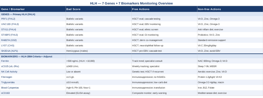
7 Biomarkers That Reveal HLH Severity and Guide Treatment
The eight diagnostic criteria for HLH established in the HLH-2004 guidelines (Henter et al., 2007) include several laboratory values that function as both diagnostic markers and ongoing disease activity monitors. A diagnosis requires five of eight criteria, and several of those are quantitative biomarkers with defined thresholds. Tracking them longitudinally — not just at diagnosis — gives clinicians and patients the clearest signal of whether treatment is working, whether remission is holding, or whether the disease is beginning to rebound.
The seven biomarkers below are organized by clinical importance. Each one is explained in terms of what it measures, what its levels reveal, how to obtain the test and at what cost, and what actionable steps exist when values fall outside target range.
Biomarker 1: Ferritin
Why it matters: Ferritin is the most immediately accessible and diagnostically powerful initial screening marker for HLH. It is released in massive quantities by activated macrophages and by cells undergoing damage during the cytokine storm. The HLH-2004 criterion sets a threshold of ≥500 ng/mL, but in active disease, levels routinely reach 10,000 to 100,000 ng/mL — and sometimes far higher.
Research published in Pediatric Blood & Cancer found that a ferritin level above 10,000 ng/mL had a sensitivity of approximately 90% and a specificity of 96% for HLH among pediatric patients, making it one of the most clinically meaningful single-lab values in the field — Allen et al., 2008. That threshold has since become a widely used clinical reference point even though HLH can occur with lower values, particularly in secondary disease caught early.
How to measure it: Serum ferritin is a standard laboratory test available at virtually any hospital or diagnostic lab. Cost ranges from $20 to $60 without insurance in the US. It can be ordered as part of an iron panel or as a standalone test. During active treatment, repeated measurements every 48 to 72 hours are standard practice. In remission monitoring, monthly checks for the first six months, then quarterly, are a reasonable baseline.
If the level is high: the plan without supplements
The first imperative is establishing the cause. Ferritin at HLH levels almost never reflects iron overload alone — it signals acute macrophage activation, infection, or a hyperinflammatory state. Urgent hematology or rheumatology consultation is warranted. During treatment, track the trend rather than the absolute value: a consistently declining ferritin on dexamethasone plus etoposide is a favorable prognostic sign. A plateauing or rising ferritin despite two weeks of therapy is a red flag for treatment resistance, an unrecognized trigger, or early recurrence.
During remission, ferritin should normalize below 500 ng/mL and ideally below 200 ng/mL. Persistently elevated levels (above 1000 ng/mL) without an obvious explanation warrant re-evaluation.
If the level is high: the plan with supplements or equipment
These approaches apply only during the recovery or remission phase — never during active HLH, where medical treatment takes absolute priority.
- NAC (N-acetylcysteine): 600 mg twice daily. Antioxidant with evidence for reducing oxidative stress in macrophage-driven inflammation in experimental models. Discuss with the treating physician. Generally well-tolerated; avoid in patients with active bronchospasm. - Omega-3 fatty acids (EPA + DHA): 2 to 4 g/day from pharmaceutical-grade fish oil. Anti-inflammatory through resolvin and protectin pathways. Continuous use is appropriate; recheck lipid panel and inflammatory markers at 3 months. - Vitamin D3: 2000 to 4000 IU/day if testing shows deficiency. Vitamin D modulates macrophage polarization toward less inflammatory phenotypes. Test serum 25-OH vitamin D before supplementing. Side effects at this dose are minimal; avoid exceeding 10,000 IU/day without monitoring.
Biomarker 2: Soluble CD25 (sIL-2Rα)
Why it matters: Soluble CD25, also called soluble interleukin-2 receptor alpha (sIL-2Rα), is shed from the surface of activated T cells during the cytokine storm. It is one of the most direct measurable reflections of T cell hyperactivation — the driving force behind the immune cascade in HLH. The HLH-2004 criterion uses a threshold of ≥2400 U/mL, though reference ranges vary by laboratory. In severe HLH, values can reach tens of thousands of units per milliliter.
sCD25 is particularly useful for distinguishing HLH from other high-ferritin conditions such as sepsis or iron overload. Because it reflects T cell activation specifically rather than general inflammation, it adds specificity that ferritin alone cannot provide.
How to measure it: Measured via ELISA. Not available at all community labs — often requires a tertiary center or specialized immunology laboratory. Cost ranges from $100 to $300 depending on facility. Results typically take 24 to 72 hours. Some centers include sCD25 in their standard HLH workup panel; others require an explicit order.
If the level is high: the plan without supplements
Elevated sCD25 in the context of fever, cytopenias, and high ferritin strongly supports the HLH diagnosis and should prompt urgent specialist involvement. During treatment, track sCD25 weekly or biweekly — declining values correlate reliably with treatment response. If sCD25 remains elevated or increases after two weeks of standard therapy, escalation (including emapalumab or anakinra in some centers) may be warranted. Establishing a baseline sCD25 value during remission helps define the patient's personal normal range, making future surveillance more meaningful.
If the level is high: the plan with supplements or equipment
No supplement directly suppresses sCD25 in a clinically meaningful way during active disease. During remission, strategies that reduce chronic T cell activation include:
- Consistent sleep (7 to 9 hours per night): Growth hormone secretion during deep sleep plays a role in restoring lymphocyte homeostasis. Sleep fragmentation is one of the most reliably documented drivers of inflammatory T cell activity. No cost. No cycling needed. - Daily mindfulness or breathwork (20 minutes): Mindfulness-based stress reduction (MBSR) has demonstrated reductions in IL-6 and other inflammatory markers in human trials, reflecting downstream effects on T cell activity. See the complementary approaches section below for more detail.
Biomarker 3: NK Cell Activity
Why it matters: Natural killer (NK) cell dysfunction is not just a feature of HLH — it is the central mechanism that allows the disease to persist. NK cells are supposed to kill infected, transformed, or abnormal cells and then serve as a brake on immune activation. When NK cell function is genetically impaired (as in primary HLH) or functionally exhausted (as in secondary HLH), the immune system has no shutdown signal. T cells and macrophages continue activating unchecked.
The HLH-2004 criterion is "low or absent NK cell activity." This can be measured functionally via a cytotoxic killing assay or phenotypically via flow cytometry counting NK cell numbers. Functional testing is meaningfully more informative: a patient can have normal NK cell numbers but profoundly impaired killing capacity — the combination that most clearly points toward a genetic etiology.
How to measure it: NK cell functional testing is done via chromium-51 release assay or newer flow-based cytotoxicity assays. Not available at community labs; requires a specialized HLH center or academic immunology program. Cost: $200 to $500. NK cell phenotyping by flow cytometry (CD3−CD56+ counting) is more widely available at $100 to $200 and is a useful first step.
If the level is low: the plan without supplements
Absent or profoundly reduced NK cell function — especially in children or young adults with recurrent disease episodes — is the strongest prompt for genetic testing of primary HLH mutations. This single result can redirect the entire treatment pathway. Functional NK testing should also be repeated during remission to assess recovery. Persistent impairment after HLH treatment completion, particularly in combination with a pathogenic variant, strengthens the case for hematopoietic stem cell transplant consultation.
If the level is low: the plan with supplements or equipment
NK cell enhancement during remission has meaningful evidence at the lifestyle level:
- Moderate-intensity aerobic exercise: 150 minutes per week. Multiple human studies document increases in both NK cell number and cytotoxic killing capacity with sustained aerobic exercise. This is the single best-supported non-pharmacological NK cell intervention. No cycling needed; monitor for fatigue during recovery. - Zinc: 15 to 30 mg/day elemental zinc. Zinc deficiency is well documented to impair NK cell function. Measure serum zinc before supplementing. Cycle at 3 months on with 1 to 2 mg/day copper co-supplementation to prevent copper depletion. - Vitamin D3: 2000 to 4000 IU/day if deficient. Vitamin D receptors are expressed on NK cells and modulate both their development and cytotoxic capacity. Test before starting; side effects minimal at this dose.
Biomarker 4: Fibrinogen
Why it matters: Fibrinogen is a coagulation protein that falls sharply in HLH because activated macrophages consume it during phagocytosis, and because the cytokine storm broadly disrupts the coagulation cascade. The HLH-2004 criterion is fibrinogen ≤1.5 g/L (150 mg/dL). Hypofibrinogenemia in this context reflects both the severity of macrophage activation and a meaningful bleeding risk — particularly at values below 0.5 g/L.
Clinically, fibrinogen serves simultaneously as a diagnostic criterion and a safety monitor throughout treatment. It is also one of the faster-responding biomarkers: as immunosuppression takes effect and macrophage activity declines, fibrinogen typically recovers within 7 to 14 days.
How to measure it: Fibrinogen is a standard component of a coagulation panel (alongside PT and aPTT). Available at all hospital labs and most outpatient facilities. Cost: $20 to $50. Results are available within a few hours. During active HLH, monitoring every 2 to 3 days is appropriate.
If the level is low: the plan without supplements
Low fibrinogen in HLH context is primarily a medical management issue. Treating the underlying immune activation is the only mechanism that restores fibrinogen reliably. If levels fall below 0.5 g/L or if active bleeding develops, cryoprecipitate transfusion provides a temporary bridge. Avoid NSAIDs and anticoagulants unless medically necessary. Implement bleeding precautions (fall prevention, gentle handling, avoiding invasive procedures unless essential) until the coagulation panel normalizes.
If the level is low: the plan with supplements or equipment
During recovery, when fibrinogen is normalizing but not yet at optimal levels:
- Protein intake optimization: Fibrinogen is a glycoprotein synthesized by the liver. Adequate dietary protein (1.2 to 1.6 g/kg body weight per day) supports hepatic protein synthesis during recovery. A registered dietitian consultation during HLH recovery is underutilized and valuable. - Vitamin K2 (MK-7): 100 to 200 mcg/day. Supports normal coagulation factor carboxylation. Not a direct fibrinogen replacement, but contributes to broader coagulation system normalization during recovery.
Biomarker 5: Triglycerides
Why it matters: Hypertriglyceridemia in HLH occurs because interferon-gamma (IFN-γ) — the cytokine at the center of HLH pathophysiology — inhibits lipoprotein lipase, the enzyme responsible for clearing triglycerides from the blood. The HLH-2004 criterion is fasting triglycerides ≥3.0 mmol/L (approximately 265 mg/dL). This is regularly misattributed to metabolic syndrome or dietary causes when it actually reflects active cytokine storm.
Triglyceride levels track disease activity longitudinally and serve as a useful surrogate treatment response marker alongside ferritin and sCD25. Normalization as treatment proceeds is reassuring.
How to measure it: Standard fasting lipid panel. Available everywhere, cost $20 to $50. Patient must fast for 8 to 12 hours before blood draw for an accurate reading. During treatment, include in weekly monitoring. During remission, quarterly in the first year.
If the level is high: the plan without supplements
During active HLH, the triglyceride treatment is immunosuppression. No dietary adjustment normalizes triglycerides while IFN-γ is actively suppressing lipoprotein lipase. During remission, if residual elevation persists beyond three months:
- Low refined carbohydrate diet with emphasis on fiber, whole foods, and healthy fats - Elimination of alcohol (directly raises triglycerides) - Structured physical activity (150 minutes per week minimum) - Recheck fasting lipids at 3 months and again at 6 months
If the level is high: the plan with supplements or equipment
During remission only:
- Omega-3 (EPA + DHA): 2 to 4 g/day from pharmaceutical-grade fish oil. Well-documented 20 to 30% triglyceride reduction at this dose across multiple randomized trials. Continuous use is appropriate; reassess at 3-month lipid panel. Avoid if on anticoagulants without physician clearance. - Extended-release niacin: 500 to 2000 mg/day. Potent triglyceride reduction through multiple mechanisms, but flushing, liver monitoring, and glucose effects require medical supervision. Only consider under prescribing physician oversight.
Biomarker 6: Blood Count Cytopenias
Why it matters: Cytopenias affecting two or more blood cell lines are among the most visible and consistent findings in HLH. Macrophages directly engulf red blood cells, white blood cells, and platelets in the bone marrow and spleen — a process called hemophagocytosis that gives the condition its name. The HLH-2004 criteria specify: hemoglobin below 9 g/dL, platelets below 100 × 10⁹/L, or neutrophils below 1 × 10⁹/L.
A complete blood count is the most accessible, cost-effective, and frequently repeatable window into HLH disease activity, bone marrow suppression, and recovery trajectory.
How to measure it: Standard CBC with differential. Available at any lab or clinic. Cost: $15 to $40. During active disease, monitored every one to three days. During remission, monthly for the first six months, then quarterly. Reticulocyte count is a useful add-on to assess early bone marrow recovery.
If the values are abnormal: the plan without supplements
The primary treatment is immunosuppression to stop macrophage activity — nothing else restores counts while active hemophagocytosis continues. Blood transfusions or platelet transfusions are used for safety thresholds (typically hemoglobin below 7–8 g/dL or platelets below 10–20 × 10⁹/L with bleeding risk). Track trends carefully: rising counts after 7 to 14 days of treatment indicate response. A CBC that fails to improve — or continues to fall — after two weeks of standard therapy is a critical signal for escalation or re-evaluation of the underlying trigger, particularly for EBV-driven or malignancy-associated HLH.
If the values are abnormal: the plan with supplements or equipment
During recovery:
- Iron (if iron-deficiency component develops after treatment): 50 to 100 mg elemental iron daily as ferrous bisglycinate (gentler on the gastrointestinal tract). Reassess at 6 weeks with CBC and reticulocyte count. Do not supplement iron during active HLH — this can fuel pathogenic organisms. - Vitamin B12 and folate: Test levels. Both are essential for red blood cell maturation, and deficiency in either impairs recovery. Supplement if deficient: methylcobalamin 1000 mcg/day for B12, methylfolate 400 to 800 mcg/day for folate. - Nutritional support: Adequate caloric and protein intake is critical for bone marrow recovery after chemotherapy-level immunosuppression. This is consistently underaddressed. A registered dietitian who works with hematology patients is an underused asset.
Biomarker 7: Soluble CD163 (sCD163)
Why it matters: sCD163 is a hemoglobin scavenger receptor shed from the surface of activated macrophages into the bloodstream. It is the most direct measurable marker of macrophage activation currently available in clinical or research use. Several studies have shown it to be more sensitive than ferritin in early HLH and in distinguishing macrophage activation syndrome (MAS, the autoimmune form of secondary HLH) from other conditions. While not yet one of the formal HLH-2004 criteria — it was not standardized in time — it is increasingly used at tertiary HLH centers as a high-sensitivity adjunct.
sCD163 is particularly valuable in rheumatology, where HLH complicates systemic juvenile idiopathic arthritis (SJIA) and adult-onset Still's disease. Identifying early macrophage activation before ferritin becomes dramatically elevated may provide a critical early warning window.
How to measure it: ELISA-based assay, requiring a specialized or academic lab. Cost: $150 to $400. Less widely available than ferritin but becoming more accessible at HLH-specialized centers and major academic medical centers.
If the level is high: the plan without supplements
Use sCD163 as part of a composite monitoring panel alongside ferritin and sCD25. An isolated early rise in sCD163 before ferritin becomes dramatically elevated may provide an earlier warning of HLH recurrence — this is its primary practical value in the monitoring phase. Establish a baseline value during confirmed remission. Discuss with the treating hematologist or rheumatologist about incorporating it into routine surveillance, particularly for patients with autoimmune-associated secondary HLH.
If the level is high: the plan with supplements or equipment
sCD163 responds to the same foundational anti-inflammatory lifestyle measures as ferritin and sCD25 in remission. The evidence base centers on macrophage polarization:
- Mediterranean dietary pattern: Rich in olive oil, oily fish, vegetables, and legumes. Multiple human intervention studies document reductions in macrophage inflammatory markers with sustained Mediterranean-style eating. No cycling; this is a sustainable baseline approach. - Exercise (moderate-intensity, 3 to 5 sessions per week): Physical activity shifts macrophage polarization toward an M2 (anti-inflammatory) phenotype in human tissue studies. Sustained aerobic activity is the single best-supported lifestyle lever for macrophage activation markers. - Sleep hygiene: Consistent 7 to 9 hours of uninterrupted sleep. Macrophage inflammatory activity is measurably higher in sleep-restricted individuals across multiple human studies.
The Genetic Blueprint of Primary HLH
Understanding biomarkers tells you where the disease stands right now. Understanding genetics tells you why it is happening at the fundamental cellular level — and whether other family members are at risk. Primary HLH (also called familial HLH, or fHLH) is caused by inherited mutations in genes that encode the cytotoxic machinery of NK cells and cytotoxic T lymphocytes (CTLs). These cells are supposed to kill infected targets and then trigger the immune shutdown. When their killing mechanism is broken, the shutdown never comes.
Seven genes account for the vast majority of primary HLH cases and HLH-associated syndromes. For each one, the mechanism of failure follows the same basic principle — impaired granule-mediated cytotoxicity — but the molecular step that fails differs. That difference has implications for diagnosis, prognosis, and transplant planning.
Gene 1: PRF1 (Perforin 1) — FHL2
What it does: PRF1 encodes perforin, the pore-forming protein stored in cytotoxic granules. When a CTL or NK cell contacts a target, perforin is released into the immunological synapse, punctures holes in the target cell membrane, and allows granzymes to enter and trigger apoptosis. Without functional perforin, cytotoxic cells cannot kill infected cells and cannot shut off the immune response.
PRF1 mutations are the most common cause of familial HLH (designated FHL2), accounting for 20 to 40% of primary HLH cases in most cohorts. Biallelic loss-of-function mutations cause complete perforin deficiency. Hypomorphic mutations may cause partial deficiency, with a later and sometimes more indolent disease course.
What to do if a PRF1 mutation is identified: Biallelic pathogenic variants in PRF1 in a child with HLH should prompt immediate evaluation for hematopoietic stem cell transplant (HSCT), which remains the only curative treatment for primary HLH. Heterozygous carriers may have subtly reduced perforin expression and may be more susceptible to secondary HLH triggers — this is not fully established but merits clinician awareness.
If the gene is pathogenic: the plan without supplements
Genetic counseling for the patient and first-degree family members. Cascade genetic testing of siblings (who have a 25% chance of also being biallelic if parents are both carriers). Infection prevention — particularly aggressive EBV exposure avoidance in young children, since EBV is the most common HLH trigger in PRF1-deficient patients. Establish care at an HLH center with HSCT capability. Discuss prophylactic HSCT timing with the treating immunologist in confirmed biallelic cases.
If the gene is pathogenic: the plan with supplements or equipment
No supplement restores perforin function. During the interval between diagnosis and HSCT, or during remission in hypomorphic cases:
- Infection surveillance and prophylaxis: EBV PCR monitoring every 3 months. Antiviral prophylaxis (acyclovir) discussed with physician. - Supportive immune nutrition: Vitamin D (2000 to 4000 IU/day if deficient), zinc (15 to 25 mg/day with copper), omega-3 (2 g/day). These support residual immune function and reduce systemic inflammation without contraindication in this context.
Gene 2: UNC13D (Munc13-4) — FHL3
What it does: UNC13D encodes Munc13-4, a protein required for priming cytotoxic granules before they dock at the plasma membrane for release. Without functional Munc13-4, granules accumulate correctly inside the cell but cannot be prepared for fusion — the equivalent of loading a weapon that cannot be fired. FHL3 (caused by UNC13D mutations) accounts for approximately 15 to 30% of familial HLH in European cohorts and is among the most common causes overall.
What to do if a UNC13D mutation is identified: Same HSCT pathway as PRF1 for biallelic cases. UNC13D mutations are notable for sometimes presenting slightly later in childhood compared to PRF1, and heterozygous mutations have been described in some cases of secondary HLH (including MAS in SJIA), suggesting partial haploinsufficiency in certain contexts.
If the gene is pathogenic: the plan without supplements
HSCT evaluation for biallelic cases. EBV monitoring given the high rate of EBV-triggered HLH in UNC13D-deficient patients. Surveillance for features of MAS in any patient with an underlying inflammatory condition who also carries a UNC13D variant.
If the gene is pathogenic: the plan with supplements or equipment
Same supportive framework as PRF1: vitamin D, zinc, omega-3 at standard doses during remission. No supplement bypasses the granule-priming defect.
Gene 3: STX11 (Syntaxin 11) — FHL4
What it does: STX11 encodes Syntaxin-11, a SNARE protein involved in the fusion of secretory vesicles with the plasma membrane. In STX11-deficient patients, NK cells have normal granule content, normal granule polarization toward the immunological synapse — but the final fusion step fails. The mechanism of failure is more distal in the exocytosis pathway than in FHL2 or FHL3.
FHL4 is particularly prevalent in Kurdish and Turkish patient populations and accounts for a significant proportion of familial HLH in those ethnic groups. Recognition of this gene's geographic distribution can guide clinical suspicion.
If the gene is pathogenic: the plan without supplements
HSCT evaluation for biallelic cases. Ethnicity-informed genetic screening: in Kurdish or Turkish patients with HLH, STX11 should be prioritized in the genetic workup. Family cascade testing for siblings.
If the gene is pathogenic: the plan with supplements or equipment
Same supportive remission framework as above: sleep optimization, moderate exercise, anti-inflammatory diet, and standard-dose micronutrient support (D3, zinc, omega-3). Adjust all supplementation in collaboration with the treating team.
Gene 4: STXBP2 (Munc18-2) — FHL5
What it does: STXBP2 encodes Munc18-2, a chaperone protein that binds to Syntaxin-11 and stabilizes it. Without functional Munc18-2, Syntaxin-11 is rapidly degraded — meaning FHL5 effectively phenocopies FHL4 through a different mechanism. The two genes are biochemically linked: Munc18-2 and Syntaxin-11 work as a functional pair.
FHL5 is notable for sometimes presenting with additional features including hypopigmentation and inflammatory bowel disease-like symptoms, reflecting broader vesicle trafficking roles of STXBP2 beyond the immune system.
If the gene is pathogenic: the plan without supplements
HSCT evaluation. Gastrointestinal monitoring given the IBD-like presentations described in FHL5. Nutritional assessment in patients with active gut involvement.
If the gene is pathogenic: the plan with supplements or equipment
In patients with gut involvement: consider probiotic support (specific strains with gut barrier evidence: Lactobacillus rhamnosus GG, Bifidobacterium longum) alongside standard remission supplementation. Evidence for gut-specific benefit in this context is preliminary but theoretically grounded.
Gene 5: RAB27A — Griscelli Syndrome Type 2
What it does: RAB27A encodes a small GTPase that acts as a molecular switch for transporting cytotoxic granules from the cell interior to the immunological synapse. Without functional Rab27a, granules cannot be moved to where they need to go. Mutations cause Griscelli syndrome type 2, characterized by partial albinism (silver-grey hair, pale skin with melanin clumping in hair shafts) combined with immune deficiency and HLH risk.
The silvery hair phenotype is a diagnostic clue that can direct genetic workup before HLH onset. Any child presenting with silvery hair and recurrent infections warrants immunological evaluation including NK cell functional testing and RAB27A sequencing.
If the gene is pathogenic: the plan without supplements
Dermatology and immunology co-management. Confirm diagnosis with electron microscopy of hair shafts showing melanin clumping. HSCT is the standard of care for Griscelli syndrome type 2 and resolves the immune deficiency, though pigmentation abnormalities persist.
If the gene is pathogenic: the plan with supplements or equipment
Standard remission support framework. No supplement addresses the cytoskeletal transport defect.
Gene 6: LYST — Chediak-Higashi Syndrome
What it does: LYST encodes the lysosomal trafficking regulator, a large protein that controls the size and trafficking of lysosome-related organelles including cytotoxic granules. LYST mutations cause Chediak-Higashi syndrome: partial oculocutaneous albinism, giant abnormal lysosomes in multiple cell types, recurrent infections, progressive neurological deterioration, and a highly elevated risk of HLH (called the "accelerated phase" of Chediak-Higashi).
The giant lysosomes are visible on peripheral blood smear as large azurophilic granules in neutrophils — a pathognomonic finding that can be identified on routine laboratory testing.
If the gene is pathogenic: the plan without supplements
Peripheral blood smear review for giant granules in neutrophils. Ophthalmology evaluation for albinism-related visual impairment. Neurological follow-up. HSCT resolves the HLH susceptibility but does not reverse the neurological course. Early HSCT — before accelerated phase onset — is associated with better neurological outcomes.
If the gene is pathogenic: the plan with supplements or equipment
High-dose ascorbic acid (vitamin C, 20 mg/kg/day in children, 1–2 g/day in adults) has been historically studied in Chediak-Higashi as a way to partially improve neutrophil function. Evidence is limited but the intervention is low-risk. This should be discussed with the treating team.
Gene 7: SH2D1A (SAP) — X-Linked Lymphoproliferative Disease Type 1
What it does: SH2D1A encodes the SAP adapter protein, expressed in T cells and NKT cells. SAP is required for NKT cell-mediated regulation of B cell and T cell responses during viral infection — particularly EBV. Without SAP, EBV infection cannot be controlled, triggering a catastrophic HLH response. X-linked lymphoproliferative disease type 1 (XLP1) affects males almost exclusively (X-linked inheritance). An affected boy may appear completely healthy until first EBV exposure, at which point the disease can present as fatal HLH, aplastic anemia, or lymphoma.
A closely related gene, XIAP/BIRC4 (causing XLP2), presents similarly but with additional splenomegaly and inflammatory bowel disease features and responds less predictably to HSCT.
If the gene is pathogenic: the plan without supplements
In male children identified as SH2D1A-deficient before EBV exposure: rituximab prophylaxis or HSCT before EBV acquisition are discussed in specialized centers. EBV-seronegative status should be confirmed and protected. Prophylactic IVIG has been used in some centers. This is a condition where proactive genetic family screening of maternal relatives (sisters, maternal uncles) is critical.
If the gene is pathogenic: the plan with supplements or equipment
Pre-EBV-exposure: standard vitamin D and zinc supplementation to support residual immune function. Aggressive avoidance of EBV exposure (avoiding shared utensils, saliva contact in daycare settings). After HSCT: standard post-transplant nutritional support protocol under the transplant team's guidance.
What Researchers in Immune Hyperactivation Would Tell You: 10 Things That Change How You Think About HLH
The deeper you go into the HLH literature, the more you encounter ideas that directly challenge the conventional clinical framing. Biomarker researchers, immunologists, and clinicians who specialize in cytokine storm syndromes have developed insights that most patients — and many non-specialist physicians — haven't encountered. Drawing on decades of published research and the work of leading experts in immune dysregulation, biomarker tracking (including researchers like Michael Jordan at Cincinnati Children's Hospital, Carl Allen at Texas Children's, and broader cytokine storm scientists who emerged prominently through COVID-19 research), the following ten points represent the clearest departures from oversimplified thinking about this condition.
1. Ferritin Is Not Just a Diagnostic Threshold — It Is a Disease Activity Meter
Most clinicians treat ferritin as a binary: above 500, the HLH criterion is met. But research shows that ferritin's kinetics — how fast it rises or falls — carry prognostic information that the absolute value doesn't. A ferritin dropping 50% within a week of treatment is a meaningful positive sign. A ferritin that plateaus at 3000 ng/mL despite two weeks of therapy is a warning. Track the slope, not just the number.
2. Secondary HLH Has Triggers That Must Be Treated Simultaneously
One of the most consequential errors in HLH management is treating the immune hyperactivation while leaving the trigger unresolved. EBV-driven HLH will persist or recur if EBV replication is not controlled. Malignancy-associated HLH will not remit sustainably until the malignancy is addressed. Treating HLH without aggressively pursuing the trigger is like cooling a fever without addressing the infection.
3. The HScore Is a Practical Probabilistic Tool — Use It
The HScore (Fardet et al., 2014, Arthritis & Rheumatology) is a validated scoring tool that uses nine variables — including ferritin, triglycerides, fibrinogen, transaminases, and bone marrow findings — to generate a probability estimate for reactive HLH. A score above 169 carries greater than 93% probability of HLH in adult patients. It is freely available online and clinically validated. Many patients are diagnosed (or delayed in diagnosis) without it ever being calculated.
4. NK Cell Functional Testing Is Not Optional for Recurring Disease
In patients who have had two or more episodes of HLH, or who have a first-degree relative with the diagnosis, NK cell functional testing is not an optional add-on — it is a critical diagnostic step that determines whether a lifelong genetic vulnerability exists. Without it, the distinction between primary and secondary HLH cannot be reliably made, and treatment planning defaults to inadequate.
5. Heterozygous Genetic Variants Are Clinically Relevant
Carrying one pathogenic variant in PRF1, UNC13D, or STXBP2 — heterozygous, not biallelic — is not benign in every case. Accumulating evidence suggests that some heterozygous carriers have subtly impaired cytotoxic function and are more susceptible to developing secondary HLH under strong inflammatory triggers (severe EBV, sepsis, autoimmune disease). This remains an area of active investigation, but the implication is that a "carrier" status warrants clinical context rather than automatic reassurance.
6. Macrophage Polarization Is Modifiable During Remission
Macrophages exist on a spectrum between pro-inflammatory (M1) and anti-inflammatory (M2) activation states. Exercise, sleep, dietary composition, and chronic stress all shift macrophage polarization in measurable ways in human studies. During remission, these levers are not trivial — they form the biological basis for why lifestyle factors meaningfully affect relapse susceptibility in autoimmune-associated HLH.
7. sCD163 May Catch Macrophage Activation Earlier Than Ferritin
Multiple studies comparing sCD163 to ferritin in MAS/secondary HLH show that sCD163 rises earlier in disease flares and normalizes faster in remission. Using it as an early warning marker — establishing a personal baseline during confirmed remission and tracking it quarterly — could identify brewing immune activation before the clinical picture becomes critical.
8. Gut Microbiome Disruption Amplifies Systemic Inflammation
Emerging research on the gut-immune axis demonstrates that microbiome dysbiosis amplifies systemic inflammatory signaling, partly through increased intestinal permeability and partly through altered immune cell education in gut-associated lymphoid tissue. Patients who receive multiple courses of broad-spectrum antibiotics — which is common in HLH workups — often emerge with significant microbiome disruption. Deliberate microbiome restoration during remission is not speculative; it has mechanistic grounding and is discussed in more detail in the complementary approaches section below.
9. Emapalumab Changes the Equation for Refractory Primary HLH
Emapalumab, an anti-IFN-γ monoclonal antibody, received FDA approval in 2018 for primary HLH refractory to conventional treatment. Its mechanism — directly neutralizing the central cytokine driving HLH — represents a fundamentally different approach from etoposide-based immunosuppression. For patients who do not respond to HLH-2004 protocol, emapalumab has produced response rates sufficient to allow bridge to HSCT. This option is not yet universally known and is worth asking about specifically at tertiary centers.
10. HSCT Outcomes Have Improved Substantially — and Timing Matters
Hematopoietic stem cell transplant for primary HLH had historically poor outcomes due to transplant-related toxicity. Reduced-intensity conditioning regimens have significantly improved five-year survival in more recent cohorts. The principle that transplant should be delayed until a patient is "more stable" is increasingly being challenged — earlier transplant in controlled (not necessarily full remission) disease is associated with better outcomes in recent series. Asking the transplant team about timing explicitly is not premature.
Complementary and Supportive Approaches During Recovery
The following approaches should be understood as supportive care for patients in remission or in the recovery phase of HLH — not as alternatives to medical treatment during active disease. Three modalities from the evidence base have meaningful human clinical support relevant to HLH's underlying mechanisms: immune dysregulation, chronic inflammation, and autoimmune-associated disease activity.
Mindfulness Meditation and MBSR
Mindfulness-Based Stress Reduction (MBSR) is an 8-week structured program combining meditation, body scan, and mindful movement developed at the University of Massachusetts Medical School. Its relevance to HLH lies in the well-documented relationship between psychological stress and immune dysregulation. Chronic stress activates the HPA axis and the sympathetic nervous system, sustaining elevated cortisol and catecholamines that paradoxically increase pro-inflammatory cytokine production (including IL-6 and TNF-α) in the long run — the same cytokine environment that characterizes HLH vulnerability.
A randomized controlled trial published in Brain, Behavior, and Immunity (Creswell et al., 2012) demonstrated that MBSR reduced circulating inflammatory biomarkers including IL-6 in stressed adults compared to an active control condition. Multiple subsequent meta-analyses have confirmed reductions in inflammatory markers with sustained mindfulness practice in chronic disease populations.
For HLH patients in remission — particularly those with autoimmune-associated secondary HLH where psychological stress is a documented trigger of flares — a standard 8-week MBSR program (available in-person or online) practiced 5 to 6 days per week is a realistic, cost-effective, and evidence-grounded component of remission maintenance. No cycling required; sustainability is the goal. No known contraindications.
Microbiome-Directed Therapies
The gut microbiome plays a central role in educating and calibrating the immune system, particularly immune tolerance and the balance between pro- and anti-inflammatory signaling. In HLH, this matters for two reasons. First, repeated courses of broad-spectrum antibiotics — which most HLH patients receive during the diagnostic workup — cause substantial and sometimes persistent microbiome disruption. Second, secondary HLH is often associated with autoimmune conditions (SJIA, SLE, adult Still's disease) in which gut dysbiosis is a documented contributing factor to systemic inflammation.
A 2022 study in Nature Communications documented that microbiome diversity is inversely associated with systemic inflammatory cytokine levels in immune-mediated disease, supporting the mechanistic rationale for microbiome restoration in the post-HLH recovery context. Dietary approaches — high fiber intake, fermented foods, prebiotic-rich vegetables, reduced ultra-processed food — have the strongest human evidence base for sustained microbiome restoration.
In practical terms: during recovery, prioritize 30+ different plant foods per week (associated with higher microbiome diversity in human dietary studies), daily fermented foods (yogurt, kefir, sauerkraut, kimchi), and 25 to 35 g/day of dietary fiber. If pharmacological probiotic use is considered, discuss with the treating physician — certain immunocompromised states warrant caution with live organisms, though the recovery phase of HLH typically does not carry this risk. Avoid unnecessary antibiotic courses; advocate for culture-directed treatment when infection is suspected.
The Autoimmune Protocol (AIP)
The Autoimmune Protocol, developed and systematized by Dr. Sarah Ballantyne in her book The Paleo Approach, is a dietary and lifestyle elimination protocol specifically designed for autoimmune and autoinflammatory conditions. Its relevance to HLH is most direct in patients with secondary HLH arising from an autoimmune trigger — SJIA, SLE, rheumatoid arthritis, or adult-onset Still's disease — where dietary inflammatory burden is a modifiable factor in systemic immune activation.
The AIP eliminates known gut-irritating and pro-inflammatory foods (grains, legumes, dairy, nightshades, eggs, alcohol, seed oils, NSAIDs, and processed foods) during an elimination phase of 30 to 90 days, followed by systematic reintroduction to identify individual triggers. A 2017 pilot study published in Inflammatory Bowel Diseases (Konijeti et al.) documented significant clinical remission rates in Crohn's disease using the AIP — the first prospective human evidence supporting the protocol. While direct HLH-specific trials do not exist, the mechanism is sound: reducing dietary inflammatory load and gut barrier disruption to lower the systemic inflammatory milieu that sustains autoimmune HLH triggers.
For practical application: the AIP elimination phase requires significant dietary restructuring and is most sustainable with structured support (Ballantyne's book, the Autoimmune Wellness community). It is not appropriate during active HLH treatment, where nutritional adequacy is the first priority. In remission, particularly in patients with confirmed autoimmune-associated HLH, it represents the most structured dietary approach available with a reasonable evidence-base and clear clinical reasoning.
Conclusion
Hemophagocytic lymphohistiocytosis is one of the few conditions where knowing more can genuinely change what happens next. The biomarkers covered here — ferritin, sCD25, NK cell activity, fibrinogen, triglycerides, blood count cytopenias, and sCD163 — are not abstract concepts. They are measurable values that reveal the disease's current state, its trajectory under treatment, and the stability of remission. The genes covered — PRF1, UNC13D, STX11, STXBP2, RAB27A, LYST, and SH2D1A — can explain why a person developed HLH in the first place and what it means for their family.
The next smart step depends on where you are in the journey. If you are newly diagnosed or supporting someone who is: advocate for comprehensive biomarker tracking from day one, ask specifically about NK cell functional testing, and raise the question of genetic workup if there is any family history or recurrent disease. If you are in remission: establish baseline values for ferritin, sCD25, and if possible sCD163, and build a monitoring schedule with the treating team. If you have an identified genetic mutation: ensure cascade family testing happens, and discuss HSCT timing openly with an HLH-specialized center. None of this replaces the clinical team — but it makes every conversation with that team more precise and more productive.