This article contains AI generated content.
· UpdatedHemophilic Pseudotumor - 5 Genes And 6 Biomarkers To Track
Introduction
Living with hemophilia means managing a condition that most of the world, including most general practitioners, has never seen up close. Hemophilic pseudotumor — an encapsulated, progressively expanding mass of blood that forms when repeated bleeding goes unresolved — is one of the rarest and most serious complications that hemophilia can produce. It develops quietly. A pseudotumor can grow for months or years, eroding bone, compressing nerves, and threatening surrounding structures while standard monitoring focuses on factor levels and joint scores. By the time it becomes visible on imaging, the structural damage is often already significant.
The standard guidance around hemophilia management — maintain factor levels, avoid trauma, adhere to prophylaxis — is correct but incomplete once pseudotumors enter the picture. When a pseudotumor forms, the equation changes. Inhibitor status, local inflammation, bone metabolism, iron handling, and hemosiderin accumulation all begin to matter in ways that general hemophilia protocols were never designed to address individually. Without tracking the right signals, it is very difficult to know whether a pseudotumor is stable, actively expanding, or responding to treatment.
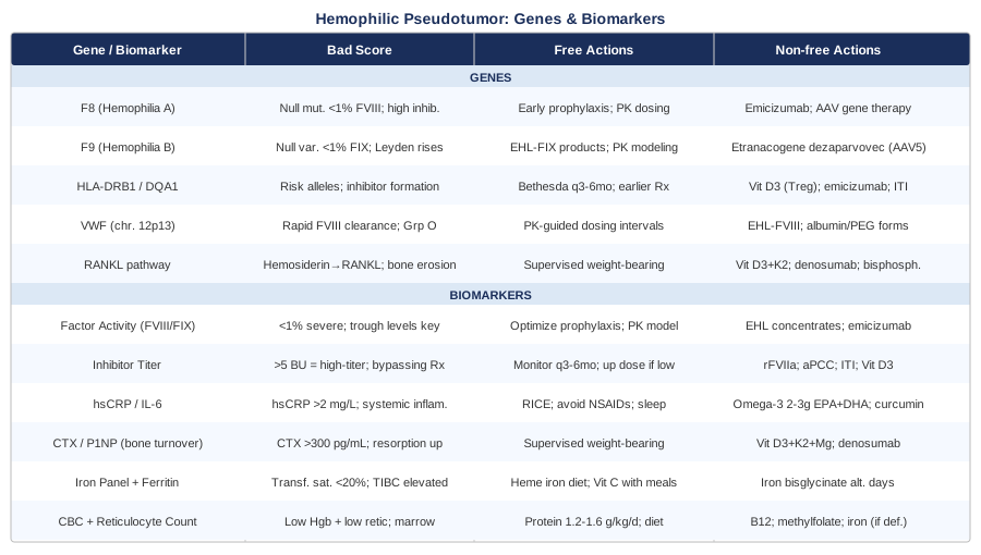
This article was written to fill that gap. It brings together two complementary frameworks for understanding hemophilic pseudotumor at a deeper level. The first is biomarker-based: six laboratory values that together create a real-time picture of clotting function, inflammatory burden, bone breakdown, and the systemic effects of chronic bleeding. The second is genetic: five genes whose variants influence disease severity, inhibitor development risk, and the bone remodeling pathways that make some pseudotumors more destructive than others.
Neither framework promises a cure or replaces the specialized care that this condition demands. What they offer is a clearer map. With the right information, conversations with hematologists and hemophilia treatment center teams become more targeted, imaging intervals become better justified, and decisions about prophylaxis intensity, adjunct therapies, and lifestyle modification become more informed. That is the kind of clarity this article is designed to provide.
6 Biomarkers That Matter Most in Hemophilic Pseudotumor
Tracking the right laboratory values is the most direct way to understand what is happening inside a pseudotumor and how the body is responding overall. The six biomarkers below are not a generic hemophilia panel — each one was chosen because it illuminates a specific mechanism relevant to pseudotumor formation, expansion, or systemic consequences. Together, they form a monitoring framework that is both practical and genuinely actionable.
1. Factor Activity Level: Your Baseline Risk Signal
Why it matters
Factor VIII or Factor IX activity level is the single most fundamental number in hemophilia care. It determines disease severity — severe (under 1% activity), moderate (1–5%), or mild (6–40%) — and is the primary driver of bleeding frequency and intensity. Hemophilic pseudotumors occur almost exclusively in patients with severe hemophilia, where spontaneous bleeding into muscles and bones is common and a single poorly-resolved hematoma can initiate the pseudotumor cascade. Factor activity is not static in patients on prophylaxis: there are peaks after infusion and troughs at the end of a dosing interval. The trough level — the lowest point before the next dose — is often what matters most for tissue protection, because unprotected hours are when bleeding occurs.
How to measure it
Factor activity is measured via a one-stage clotting assay or a chromogenic assay, both performed at a hospital coagulation laboratory or hemophilia treatment center. Chromogenic assays are preferred for patients on certain extended half-life (EHL) products, where one-stage assays may over- or underestimate true levels. Turnaround is typically 24–48 hours. Cost ranges from $80 to $200 depending on location and insurance. For severe hemophilia patients with a known pseudotumor, most specialized centers now target trough factor levels above 10–15% rather than the historical 1–2% minimum, and some aim higher during active pseudotumor management.
If the score is bad — the plan without supplements
The primary intervention is adjusting the prophylaxis schedule. Moving from twice-weekly to every-other-day or daily dosing reduces unprotected trough windows. Switching to extended half-life concentrates (EHL-FVIII or EHL-FIX) can maintain better troughs with fewer infusions. For Hemophilia A patients without inhibitors, emicizumab — a subcutaneous antibody that functionally mimics FVIII — provides highly stable protection without peaks and troughs, and has become an important option for pseudotumor-prone patients. All prophylaxis changes should be made with the hemophilia treatment center using pharmacokinetic modeling when possible.
If the score is bad — the plan with supplements or equipment
No supplement raises endogenous factor VIII or IX production in severe hemophilia. Desmopressin (DDAVP) releases stored endothelial FVIII and is useful in mild Hemophilia A, but has no role when baseline levels are near zero. From an equipment perspective, well-fitted joint-protection bracing during physical activity may reduce micro-trauma bleeds in vulnerable areas — consult a physiotherapist experienced in hemophilia for appropriate options. Early-stage research on N-acetylcysteine (NAC) as a molecular chaperone that may support FVIII folding stability is interesting but remains preclinical; not recommended as a clinical strategy at this time.
2. Factor Inhibitor Titer: The Most Underestimated Variable
Why it matters
Inhibitors are antibodies that neutralize infused factor VIII or IX, rendering standard replacement therapy ineffective. They are the single greatest risk amplifier for hemophilic pseudotumor. A patient with an inhibitor who experiences muscle or bone bleeding faces a far longer, harder-to-treat hematoma — and it is often an undertreated, inhibitor-complicated bleed that initiates a pseudotumor. Inhibitors affect roughly 25–30% of patients with severe Hemophilia A and 3–5% of those with severe Hemophilia B. They are measured in Bethesda Units (BU): below 5 BU is low-titer, above 5 BU is high-titer.
How to measure it
The Nijmegen-Bethesda assay is the current gold standard, offering improved specificity over the older Bethesda assay by reducing false positives. Blood is drawn and serial dilutions of patient plasma are mixed with pooled normal plasma to quantify inhibitory activity. This test requires a specialized coagulation laboratory — hemophilia treatment centers reliably offer it — and costs roughly $100 to $350. Monitoring frequency should increase during periods of intensive factor exposure (surgery, heavy bleeding episodes, new product switches).
If the score is bad — the plan without supplements
For low-titer inhibitors, treating through with higher doses of factor concentrate is sometimes feasible. For high-titer inhibitors, bypassing agents are used: recombinant activated factor VII (rFVIIa / NovoSeven) and activated prothrombin complex concentrate (aPCC / FEIBA) work around the inhibitor. Emicizumab has transformed care for Hemophilia A patients with inhibitors, providing substantially better bleed protection than bypassing agents alone. Immune tolerance induction (ITI) — daily high-dose factor infusions over 12–24+ months — eradicates inhibitors in roughly 60–70% of Hemophilia A patients and remains the primary strategy for long-term inhibitor elimination.
If the score is bad — the plan with supplements or equipment
No supplement eliminates inhibitors. However, vitamin D3 at levels maintaining 25-OH vitamin D above 40 ng/mL may support favorable immune modulation — vitamin D is involved in regulatory T-cell (Treg) differentiation, and Treg activity plays a role in immune tolerance. Dose: 2000–5000 IU D3 daily with 100 mcg MK-7 vitamin K2. Testing: every 6 months to adjust dose. No cycling required. Side effects: rare at these doses; avoid exceeding 10,000 IU without monitoring. Rituximab (anti-CD20) has been used as an adjunct to ITI in select refractory cases — this is a prescription medication administered under specialist supervision, not a supplement strategy.
3. High-Sensitivity CRP and Interleukin-6: The Inflammation Window
Why it matters
Hemophilic pseudotumors are not simply static collections of blood. They are actively inflammatory structures. Blood trapped within a pseudotumor degrades over time, releasing hemosiderin, pro-inflammatory cytokines, and tissue breakdown products. Interleukin-6 (IL-6) and TNF-alpha drive local and systemic inflammation, promote capsule expansion, and activate osteoclasts in nearby bone. High-sensitivity CRP (hsCRP) is an acute-phase protein produced by the liver in direct response to IL-6, making it a practical, inexpensive proxy for systemic inflammatory burden. Tracking hsCRP over time — rather than as a one-time snapshot — reveals whether inflammation is escalating, stable, or responding to treatment decisions.
How to measure it
hsCRP is a standard blood test available at any clinical laboratory, requiring no special preparation. Cost: $20 to $60. IL-6 itself (serum or plasma) is more specific and informative but costs $80–$200 and is ordered less routinely; it is worth requesting when hsCRP is borderline or when a more granular picture of cytokine activity is needed. Peter Attia, who tracks hsCRP consistently in his longevity medicine framework, recommends a target below 0.5 mg/L for optimal long-term health — a useful reference point. In hemophilic disease, values below 2 mg/L in the absence of an acute bleed or known infection are a reasonable intermediate target.
If the score is bad — the plan without supplements
The most effective way to reduce inflammatory burden in hemophilic pseudotumor is to address the source: optimize factor prophylaxis to minimize new bleeding events and their inflammatory aftermath. For acute bleeds, RICE principles — rest, ice (with protective layer to avoid frostbite), compression, elevation — reduce early inflammatory triggers. NSAIDs must be avoided in hemophilia unless specifically authorized by the treating hematologist: they impair platelet aggregation and significantly increase bleeding risk, eliminating their anti-inflammatory benefit in this context.
If the score is bad — the plan with supplements or equipment
Omega-3 fatty acids (EPA + DHA combined) have the most substantial evidence base for reducing systemic inflammation via suppression of prostaglandin E2, thromboxane A2, and IL-6 signaling. Dose: 2–3g combined EPA+DHA daily with meals. Important caution for hemophilia: at doses above 3–4g, omega-3 can meaningfully reduce platelet aggregation — explicitly discuss with your hematologist before starting. Cycling: not required; this is a long-term supplement. Side effects: fishy reflux (use enteric-coated, take with food), mild GI upset at higher doses. Curcumin (as BCM-95 or with piperine): 500 mg twice daily has documented NF-kB and IL-6 inhibitory effects, but carries mild anti-platelet activity — use cautiously, discuss with your care team. Cycling: 8 weeks on, 2 weeks off if GI effects occur.
4. Bone Turnover Markers — CTX and P1NP: Watching Bone Destruction in Real Time
Why it matters
Intraosseous hemophilic pseudotumors — those forming within bone — are among the most destructive manifestations of this condition. They expand by eroding cancellous and cortical bone, driven in large part by hemosiderin-activated osteoclasts. Hemosiderin deposits trigger upregulation of RANKL (receptor activator of nuclear factor-kappa B ligand) in local macrophages and fibroblasts, which in turn recruits and activates osteoclasts — the cells responsible for bone resorption.
CTX (C-terminal telopeptide of type I collagen) is released into the bloodstream when osteoclasts degrade bone collagen — elevated CTX signals accelerated bone resorption. P1NP (procollagen type 1 N-terminal propeptide) reflects bone formation activity by osteoblasts. The ratio of CTX to P1NP reveals whether bone loss is outpacing repair — a critical insight for intraosseous pseudotumors. Additionally, multiple studies have documented significantly lower bone mineral density in patients with severe hemophilia compared to the general population, driven by reduced weight-bearing activity, chronic inflammation, and suboptimal micronutrient status.
How to measure it
Both CTX and P1NP are measured from a blood draw — CTX is ideally drawn fasting in the morning, as it shows significant diurnal and food-related variation. Both are available through large reference laboratories and cost $60–$150 each. Some hemophilia treatment centers include these in annual monitoring panels for patients with known bone involvement. Optimal fasting CTX in adults: under 300 pg/mL. P1NP optimal range: approximately 16–96 mcg/L, though longitudinal trends are more informative than any single value.
If the score is bad — the plan without supplements
Appropriate physical activity is the most accessible non-pharmacological intervention for bone health. Weight-bearing exercise stimulates osteoblast activity and suppresses osteoclast-driven resorption. For hemophilia patients, low-impact weight-bearing activities — walking, aquatic exercise with progressive return to land, supervised resistance training with joint protection — are the most practical. All exercise programs must be designed with a physiotherapist experienced in hemophilia care. When bone destruction is severe, bisphosphonates (zoledronic acid IV annually, or alendronate orally weekly) or denosumab (anti-RANKL antibody, subcutaneous every 6 months) may be appropriate — specialist prescription and monitoring required.
If the score is bad — the plan with supplements or equipment
Vitamin D3: 2000–5000 IU daily. Purpose: supports calcium absorption, modulates osteoclast activity, supports immune function. Test 25-OH vitamin D every 6 months; target 40–60 ng/mL. Side effects: rare at these doses. Vitamin K2 (MK-7): 100–200 mcg daily. Purpose: activates osteocalcin to direct calcium into bone matrix; suppresses osteoclast formation. No meaningful interaction with hemophilia clotting cascade at these doses. Cycling: not required. Calcium citrate: 500–600 mg twice daily with food if dietary intake is insufficient (under 700 mg/day from food). Cycling: not required. Magnesium glycinate: 300–400 mg at bedtime. Supports bone matrix formation, cofactor for vitamin D activation. Side effects: loose stool at higher doses; glycinate is the most tolerable form. Collagen peptides: 10–15g daily. Type I collagen is the structural scaffold of bone; hydrolyzed collagen peptides support collagen synthesis. Cycling: not required.
5. Iron Panel and Serum Ferritin: The Hidden Consequence of Chronic Bleeding
Why it matters
Every episode of bleeding deposits red blood cells into tissue. Over time, those cells degrade and release their iron content — in the form of hemosiderin — which accumulates at the bleeding site. In hemophilic pseudotumors, hemosiderin deposition is extensive and plays a direct role in driving local inflammation and osteoclast activation through the RANKL pathway. Systemically, repeated blood loss depletes iron stores, leading to iron deficiency anemia that can be masked by the inflammatory component of the condition.
The challenge with serum ferritin alone is that it is an acute-phase reactant: it rises with inflammation regardless of true iron stores, making it unreliable as a standalone measure in an active inflammatory state. A full iron panel — ferritin, serum iron, total iron-binding capacity (TIBC), and transferrin saturation — provides a much more accurate picture. Transferrin saturation below 20% with elevated TIBC reliably indicates true iron deficiency even when ferritin appears normal or elevated due to inflammation.
How to measure it
Standard fasting blood draw; serum iron and TIBC are best drawn fasting. Full iron panel cost: $30–$80. Ferritin alone is lower cost and widely available. Optimal targets: ferritin 50–150 ng/mL for most adults; transferrin saturation 20–45%; serum iron 60–170 mcg/dL. Thomas Dayspring, a leading lipidologist and metabolic diagnostics expert, consistently emphasizes interpreting ferritin within a full iron panel and inflammatory context rather than as an isolated number — a principle that directly applies here.
If the score is bad — the plan without supplements
The foundational intervention is optimizing factor prophylaxis to reduce the frequency and volume of bleeding events — this simultaneously addresses the source of hemosiderin accumulation locally and systemic iron loss. For dietary iron, heme iron sources (red meat, organ meats, dark poultry) are 2–3 times more bioavailable than plant-based non-heme iron. Pairing non-heme iron foods with vitamin C-rich foods substantially improves absorption. Tea, coffee, and calcium-rich foods consumed in the same meal reduce non-heme iron absorption and should be spaced apart.
If the score is bad — the plan with supplements or equipment
Iron supplementation when transferrin saturation confirms true deficiency: ferrous bisglycinate (iron bisglycinate) at 25–50 mg elemental iron on alternate days. Research supports that alternate-day dosing improves net absorption compared to daily dosing, because daily iron triggers hepcidin upregulation that blocks intestinal absorption. Cycling: treat for 8–12 weeks and retest the full panel. Side effects: constipation, nausea — bisglycinate is substantially gentler than ferrous sulfate; taking on an empty stomach with vitamin C (500 mg) maximizes absorption. Critical caution: do not supplement iron without confirming deficiency via iron panel — hemosiderin overload in the pseudotumor itself is already a concern, and indiscriminate supplementation does not address local iron accumulation and may worsen systemic iron burden.
6. Complete Blood Count with Reticulocyte Count: Tracking the Cost of Ongoing Blood Loss
Why it matters
A complete blood count (CBC) provides a snapshot of systemic blood loss and bone marrow response. Hemoglobin and hematocrit reflect the degree of anemia; in patients with active or large pseudotumors, ongoing slow bleeding into the expanding mass can create a chronic blood loss pattern that eventually depletes hemoglobin without any single dramatic bleeding event to signal the decline.
The reticulocyte count — the proportion of immature red blood cells — shows whether the bone marrow is mounting an adequate compensatory response. A low reticulocyte count in the presence of anemia suggests suppressed marrow function, nutritional deficiency (iron, B12, or folate), or both. A high reticulocyte count signals the marrow is working hard to replace ongoing losses. Platelet count, also part of the CBC, is relevant when aspirin or other platelet-affecting medications are co-prescribed — tracking trends over time provides important context.
How to measure it
CBC is one of the most widely available and inexpensive blood tests: $20 to $50 in most settings. Adding a reticulocyte count adds minimal cost and is standard in most hemophilia monitoring panels. Normal hemoglobin: 13.5–17.5 g/dL for adult men, 12.0–15.5 g/dL for women. Reticulocyte percentage: 0.5–2.5% is normal; below 0.5% in the presence of anemia is a flag for further investigation.
If the score is bad — the plan without supplements
The primary intervention is optimizing prophylaxis to reduce ongoing blood loss into the pseudotumor. For hemoglobin below 8–9 g/dL in an acute context, red cell transfusion may be indicated — a clinical decision made with the treating team. For borderline anemia, addressing the underlying nutritional deficiency is the first step. Protein adequacy — 1.2–1.6g per kilogram of body weight daily — is often overlooked in hemophilia care and is essential for erythropoiesis and all tissue repair processes.
If the score is bad — the plan with supplements or equipment
Iron supplementation if iron deficiency is confirmed (as described above). Vitamin B12: 500–1000 mcg methylcobalamin sublingually daily if serum B12 is below 400 pg/mL. Cycling: treat for 3 months and retest. Side effects: essentially none at these doses. Folate: 400–800 mcg methylfolate daily if deficiency is confirmed (especially relevant if MTHFR variants are present — methylfolate bypasses the enzyme impairment that makes synthetic folic acid less effective in this population). Cycling: treat until CBC normalizes. For patients who maintain an inadequate protein intake despite dietary efforts, whey protein or collagen peptide supplementation (15–20g once or twice daily) provides a practical pathway to meeting erythropoiesis and tissue repair demands.
The Genetic Factors Behind Hemophilic Pseudotumor
Biomarkers tell you what is happening right now. Genetics tells you why certain patients are more vulnerable to pseudotumor formation in the first place, and which biological systems need the most proactive support. Understanding the five genes below does not require a genetics degree — it requires knowing what each mutation means for treatment strategy and which adjunct interventions are most rational given the underlying biology.
The F8 Gene: Mutation Type Determines Everything in Hemophilia A
The F8 gene on the X chromosome (Xq28) encodes Factor VIII. It spans approximately 186 kilobases across 26 exons and has generated over 3,000 documented pathogenic mutations. The type of mutation — not just the diagnosis of Hemophilia A — determines disease severity, inhibitor risk, and pseudotumor likelihood.
The most consequential mutation is the intron 22 inversion, present in approximately 45% of severe Hemophilia A cases. This large chromosomal rearrangement completely disrupts F8 transcription, resulting in less than 1% factor activity and — because the protein produced is structurally foreign — the highest rate of inhibitor formation of any mutation class. Large deletions carry a similarly high inhibitor risk. Nonsense and frameshift mutations are generally severe. Missense mutations more commonly cause moderate or mild phenotype, with substantially lower pseudotumor risk.
The clinical implication is direct: patients with null mutations (inversions, large deletions) have both the most severe bleeding phenotype and the highest inhibitor incidence — creating the double vulnerability most associated with pseudotumor formation.
If the gene is bad — the plan without supplements
Primary prophylaxis beginning in early childhood — before the first major bleed — is the most powerful preventive intervention for severe F8 mutation carriers. Pharmacokinetic-guided individualized prophylaxis using tools such as WAPPS-Hemo achieves substantially better protection than standardized weight-based protocols by mapping each patient's unique factor half-life. Emicizumab is increasingly used in severe Hemophilia A regardless of inhibitor status, offering stable subcutaneous dosing (weekly, biweekly, or monthly). AAV-based F8 gene therapy has demonstrated sustained factor expression in clinical trials, with some patients achieving near-normal levels — this is the most promising long-term solution currently available.
If the gene is bad — the plan with supplements or equipment
No supplement changes F8 gene function. Supporting the downstream consequences of severe bleeding — bone health, inflammation, iron metabolism — is where supplementation is actionable. Vitamin D3 (2000–5000 IU daily, target 40–60 ng/mL): relevant for bone density, immune modulation, and potentially inhibitor risk. Test every 6 months; no cycling required. Omega-3 (2–3g EPA+DHA, with hematologist clearance): reduces systemic inflammatory burden from chronic micro-bleeds. Long-term. Joint-protection equipment — well-fitted knee, ankle, and elbow braces during physical activity — reduces micro-trauma bleeds without the bleeding risk of invasive interventions.
The F9 Gene: Hemophilia B and the Leyden Phenomenon
The F9 gene (Xq27.1) encodes Factor IX, the missing or dysfunctional protein in Hemophilia B. At approximately 33 kilobases and 8 exons, it is smaller than F8 but genetically diverse — more than 1,000 pathogenic variants are documented. Clinical presentation in severe cases overlaps significantly with Hemophilia A, including pseudotumor risk.
One uniquely important variant category is Hemophilia B Leyden: mutations in the F9 gene promoter region that cause severe or moderate disease in childhood, but where factor IX levels rise spontaneously at puberty due to androgen-responsive elements in the promoter. Patients in this category face high pseudotumor risk during childhood and adolescence, then often improve substantially in adulthood — a phenomenon that has illuminated the hormonal regulation of clotting factor expression.
Hemophilia B patients who develop inhibitors face unique risks: inhibitors against FIX are associated with a higher rate of anaphylactic reactions to FIX infusions and with development of nephrotic syndrome following immune tolerance induction — a complication essentially absent in Hemophilia A.
If the gene is bad — the plan without supplements
Extended half-life FIX products — nonacog beta pegol and albutrepenonacog alfa — offer substantially longer dosing intervals (weekly to every-10-day) while maintaining adequate troughs, a major practical advantage over standard FIX concentrates. Etranacogene dezaparvovec (AAV5-based FIX gene therapy) has demonstrated high sustained factor IX expression from a single infusion in clinical trials and is approved in some markets — the most transformative option currently available for eligible Hemophilia B patients.
If the gene is bad — the plan with supplements or equipment
Similar framework to Hemophilia A: vitamin D3 for bone and immune support, omega-3 for inflammation management (with hematologist clearance), and joint-protection equipment. For Leyden patients specifically, no supplements accelerate the puberty-related improvement in FIX levels, but ensuring adequate nutrition during adolescence — protein (1.2–1.5g/kg/day), calcium, vitamin D — supports bone health during the critical transition period when pseudotumor risk is highest.
HLA-DRB1 and HLA-DQA1: The Inhibitor Risk Genes
Human leukocyte antigen (HLA) genes on chromosome 6p21 encode surface proteins that present antigens to T-cells — a critical step in determining whether the immune system tolerates infused factor concentrates or mounts an antibody response against them. Certain HLA alleles substantially increase inhibitor risk in hemophilia, particularly when combined with high-risk F8 mutations.
Key associations: HLA-DRB1*15:01 and HLA-DQA1*01:02 are linked to higher inhibitor risk in Hemophilia A patients with intron 22 inversions. HLA-DQA1*05:01 has been consistently associated with inhibitor development across multiple cohort studies. The CANAL study, Malmo International Brothers Study (MIBS), and PedNet cohort have collectively mapped how HLA alleles interact with mutation severity and treatment history to predict inhibitor formation — the highest-risk patients are those with null F8 mutations plus inhibitor-promoting HLA profiles.
If the gene is bad — the plan without supplements
Genetic HLA profiling at diagnosis, now increasingly available through hemophilia treatment centers, can guide prophylaxis strategy. High-inhibitor-risk HLA profiles may support: starting primary prophylaxis earlier to minimize intensive factor antigen exposure during peak immune sensitization windows; considering emicizumab earlier to reduce cumulative factor antigen load; and implementing a rigorous Bethesda assay monitoring protocol (every 3–6 months during early treatment years, especially through the first 50 exposure days when inhibitor risk is highest).
If the gene is bad — the plan with supplements or equipment
Vitamin D3 at levels maintaining 25-OH vitamin D above 40 ng/mL is the most accessible immune-modulating supplement with a sound mechanistic rationale. Vitamin D supports regulatory T-cell (Treg) differentiation, and Treg activity is central to immune tolerance — the mechanism that ITI attempts to exploit. Human trial data in hemophilia-specific inhibitor prevention remains limited, but the safety profile is excellent and the biological rationale is clear. Dose: 2000–5000 IU D3 + 100 mcg MK-7 K2 daily. Test every 6 months. No cycling required. Side effects: rare at these doses.
The VWF Gene: The FVIII Guardian
Von Willebrand factor (VWF) is a carrier protein for FVIII in the circulation, protecting it from premature degradation by proteases and delivering it to sites of injury. VWF is encoded by the VWF gene on chromosome 12p13. Variants in this gene affect FVIII's effective half-life — meaning two patients with identical F8 mutations can have meaningfully different circulating FVIII protection based on their VWF genetics.
Blood group O is strongly associated with lower VWF levels due to accelerated clearance; patients with blood group O and Hemophilia A may have effectively shorter FVIII protection windows than blood group A, B, or AB patients. VWF clearance-accelerating variants — including the VWF p.Arg1205His (Vicenza) variant — further reduce FVIII protection. This explains why some patients have worse-than-expected bleeding despite what appears to be adequate prophylaxis, and why the same dosing protocol works very differently in different individuals.
If the gene is bad — the plan without supplements
Pharmacokinetic modeling using population PK tools like WAPPS-Hemo is particularly valuable for patients with rapid FVIII clearance driven by VWF variants — it quantifies the individual half-life curve and allows dosing intervals to be precisely tailored rather than estimated from population averages. Extended half-life factor concentrates that use albumin fusion or PEGylation to slow clearance independent of VWF are especially relevant in this group.
If the gene is bad — the plan with supplements or equipment
Desmopressin (DDAVP) releases stored endothelial VWF and FVIII and is effective in mild Hemophilia A and von Willebrand disease, but has no role in severe hemophilia where stores are already depleted. No supplement meaningfully slows VWF-driven FVIII clearance. Adequate hydration supports plasma volume and may minimally influence clearance kinetics — a simple and zero-risk baseline habit.
RANKL Pathway Genes: Bone Destruction at the Molecular Level
Bone remodeling is governed by a molecular triad: RANK (receptor activator of nuclear factor-kappa B, encoded by TNFRSF11A), RANKL (its ligand, encoded by TNFSF11), and OPG (osteoprotegerin, a decoy receptor, encoded by TNFRSF11B). When RANKL binds RANK on osteoclast precursors, it drives their maturation and activation, causing bone resorption. OPG neutralizes free RANKL and functions as a natural brake on this process.
In hemophilic pseudotumors affecting bone, hemosiderin deposits directly activate RANKL expression in local fibroblasts and macrophages — overwhelming the OPG brake and driving progressive osteoclast-mediated destruction. Variants in RANKL and OPG genes influence the baseline sensitivity of this pathway: patients with naturally elevated RANKL signaling or reduced OPG expression may face faster and more extensive bone destruction when hemosiderin accumulates. This pathway is increasingly recognized in the closely related context of hemophilic arthropathy and is emerging as a relevant target in pseudotumor bone involvement.
If the gene is bad — the plan without supplements
Denosumab (Prolia or Xgeva) is a fully human monoclonal antibody that binds and neutralizes RANKL, preventing osteoclast maturation. Administered subcutaneously every 6 months (Prolia formulation for bone health), it is currently the most potent anti-resorptive therapy available and has been used in hemophilia-related bone destruction contexts. Bisphosphonates (zoledronic acid annually IV, or alendronate weekly orally) provide an alternative by accumulating in bone and directly impairing osteoclast function. Both require specialist prescription and monitoring — including dental assessment before bisphosphonate initiation.
If the gene is bad — the plan with supplements or equipment
Vitamin D3 (2000–5000 IU daily, target 40–60 ng/mL) directly downregulates RANKL expression in osteoblasts and upregulates OPG — making it the most mechanistically on-target supplement for this pathway. Vitamin K2 (MK-7) (100–200 mcg daily) also inhibits RANKL expression and activates OPG, providing complementary RANKL suppression. These two supplements work synergistically and have an excellent safety profile at standard doses. Cycling: not required. Magnesium glycinate (300–400 mg at bedtime): cofactor for vitamin D activation (the renal 25-OH to 1,25-OH conversion requires magnesium) and supports bone matrix enzymes. Side effects: loose stool at high doses; glycinate minimizes this. Collagen peptides (10–15g daily): support the collagen matrix that RANKL-driven resorption degrades. Cycling: not required.
What "Outlive" by Peter Attia Reveals About Long-Term Management
Peter Attia's 2023 book Outlive: The Science and Art of Longevity is not a hemophilia book. It is a systematic, evidence-grounded framework for extending healthspan — the quality years of life — through proactive biomarker tracking, individualized medicine, and deliberate lifestyle interventions. But the framework Attia builds translates directly to the challenges of managing hemophilic pseudotumor's long-term consequences, in ways that most specialist care — necessarily focused on acute clotting management — does not address. Ten of the most impactful ideas from the book apply directly to this condition.
Medicine 3.0: From Reactive to Proactive
Attia's central argument is that conventional medicine ("Medicine 2.0") waits for disease to become diagnosable before acting. Medicine 3.0 intervenes years or decades before that point, using biomarkers to detect unfavorable trajectories and correct them while correction is still simple. For pseudotumor patients, this means not waiting for imaging to confirm progression before acting on elevated CTX, rising hsCRP, or declining hemoglobin — each is a signal that the system is under stress before structural damage is visible.
Inflammation Is a Driver, Not Just a Symptom
Attia dedicates significant attention to the role of chronic low-grade inflammation as a root driver of tissue damage, not merely a byproduct. In hemophilic pseudotumor, the inflammatory environment within and around the mass is both a consequence of bleeding and a driver of expansion. Managing CRP and IL-6 proactively — through prophylaxis optimization, omega-3 intake (with hemophilia-specific cautions), and sleep quality — is not secondary care; it is primary disease management.
Bone Is a Metabolically Active Organ
Attia challenges the common framing of bone as passive scaffolding. Bone is constantly remodeled, responds to mechanical load, and is deeply connected to metabolic and inflammatory status. His emphasis on bone mineral density tracking and vitamin D, K2, and calcium optimization maps directly onto the bone marker monitoring framework in this article — particularly relevant for patients whose pseudotumors involve osseous structures.
Zone 2 Training Reduces Inflammatory Burden
Attia strongly advocates for Zone 2 cardio (moderate-intensity sustained aerobic exercise at roughly 60–70% of maximum heart rate) as one of the most powerful systemic anti-inflammatory interventions available. For hemophilia patients, supervised low-impact Zone 2 exercise — cycling, swimming, elliptical — is achievable with appropriate joint protection and hematologist clearance, and carries meaningful benefits for CRP reduction and metabolic health. Attia recommends 3–4 hours per week for long-term health.
Strength Training Is Medicine for Bone and Tissue
Beyond aerobic work, Attia makes a compelling case for progressive resistance training as one of the highest-return health interventions available — particularly for bone density, muscle mass as a buffer against injury, and insulin sensitivity. For hemophilia patients, supervised resistance training with appropriate joint protection and avoidance of high-impact loading is well supported by hemophilia physiotherapy guidelines and directly addresses the bone density deficit described in this article.
Sleep Is Underrated in Tissue Repair
Attia dedicates an entire chapter to sleep as a biological necessity for tissue repair, immune regulation, and inflammatory resolution. Chronic sleep deficiency elevates CRP and IL-6, accelerates bone resorption via cortisol, and impairs erythropoiesis. For pseudotumor patients — many of whom experience pain-disrupted sleep — optimizing sleep hygiene (consistent schedule, cooler room temperature, darkness, limiting blue light, and addressing pain management adequately before bed) is not optional self-care but a direct component of disease management.
Protein Intake Is Almost Always Insufficient
Attia's protein targets — 1.6g per kilogram of body weight daily, prioritizing leucine-rich complete protein sources — are substantially above conventional recommendations. In the context of hemophilic pseudotumor, adequate protein is necessary for erythropoiesis, collagen synthesis, bone matrix formation, immune function, and wound healing. Most patients with chronic disease consume significantly less protein than optimal. Meeting this target through food (meat, fish, eggs, dairy) or supplemented with whey or collagen peptides is a high-return, low-risk intervention.
Individualize Biomarker Targets
Attia consistently argues against using population-average reference ranges as personal health targets. Optimal for an individual may differ substantially from "normal." This applies directly here: a "normal" ferritin of 15 ng/mL is clearly suboptimal for a patient with chronic blood loss; a "normal" 25-OH vitamin D of 22 ng/mL leaves substantial benefit on the table. The biomarker targets in this article reflect this individualization philosophy.
Metabolic Health Affects Every System
Insulin resistance and metabolic dysfunction impair every system implicated in pseudotumor management: they elevate CRP, worsen bone resorption, impair immune function, and reduce tissue healing. Attia's framework for metabolic health — limiting refined carbohydrates, prioritizing protein, maintaining muscle mass, and tracking fasting glucose and insulin — applies meaningfully to hemophilia patients, particularly as they age and the risk of compounding metabolic dysfunction rises.
Compounding Small Improvements Has Outsized Long-Term Impact
Perhaps the most practically useful insight in Outlive for this context: Attia's framework consistently emphasizes that small, consistent improvements in multiple domains compound into large health differences over years. For a pseudotumor patient, optimizing vitamin D by 20 ng/mL, adding omega-3, improving protein intake, and sleeping 30 minutes more each night may each seem marginal — but their combined effect on CRP, bone turnover, anemia, and immune regulation across years is substantial. This is the philosophy that should frame the supplementation and lifestyle strategies described throughout this article.
Complementary Approaches Worth Considering
Beyond biomarker tracking, genetic understanding, and lifestyle optimization, certain well-studied non-pharmacological approaches can meaningfully support quality of life and pain management in hemophilic pseudotumor. The approaches below were selected because they are non-invasive — carrying no bleeding risk — and have meaningful human evidence in the pain management and chronic condition contexts most relevant here.
Mindfulness-Based Stress Reduction (MBSR)
Mindfulness-based stress reduction (MBSR) is an 8-week structured program combining mindfulness meditation, body scan, and gentle movement developed by Jon Kabat-Zinn at the University of Massachusetts. Chronic pain, which is common in hemophilic pseudotumor due to nerve compression, bone involvement, and the persistent inflammatory environment, is one of the most well-studied applications of MBSR. Chronic stress also elevates cortisol, which accelerates bone resorption — creating a direct pathway through which stress management affects pseudotumor biology.
A meta-analysis published in JAMA Internal Medicine (2015) found that mindfulness meditation programs produced moderate reductions in pain, psychological distress, and functional impairment in patients with chronic pain conditions. The effect size for pain was comparable to active pharmacological interventions without adverse effects.
Realistic application: the standard 8-week MBSR program is widely available in-person and via validated online platforms (MBSR training through UMASS Center for Mindfulness or equivalent). Daily practice of 20–45 minutes is recommended during the program; 10–20 minutes of maintenance practice afterward sustains benefit. For pseudotumor patients, body scan and breath-focused meditation are the most accessible modalities and can be practiced lying down during periods of reduced mobility.
Biofeedback for Pain and Stress Regulation
Biofeedback uses real-time physiological monitoring — heart rate variability (HRV), skin conductance, respiration — to teach conscious regulation of normally automatic bodily functions. By learning to reduce physiological stress markers, patients can directly modulate pain perception pathways and lower cortisol-driven inflammatory burden. For pseudotumor patients experiencing chronic pain, biofeedback provides a tool for active pain self-regulation without any pharmacological or bleeding risk.
A systematic review in Applied Psychophysiology and Biofeedback (2019) found clinically meaningful reductions in chronic pain intensity and pain-related disability with HRV biofeedback training across multiple chronic pain populations. HRV biofeedback specifically has shown anti-inflammatory effects through vagal nerve activation, which is directly relevant to the inflammatory burden of pseudotumor.
Practical protocol: HRV biofeedback using a validated consumer device (such as the HeartMath Inner Balance sensor or a medical-grade HRV monitor) with 20 minutes of resonance-frequency breathing practice daily (typically at 5–6 breaths per minute, individualized). 8-week training programs with a biofeedback practitioner or through supervised software are the most evidence-backed entry point.
Breathing-Based Therapies
Controlled breathing techniques — including diaphragmatic breathing, coherence breathing (5–6 breaths per minute), and 4-7-8 breathing — activate the parasympathetic nervous system through the vagal nerve, reducing cortisol output, lowering systemic inflammatory markers including CRP, and shifting the body away from the chronic sympathetic activation that accompanies pain and chronic disease. For hemophilic pseudotumor, breathing-based therapies are one of the simplest, most accessible, and lowest-risk tools available.
Research published in Frontiers in Human Neuroscience has demonstrated that slow, controlled breathing at approximately 6 breaths per minute significantly increases HRV, reduces salivary cortisol, and decreases inflammatory cytokine levels in repeated studies. This anti-inflammatory effect is mechanistically relevant to any condition in which chronic inflammation plays a maintaining role.
A practical starting protocol: 10–15 minutes of coherence breathing (5 seconds inhale, 5 seconds exhale) twice daily — morning and evening. No equipment required; free breathing apps (such as Breathe2Relax or Paced Breathing) can guide pacing. For patients with pain-disrupted sleep, a 10-minute session before bed combined with progressive muscle relaxation consistently improves sleep onset latency.
Music Therapy for Pain and Procedural Anxiety
Music therapy uses structured listening or active music-making under the guidance of a certified music therapist to modulate pain perception, reduce procedural anxiety, and lower stress-related inflammatory responses. For hemophilia patients, who face frequent medical procedures — infusions, imaging, and in some cases aspiration or surgical management of pseudotumors — procedural anxiety is a real and underaddressed burden that amplifies pain perception and stress responses.
A Cochrane systematic review (2016) of music-based interventions for pain found significant pain intensity reductions and reduced analgesic requirements across 73 randomized trials, with effects present across surgical, procedural, and chronic pain contexts. The mechanism involves endorphin release and modulation of the anterior cingulate cortex, reducing the affective dimension of pain.
Practical application: for pseudotumor patients, receptive music therapy — listening to carefully chosen, personally meaningful music during infusions, imaging studies, or periods of heightened pain — is the most accessible form. Certified music therapists (locate through the American Music Therapy Association) can design individualized protocols. Independent self-guided use with personally curated playlists at approximately 60–80 beats per minute (corresponding to resting heart rate) provides a lower-cost starting point.
Guided Imagery for Pain Management
Guided imagery uses directed mental visualization — typically narrated audio scripts leading the listener through calming, healing imagery — to reduce pain intensity, anxiety, and the anticipatory suffering that often accompanies chronic conditions like hemophilic pseudotumor. It engages the same neural pathways activated by real sensory experience, allowing the brain to practice a pain-reduced state through imagination.
A randomized controlled trial in pediatric and adult hemophilia patients (examining guided imagery specifically for procedure-related pain) found significant reductions in self-reported pain and anxiety compared to standard care alone. More broadly, a systematic review in Pain Management Nursing (2019) found guided imagery produced consistent reductions in chronic and procedural pain across multiple patient populations with no adverse effects.
A practical starting protocol: 15–20 minutes of guided imagery practice daily using validated audio programs (available through apps like Insight Timer, Belleruth Naparstek's Health Journeys library, or through a licensed clinical hypnotherapy practitioner). For pseudotumor patients, imagery scripts focused on immune regulation, reduced inflammation, and peaceful body awareness are the most thematically relevant. Practice immediately before anticipated painful procedures or during periods of elevated pain is the highest-yield application.
Conclusion
Hemophilic pseudotumor is a condition that demands more than standard hemophilia management. The six biomarkers described here — factor activity, inhibitor titer, hsCRP, bone turnover markers, iron panel, and CBC with reticulocytes — form a practical, genuinely informative monitoring framework that can be implemented progressively, starting with the most accessible tests and building from there. The five genes add a layer of understanding that explains individual vulnerability and informs which interventions deserve the most attention for each patient's specific profile.
None of this replaces the expert care of a hemophilia treatment center. What it does is make that care more focused, and make the patient a more informed participant in decisions that matter. The next smart step is straightforward: review which of these biomarkers you have tracked recently, identify the gaps, and bring this framework to your next hematologist appointment as a starting point for a more complete monitoring conversation.
Musculoskeletal: Bone Conditions Joint Conditions Muscle Conditions
Autoimmune: Inflammatory Conditions