This article contains AI generated content.
· UpdatedHemosiderotic Synovitis: 5 Genes And 6 Biomarkers To Track
Introduction
If you have been told you have hemosiderotic synovitis — or if you are dealing with a joint condition tied to repeated hemarthrosis, iron deposition in synovial tissue, or chronic joint inflammation — you already know how incomplete the standard conversation tends to be. Manage the bleeds. Reduce inflammation. Monitor the joint. It is not wrong advice, but it leaves out the most important question: why is your body accumulating iron in the joint space the way it does, and what is driving the severity?
Hemosiderotic synovitis sits at the intersection of iron metabolism, local inflammatory biology, and genetic predisposition. Two people with nearly identical joint bleed histories can have very different outcomes. One develops manageable chronic synovitis; the other progresses toward significant cartilage degradation and hemophilic arthropathy within years. A significant part of that difference is encoded in their genes and reflected in specific blood markers that most practitioners do not routinely order or interpret with sufficient depth.
Standard lab reference ranges were designed for the general population, not for someone trying to understand iron-driven joint oxidative stress. A ferritin of 250 ng/mL will sail through a routine review unmarked, yet in someone with synovial iron deposits and an underlying HFE mutation, it represents an amplifying force on chondrotoxicity. Generic protocols miss this entirely.
This article takes a more targeted approach. The first section covers the six most meaningful biomarkers for tracking the iron burden and inflammatory state underlying hemosiderotic synovitis — what each one reveals, how to measure it affordably, and what to do when results are concerning, both with and without supplementation. From there, you will find a section on the five iron metabolism genes most relevant to this condition, followed by a book that reframes the clinical understanding of iron overload in a way most physicians will not raise, and a look at complementary modalities backed by actual human clinical evidence. Better information does not cure anything on its own — but it does make better decisions possible.
6 Biomarkers That Reveal Your Iron Burden and Joint Inflammation Risk
Tracking hemosiderotic synovitis with imaging alone shows you damage that has already occurred. These six biomarkers let you see what is happening in real time — how much iron is circulating, whether your regulatory systems are managing it correctly, and how active the inflammatory loop has become. None of them alone is sufficient; together, they tell a coherent and actionable story.
1. Serum Ferritin — Your Iron Storage Signal
Ferritin is the intracellular protein complex that stores iron safely. Serum ferritin reflects total body iron stores and is the most widely available iron overload marker in clinical practice. In the context of hemosiderotic synovitis, elevated ferritin matters because iron accumulation in the joint does not happen in isolation — it compounds when systemic iron stores are also elevated.
Why it matters: When erythrocytes break down in the joint following hemarthrosis, synovial macrophages engulf the iron-rich debris and store it as hemosiderin. The Fenton reaction then converts stored iron into hydroxyl radicals — among the most reactive oxidants in biology — which directly damage chondrocytes, degrade cartilage extracellular matrix, and drive inflammatory gene expression in synoviocytes. Higher systemic iron stores mean more fuel for this local process.
The conventional "normal" ferritin range — up to 300 ng/mL for men, 150 ng/mL for women — was built on population distributions, not health optimization. Peter Attia and Thomas Dayspring consistently advocate for interpreting ferritin in the context of the full iron panel, with optimal levels closer to 50–100 ng/mL for most adults. Anything above 200 ng/mL, in the absence of an acute infection or inflammatory flare, warrants investigation in someone with joint iron disease. Critically, ferritin is also an acute-phase reactant — active synovitis will elevate it independent of iron stores — which is why it must never be interpreted in isolation.
How to measure it: Standard blood draw ordered as part of an iron panel. Cost: approximately $15–40. Collect fasting and, when possible, during a quiet period — not during an active inflammatory flare — to avoid artifactually elevated readings.
If ferritin is elevated — the plan without supplements
The most evidence-backed non-supplemental intervention to lower ferritin is therapeutic phlebotomy. Removing 450–500 mL of blood removes approximately 200–250 mg of iron. Blood donation every 8–12 weeks is accessible to most people and free. For formally diagnosed hemochromatosis, physician-supervised phlebotomy follows an induction protocol (weekly or biweekly draws) until ferritin normalizes, then switches to maintenance every 3–4 months.
Dietary adjustments work as a supporting measure. Reduce heme iron intake (red meat, organ meats) to 2–3 servings per week. Pair iron-containing meals with polyphenol-rich beverages — coffee, black or green tea, cacao — which reduce non-heme iron absorption by 30–60% in study conditions. Avoid vitamin C mega-doses with iron-rich meals. Retire cast-iron cookware if ferritin is significantly elevated.
If ferritin is elevated — the plan with supplements or equipment
IP6 (inositol hexaphosphate): A naturally occurring iron chelator found in rice bran and legumes. 1–2 grams taken between meals (not with meals) chelates circulating and intestinal iron. Cycling: 6 weeks on, 2 weeks off. Side effects: at higher doses, may reduce zinc and calcium absorption; not recommended in pregnancy.
Lactoferrin: A glycoprotein that binds iron in the gut and modulates inflammatory signaling in macrophages. 100–300 mg/day. Minimal side effects at these doses. Complements rather than replaces phlebotomy when ferritin is significantly elevated.
Curcumin with piperine: Curcumin has both mild iron-chelating and anti-inflammatory properties relevant to joint disease. 500–1000 mg curcuminoids daily with 5–10 mg piperine. Cycling: 8 weeks on, 2 weeks off. Side effects: mild GI upset at higher doses; avoid with blood thinners.
2. Transferrin Saturation (TSAT) — How Much Iron Is Circulating Right Now
Why it matters: Transferrin is the transport protein that carries iron through the blood. Transferrin saturation (TSAT) expresses what percentage of that transport capacity is currently occupied by iron. While ferritin reflects stores, TSAT reflects flux — the amount of iron actively moving through circulation at any given time. Thomas Dayspring, one of the most recognized clinical lipidologists in preventive medicine, consistently emphasizes that TSAT is as diagnostically important as ferritin and must be read alongside it.
In hemosiderotic synovitis, persistently elevated TSAT — above 45% — signals that iron is flooding the system and available for uptake by synoviocytes, amplifying joint oxidative stress. The standard normal range of 20–50% is too wide for clinical management; optimal for iron-related joint disease is closer to 20–35%.
How to measure it: Included in a standard iron panel (serum iron + total iron-binding capacity). Request as "iron studies" or "iron panel." Cost: $20–60. Should be done fasting.
If TSAT is elevated — the plan without supplements
Phlebotomy remains the cornerstone for sustained TSAT reduction when it reflects true iron overload. Aerobic exercise provides a complementary mechanism: regular endurance training increases erythropoiesis and hemoglobin synthesis, diverting iron from free circulation into red blood cells. Reducing red meat, avoiding alcohol (which increases intestinal iron absorption), and using tea or coffee with meals forms the dietary triad.
If TSAT is elevated — the plan with supplements or equipment
EGCG (green tea extract): 400–800 mg/day, taken between meals. EGCG has documented iron-chelating properties in both intestinal and systemic compartments. Combine with IP6 on the same cycling schedule (6 weeks on, 2 weeks off) for additive effect. Avoid taking either supplement with meals — iron chelation is only functionally relevant when taken apart from food. Side effects: high-dose EGCG can be hepatotoxic in sensitive individuals; stay within studied dose ranges and avoid combining with alcohol.
3. Hepcidin — The Master Regulator Most Physicians Do Not Order
Why it matters: Hepcidin is a small peptide hormone produced by the liver that acts as the central switch for iron homeostasis. When hepcidin is high, it degrades ferroportin — the only known cellular iron exporter — keeping iron locked inside cells. When hepcidin is low, iron flows freely from gut enterocytes and macrophages into circulation.
In hemosiderotic synovitis, hepcidin creates a specific clinical paradox. Chronic low-grade synovial inflammation drives IL-6 production from iron-loaded macrophages, and IL-6 is a potent stimulus for hepatic hepcidin synthesis. This can produce a state where standard iron labs look contradictory: ferritin may be elevated (from inflammation), serum iron may be low (hepcidin is blocking export from macrophages), yet iron continues to accumulate locally in the joint through a separate pathway. Without measuring hepcidin, clinicians may misinterpret this as iron deficiency and inadvertently worsen the condition.
How to measure it: Requires liquid chromatography-tandem mass spectrometry (LC-MS/MS). Not available in all clinical labs but increasingly accessible through reference laboratories. Cost: $80–200. Request specifically when ferritin and TSAT are elevated but serum iron is paradoxically low, or when the clinical picture does not fit classical iron deficiency or overload.
If hepcidin is dysregulated — the plan without supplements
Treating the underlying inflammatory burden is the primary lever. Hepcidin elevation driven by IL-6 will not normalize until synovitis is meaningfully controlled. Regular moderate-intensity aerobic exercise — zone 2 (conversational pace) cardio for 30–45 minutes, 4–5 days per week — temporarily suppresses hepcidin after each session and reduces baseline IL-6 over weeks. Sleep optimization (7–9 hours, consistent schedule, dark and cool environment) reduces systemic inflammatory tone and secondary hepcidin elevation.
If hepcidin is dysregulated — the plan with supplements or equipment
Vitamin D (target 40–60 ng/mL 25-OH-D): Vitamin D deficiency is associated with elevated baseline IL-6 and proinflammatory tone, which pushes hepcidin upward. Correcting deficiency with 2000–5000 IU/day (dose guided by testing) can modulate the IL-6/hepcidin axis over 3–6 months. Test 25-OH-D every 3 months while supplementing. Side effects: rare at standard doses; monitor serum calcium if taking high doses for extended periods.
NAC (N-acetyl cysteine): 600 mg twice daily supports glutathione production, reduces oxidative stress, and attenuates proinflammatory cytokine signaling including IL-6. Cycling: 8 weeks on, 2 weeks off. Side effects: mild GI upset in some individuals; rare bronchospasm in asthmatics at high doses.
4. Soluble Transferrin Receptor (sTfR) — What Your Cells Are Actually Asking For
Why it matters: Soluble transferrin receptor is shed from the surface of erythroid precursors when cells are iron-deprived. Unlike ferritin, it is not an acute-phase reactant — it rises only when there is genuine cellular iron demand, not when inflammation is simply driving ferritin upward. This makes sTfR a critical disambiguating marker in hemosiderotic synovitis.
The sTfR/log ferritin ratio — sometimes called the iron store index — helps clinicians distinguish true iron deficiency from anemia of chronic disease (ACD). In ACD, ferritin is high and sTfR is normal because cells are not iron-deprived; iron is just trapped in macrophages by high hepcidin. This distinction matters enormously before initiating any iron management protocol: treating what looks like iron overload (high ferritin) with phlebotomy when the true picture is inflammatory iron sequestration with cellular iron deficiency can cause significant harm.
How to measure it: Blood test available at most reference labs, less commonly ordered in routine care. Cost: $30–80. Often not covered by insurance without a specific clinical indication — frame the request around characterizing the iron pattern.
If sTfR is elevated with high ferritin — the plan without supplements
If sTfR is elevated alongside elevated ferritin and normal or low serum iron, the most likely diagnosis is inflammatory iron sequestration — not simple overload. In this case, the primary intervention is treating the inflammatory condition rather than restricting iron. Coordinate with your rheumatologist or hematologist before any phlebotomy or iron restriction protocol. Bringing synovitis under better control often normalizes the sTfR/ferritin pattern within months.
If sTfR is elevated with low ferritin — the plan with supplements or equipment
If sTfR is elevated alongside low ferritin and low TSAT, then true iron deficiency is confirmed. In that case, liposomal iron at 15–25 mg elemental iron every other day (rather than daily) has better intestinal absorption per recent pharmacokinetic evidence and reduces GI side effects significantly. Combine with a modest vitamin C dose (50–100 mg, not mega-dose) for absorption enhancement. Recheck ferritin and TSAT every 8–12 weeks and adjust accordingly.
5. High-Sensitivity CRP (hs-CRP) — The Inflammation Baseline Everyone Should Know
Why it matters: C-reactive protein is synthesized by the liver in response to IL-6 and other proinflammatory signals. High-sensitivity CRP detects low-grade chronic inflammation that standard CRP panels miss entirely. Peter Attia lists hs-CRP among his most consistently recommended routine biomarkers for any patient focused on long-term health outcomes. For hemosiderotic synovitis, it serves as a real-time gauge of how actively the joint's inflammatory cascade is reverberating systemically.
Target: below 1.0 mg/L for optimal metabolic and cardiovascular health. Above 3.0 mg/L indicates significant chronic inflammation requiring active intervention. Above 10 mg/L suggests acute infection or very active inflammatory disease.
How to measure it: Standard blood draw, widely available and inexpensive. Cost: $10–30. Can be combined with a lipid panel and metabolic panel in a single annual draw for minimal added cost.
If hs-CRP is elevated — the plan without supplements
Sleep quality is the single most underrated lever on CRP and one of the least discussed in chronic joint disease management. Poor sleep — under 7 hours, fragmented, or with untreated sleep apnea — elevates CRP independent of all other factors. Addressing sleep architecture (consistent sleep and wake times, cool room 65–68°F, blackout curtains, no screens 60 minutes before bed, screening for sleep apnea if symptoms are present) is the highest-leverage free action available.
A Mediterranean-style dietary pattern — high in polyphenol-rich vegetables and fruits, olive oil, fatty fish, legumes, and low in ultra-processed food and refined carbohydrates — reduces hs-CRP by clinically meaningful amounts over 3–6 months of consistent adherence.
If hs-CRP is elevated — the plan with supplements or equipment
Omega-3 fatty acids (EPA + DHA): 2–4 grams combined EPA+DHA daily from high-quality triglyceride-form fish oil or algal oil has the strongest and most replicated evidence for reducing hs-CRP across diverse populations. Long-term use is safe; no cycling required. Side effects: fishy aftertaste (take with meals), mild anticoagulant effect at doses above 3 grams/day.
Magnesium glycinate: 300–400 mg/day before bed. Magnesium deficiency is associated with elevated CRP, and correction has been shown to lower inflammatory markers in multiple studies. Side effects: loose stools at higher doses — start at 200 mg and titrate over 1–2 weeks.
6. Interleukin-6 (IL-6) — The Cytokine Driving the Entire Loop
Why it matters: IL-6 is the proinflammatory cytokine most directly upstream of both CRP synthesis and hepcidin stimulation. In hemosiderotic synovitis, synovial macrophages loaded with hemosiderin are significant sources of IL-6. This creates a self-amplifying loop: iron in the joint drives macrophage IL-6 production → IL-6 stimulates hepatic hepcidin → hepcidin traps iron in macrophages → more local iron available for Fenton chemistry. Breaking this loop requires addressing the IL-6 signal directly.
Measuring IL-6 directly, rather than inferring it from CRP alone, allows a more granular picture — particularly in distinguishing whether the inflammatory burden is primarily joint-driven or systemic.
How to measure it: ELISA-based blood test, less commonly ordered in routine care but available at most reference labs. Cost: $50–150. Target: below 3 pg/mL. Most useful for active disease tracking and treatment response monitoring rather than annual screening.
If IL-6 is elevated — the plan without supplements
Zone 2 aerobic exercise performed consistently — 30–45 minutes at conversational pace, 4–5 days per week over 8–12 weeks — reduces baseline IL-6 significantly. While acute vigorous exercise temporarily spikes IL-6 (as a myokine), regular moderate-intensity training is one of the most potent long-term anti-IL-6 interventions available without medication.
Reducing hemarthrosis frequency is the most direct intervention specific to this condition. Each bleed event triggers a macrophage response that generates weeks of elevated local IL-6. In hemophilia, consistent prophylactic factor replacement is the most important lever on joint IL-6. In non-hemophilic hemosiderotic synovitis, joint protection protocols, activity modification during recovery phases, and appropriate orthotics matter.
If IL-6 is elevated — the plan with supplements or equipment
Boswellia serrata (AKBA): 100–200 mg standardized AKBA extract (5-Loxin or similar formulations) twice daily. Human studies in inflammatory joint conditions demonstrate dose-dependent reductions in IL-6 and other proinflammatory cytokines, with onset of effect within 4–8 weeks. Cycling: 12 weeks on, 4 weeks off. Side effects: mild GI symptoms in some individuals.
Cold water immersion: 10–15 minutes at 10–15°C, 3–4 sessions per week post-exercise, has documented effects on systemic inflammatory markers including IL-6. A chest freezer converted to a cold plunge (~$200–400 one-time) is the most cost-effective long-term setup. Avoid in individuals with cardiovascular contraindications or Raynaud's phenomenon.
The table below summarizes all five genes and six biomarkers covered in this article, with key thresholds and action categories at a glance.
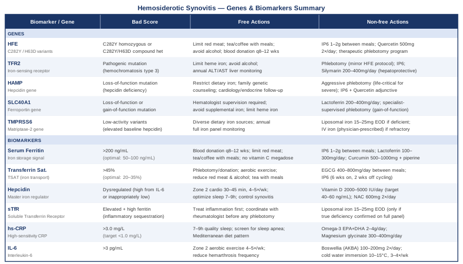
Understanding your biomarker pattern is a powerful starting point — but it becomes even more meaningful when read in the context of the underlying genetic architecture that shapes your baseline iron metabolism.
The 5 Iron Metabolism Genes That Shape Your Susceptibility
Genetic variants in iron metabolism do not cause hemosiderotic synovitis directly — but they shift the setpoint for how much iron your body absorbs, retains, and distributes, often by decades. For someone experiencing repeated joint bleeds or living with conditions that predispose to hemarthrosis, a genetic predisposition toward elevated iron absorption acts as a multiplier on local joint iron accumulation.
The following five genes are the most relevant to understanding this predisposition, based on their established roles in human iron physiology and clinical hemochromatosis research.
HFE — The Most Common Iron Overload Gene
The HFE gene, located on chromosome 6p21.3, encodes a protein that regulates hepcidin expression in response to iron sensing. Two variants are clinically meaningful: C282Y, the primary hereditary hemochromatosis mutation, and H63D, a milder variant. Individuals homozygous for C282Y have approximately 80–90% penetrance for clinical iron overload in men and 40–60% in women over a lifetime. Compound heterozygotes (C282Y/H63D) have an intermediate risk, with perhaps 2–3% developing overt iron accumulation.
In the context of hemosiderotic synovitis, HFE mutations amplify the systemic iron burden that compounds local joint accumulation. Even moderate dietary iron excesses — that would be cleared uneventfully by someone with normal HFE function — accumulate progressively in C282Y carriers. Over 10–20 years, this translates into substantially higher systemic ferritin and transferrin saturation, feeding a more reactive synovial environment.
Testing: Hemochromatosis gene panel available through most clinical genetics labs or through comprehensive wellness genetic tests. Cost: $100–300, or raw data from 23andMe can be analyzed for rs1800562 (C282Y) and rs1799945 (H63D) via third-party tools such as Genetic Genie.
According to Gary Brecka's work on gene-driven health optimization, HFE variants are among the most actionable genetic findings in iron regulation, because the intervention — phlebotomy — is straightforward once the diagnosis is established. Ali Torkamani's research on polygenic risk scores suggests that HFE variants should be interpreted in combination with other iron-pathway SNPs rather than in complete isolation.
If the gene is unfavorable — the plan without supplements
Therapeutic phlebotomy is the cornerstone and the only intervention proven to prevent organ damage in HFE hemochromatosis. The induction phase for C282Y homozygotes with elevated ferritin: 450–500 mL removed weekly or biweekly until ferritin reaches 50–100 ng/mL. Maintenance: every 3–4 months thereafter. Blood donation every 8–12 weeks serves the same purpose for those below the threshold for formal medical phlebotomy.
Dietary strategy: limit red meat to 1–2 servings per week; avoid alcohol (significantly increases intestinal iron absorption and reduces hepcidin); consume tea, coffee, or cacao with iron-rich meals; avoid cast-iron cookware; do not supplement with iron or vitamin C mega-doses.
If the gene is unfavorable — the plan with supplements
IP6 at 1–2 grams twice daily between meals provides a continuous iron chelation signal between phlebotomy sessions. Quercetin at 500 mg twice daily adds both iron chelation and anti-inflammatory properties relevant to synovial health. Cycling both: 8 weeks on, 2 weeks off. Monitor ferritin every 3 months. Side effects: IP6 reduces zinc and calcium co-absorption at higher doses; quercetin may interact with CYP3A4-metabolized medications.
TFR2 — The Iron-Sensing Receptor That Talks to Hepcidin
TFR2 encodes transferrin receptor type 2, which acts as an iron sensor on hepatocytes and contributes to hepcidin upregulation when transferrin saturation is elevated. Mutations in TFR2 cause hemochromatosis type 3 — a phenotype clinically similar to HFE disease but typically presenting earlier and potentially more aggressively, since TFR2 is an independent node in the iron-sensing pathway separate from HFE.
When TFR2 is non-functional, the liver fails to sense elevated TSAT and mount an appropriate hepcidin response. Iron continues to accumulate regardless of store levels. For hemosiderotic synovitis, this means the same amplification dynamic as HFE: a biologically uncorrectable tendency toward iron excess that must be managed through active lifestyle and clinical intervention.
If the gene is unfavorable — the plan without supplements
The phlebotomy protocol mirrors HFE hemochromatosis. Annual liver function testing (ALT, AST) is additionally important because TFR2 hemochromatosis may carry more pronounced hepatic iron accumulation. The dietary iron management strategy is identical: limit heme iron sources, use absorption inhibitors strategically, avoid alcohol.
If the gene is unfavorable — the plan with supplements
IP6, lactoferrin, and quercetin as described for HFE. Add silymarin (milk thistle) at 200–400 mg standardized silymarin extract daily as a hepatoprotective measure when liver iron accumulation is a concurrent concern. Cycling: 12 weeks on, 4 weeks off. Side effects: generally well-tolerated; rare GI symptoms; avoid in individuals with certain hormone-sensitive conditions (mild estrogenic activity).
HAMP — The Hepcidin Gene Itself
HAMP encodes hepcidin antimicrobial peptide directly. Mutations in HAMP cause juvenile hemochromatosis type 2B — a rare but severe condition presenting in the second or third decade with cardiomyopathy, hypogonadism, liver disease, and joint involvement. These mutations effectively eliminate the central brake on iron absorption, resulting in iron accumulation rates far exceeding adult-onset hemochromatosis.
More broadly, functional variants in HAMP regulatory regions that reduce (but do not eliminate) hepcidin expression are an active area of research. Gary Brecka references the hepcidin pathway as central to understanding why some individuals respond dramatically to dietary iron management while others require clinical intervention — the genetic setpoint matters. Population-level evidence for subtle HAMP variants remains preliminary, but the direction of the research is consistent.
If the gene is unfavorable — the plan without supplements
For confirmed HAMP mutations, early and aggressive phlebotomy is life-critical, not merely advisable. Genetic counseling for first-degree family members is standard. Cardiology and endocrinology follow-up are essential given multi-organ involvement in juvenile forms. Dietary interventions alone cannot compensate for severe hepcidin deficiency.
If the gene is unfavorable — the plan with supplements
For confirmed rare mutations: supplements are adjunctive only — IP6, quercetin, and lactoferrin can modestly reduce intestinal iron absorption between phlebotomy sessions, but they are not primary therapy. Experimental research is exploring synthetic hepcidin agonists and erythroferrone inhibitors for HAMP-deficient states; these are not yet commercially available. For milder promoter variants, the supplement protocol mirrors HFE.
SLC40A1 — The Ferroportin Gene and Macrophage Iron Trapping
SLC40A1 encodes ferroportin, the only known mammalian cellular iron exporter. It is the protein directly targeted by hepcidin. Mutations cause ferroportin disease (hemochromatosis type 4) in two clinically distinct forms:
Loss-of-function mutations: Ferroportin cannot export iron from macrophages. Iron accumulates inside macrophages, serum ferritin rises markedly, but TSAT may be normal or even low because iron is trapped rather than circulating. This is directly relevant to hemosiderotic synovitis: synovial macrophages are the primary cells accumulating iron after hemarthrosis. A ferroportin loss-of-function variant creates a biological environment where those macrophages are even less capable of clearing their iron burden.
Gain-of-function mutations: Ferroportin becomes resistant to hepcidin's inhibitory signal. Iron floods out of macrophages unimpeded; this phenotype resembles classical HFE hemochromatosis.
If the gene is unfavorable — the plan without supplements
Loss-of-function ferroportin disease requires specialist management. Aggressive phlebotomy can trigger anemia (because serum iron is already low) while macrophage iron remains high. The goal is gradual ferritin normalization over 12–24 months under hematologist supervision. Gain-of-function type is managed similarly to HFE hemochromatosis. Do not self-manage ferroportin disease with phlebotomy without clinical guidance.
If the gene is unfavorable — the plan with supplements
Lactoferrin at 200–400 mg/day is particularly relevant here, as it modulates iron handling specifically within macrophages — the primary iron-loaded cell type in loss-of-function ferroportin disease. Avoid all supplemental iron in any form. Reduce dietary heme iron to the minimum consistent with avoiding anemia. Monitor hemoglobin and MCV alongside ferritin.
TMPRSS6 — Iron Sensing and the Hepcidin Suppression Signal
TMPRSS6 encodes matriptase-2, a transmembrane serine protease that cleaves hemojuvelin on hepatocytes, thereby suppressing hepcidin production when the body needs more iron. Mutations that abolish TMPRSS6 function cause IRIDA (iron-refractory iron deficiency anemia), a condition where hepcidin is pathologically elevated despite depleted iron stores — oral iron supplementation fails entirely because hepcidin blocks intestinal absorption regardless of dose.
In the population-wide context, common SNPs in TMPRSS6 are among the strongest genetic determinants of baseline serum iron and hepcidin levels, according to genome-wide association studies of iron metabolism. Individuals with lower-activity TMPRSS6 variants may carry chronically higher hepcidin, which creates a paradoxical situation in hemosiderotic synovitis: systemic iron appears restricted on labs, while local joint iron accumulation continues via macrophage-mediated pathways that hepcidin does not adequately suppress at the synovial level.
If the gene is unfavorable — the plan without supplements
For IRIDA (complete loss-of-function): parenteral iron (IV iron sucrose or ferric carboxymaltose) bypasses the hepcidin blockade and is the required intervention. For common population variants with milder effects: ensure adequate dietary iron from diverse sources, monitor ferritin and TSAT annually, and interpret results in the context of the full biomarker panel rather than as isolated numbers.
If the gene is unfavorable — the plan with supplements
Standard oral iron supplementation may be partially ineffective in significant TMPRSS6 variants. Liposomal iron (15–25 mg elemental iron every other day) has modestly better efficacy across the hepcidin barrier compared to standard formulations. If iron remains refractory despite oral supplementation and confirmed deficiency, IV iron (prescribed by a physician) is the appropriate escalation. Do not initiate iron supplementation in the context of hemosiderotic synovitis without confirming iron deficiency via the full panel — supplementing into an iron-loaded joint is counterproductive.
A Book That Reframes Everything You Think You Know About Iron
Dumping Iron: How to Ditch This Secret Killer and Reclaim Your Health by P.D. Mangan is a concise, well-referenced book arguing that iron accumulation is one of the most consistently overlooked drivers of accelerated aging, cardiovascular disease, cancer risk, and metabolic dysfunction. For anyone managing a condition rooted in iron excess, it offers a perspective that most clinicians do not raise — and that challenges the implicit assumption that "normal" ferritin means safe ferritin.
10 Key Insights From Dumping Iron
1. Iron Is the Nutrient That Has No Exit
Unlike almost every other mineral, iron has no regulated excretion pathway in humans. Once absorbed, it stays. The body sheds minimal amounts through gut cell turnover, sweat, and skin cell loss — not nearly enough to compensate for excess absorption. Small daily surpluses accumulate over decades into clinically meaningful iron overload.
2. The Normal Reference Range for Ferritin Was Not Designed for Health Optimization
Mangan reviews multiple large epidemiological studies showing that the lowest quintile of ferritin — roughly 20–70 ng/mL — is associated with the lowest incidence of cardiovascular disease, type 2 diabetes, and all-cause mortality in prospective cohorts. The conventional upper limit of "normal" (up to 300 ng/mL for men) reflects population distribution, not biological optimality.
3. Iron Drives the Fenton Reaction — the Most Dangerous Chemical in Your Joints
Free iron catalyzes the conversion of hydrogen peroxide into hydroxyl radicals — among the most reactive oxidants in biology. In iron-loaded synovial tissue, this chemistry operates continuously, damaging chondrocytes and driving inflammatory gene expression. Mangan argues this mechanism connects iron overload to multiple chronic degenerative conditions through a single underlying pathway.
4. Blood Donation Is the Most Effective Iron Reduction Strategy Available Without a Prescription
Each 450–500 mL blood donation removes approximately 250 mg of iron. Donating every 8–12 weeks — the standard donation interval — can normalize ferritin in most non-genetic cases within 12–18 months. Mangan frames this as the most practical, accessible, and evidence-backed intervention available, at zero cost to the donor in most countries.
5. Alcohol Is One of the Most Powerful Iron Absorption Enhancers
Alcohol increases intestinal iron absorption through multiple mechanisms and reduces the liver's hepcidin response. Regular drinking — even at moderate levels — meaningfully elevates ferritin over time. The combination of alcohol with high-heme-iron meals is particularly problematic for iron-accumulation-prone individuals, and Mangan documents this interaction with epidemiological evidence.
6. Polyphenols in Coffee and Tea Are More Than Antioxidants
Coffee, black tea, green tea, cacao, and other polyphenol-rich foods contain compounds that bind non-heme iron in the intestine and significantly reduce its absorption. Mangan presents studies showing 30–60% reduction in iron absorption when these beverages accompany iron-containing meals. This repositions coffee and tea as practical, low-cost daily interventions rather than guilty pleasures.
7. Exercise Depletes Iron Through Several Converging Pathways
Regular aerobic exercise increases iron utilization (more hemoglobin synthesis), promotes small amounts of iron loss through sweating and GI microbleeding, and transiently suppresses hepcidin after each session. Active individuals consistently have lower ferritin than sedentary individuals with similar diets. For joint conditions, swimming and cycling achieve this benefit without adding mechanical load to the affected joint.
8. Phytic Acid in Whole Foods Deserves Rehabilitation
The nutritional establishment has long classified phytic acid (IP6) as an antinutrient because it reduces mineral absorption. Mangan argues that for iron-overloaded individuals, this same property is highly beneficial. A diet naturally rich in legumes, whole grains, and seeds provides ongoing dietary IP6 that modulates iron absorption. Supplemental IP6 builds on this baseline for those with elevated ferritin.
9. Elevated Ferritin Above 200 ng/mL May Be a Cancer Risk Signal
Mangan reviews convergent data — epidemiological, mechanistic, and from animal models — suggesting iron promotes carcinogenesis through three overlapping pathways: it drives oxidative DNA damage, supports uncontrolled cellular proliferation, and impairs immune surveillance of aberrant cells. While correlation does not equal causation, the evidence consistency across multiple cancer types is strong enough to treat moderately elevated ferritin seriously.
10. Most Physicians Are Not Trained to Treat Subclinical Iron Overload
Physicians typically act on iron only when hereditary hemochromatosis is formally confirmed — ferritin in the hundreds or thousands combined with genetic testing. In the subclinical grey zone (ferritin 150–300 ng/mL, no genetic diagnosis), most patients are reassured that their values are "within normal limits." Mangan argues this leaves millions with modifiable iron burden untreated, and that patients must understand enough to advocate for active iron management in this range.
Complementary Approaches With Clinical Evidence for Joint Inflammation
The approaches below were selected from a longer list of modalities based on the strength of human clinical evidence specifically relevant to synovial inflammation, joint pain, and the inflammatory cytokine biology underlying hemosiderotic synovitis. None replace biomarker monitoring or clinical management, but each offers a credible adjunctive benefit.
Low-Level Laser Therapy (Photobiomodulation)
Low-level laser therapy (LLLT), also called photobiomodulation (PBM), applies red and near-infrared light (typically 630–1000 nm) to tissue at non-thermal intensities. At the cellular level, PBM stimulates mitochondrial cytochrome c oxidase, increases ATP production, reduces oxidative stress, and suppresses proinflammatory cytokine expression including IL-6 and TNF-α. For hemosiderotic synovitis, this double action — addressing both oxidative stress and the inflammatory cascade — makes PBM mechanistically relevant in a way that few other physical modalities are.
A systematic review and meta-analysis by Brosseau et al. published in the Cochrane Database of Systematic Reviews found LLLT to produce statistically significant pain reduction and improved function in rheumatoid arthritis compared to placebo, with a favorable safety profile. Subsequent randomized trials in knee osteoarthritis — a condition with comparable synovial inflammatory pathology — have reproduced these findings at wavelengths between 780 and 860 nm and doses of 4–8 J/cm².
For practical application, find a physiotherapy or sports medicine clinic offering class III or IV laser therapy. Typical protocols involve 3–5 sessions per week for 3–4 weeks for an initial treatment course, followed by monthly maintenance. Home near-infrared devices (panel lights or handheld devices, 660–850 nm) are widely available at $100–500. Apply 10–15 minutes per session over the affected joint at a distance specified by the device. Avoid direct eye exposure. The evidence is strongest for knee joints and moderate for other joint sites. This modality is low-risk and worth trialing over a 4-week structured period before assessing benefit.
Tai Chi
Tai chi is a mind-body movement system combining slow, deliberate joint movements with breath coordination and directed attention. For conditions involving joint pain and functional limitation, it addresses a specific combination of deficits: proprioceptive accuracy, neuromuscular control, and reduced mechanical joint loading — all relevant to a joint that has experienced repeated hemarthrosis and synovial remodeling.
A landmark randomized controlled trial by Wang et al. published in the New England Journal of Medicine demonstrated that 12 weeks of tai chi twice weekly produced significantly greater improvements in pain, physical function, depression, and health-related quality of life in knee osteoarthritis compared to an attention control. Independent analyses have further documented reductions in systemic inflammatory markers including hs-CRP and IL-6 following sustained tai chi practice — mechanistically consistent with the cytokine management relevant to hemosiderotic synovitis.
Begin with a beginner Yang-style 24-form tai chi class — community centers, YMCA programs, and senior centers typically offer classes at $10–20 per session or less. Practice 3–5 times per week for a minimum 12-week trial before assessing benefit. Tai chi is appropriate even during the recovery phase following a mild joint bleed once acute swelling has resolved, owing to its low-impact, weight-distributed movement pattern. Avoid beginning a new tai chi practice during active significant hemarthrosis or in the presence of joint effusion.
Mindfulness-Based Stress Reduction (MBSR)
MBSR is an 8-week structured program developed by Jon Kabat-Zinn at the University of Massachusetts Medical Center. It combines formal meditation practice (body scan, sitting meditation, mindful movement) with informal daily mindfulness and group learning. For hemosiderotic synovitis, chronic pain activates the hypothalamic-pituitary-adrenal axis, elevating cortisol dysregulation over time, which impairs immune regulation and can amplify joint inflammatory tone. MBSR addresses this pathway directly.
A well-cited study by Creswell and colleagues demonstrated that an MBSR-based intervention reduced IL-6 gene expression in peripheral blood mononuclear cells in a randomized controlled design, suggesting a direct pathway from mindfulness practice to cytokine regulation at the transcriptional level. A 2019 meta-analysis reviewing mindfulness interventions across multiple populations found consistent reductions in hs-CRP and IL-6 as secondary outcomes, with effect sizes comparable to moderate pharmacological interventions for CRP.
The standard 8-week MBSR curriculum involves approximately 45 minutes of formal practice per day and one full-day retreat. Accredited online programs are available through institutions including UMass Memorial Medical Center and various university health systems, typically costing $300–500. For pain management specifically, the mechanism is well understood — neuroimaging studies demonstrate changes in how the brain processes pain signals, reducing the aversive quality without necessarily reducing pain intensity. This pain-brain remodeling is genuinely useful for individuals managing chronic joint disease, and the inflammatory benefits are a documented bonus.
Chinese Herbal Medicine — Selected Compounds With Iron-Relevant Mechanisms
Several compounds from the classical Chinese herbal medicine pharmacopoeia have well-characterized mechanisms directly relevant to iron metabolism and synovial inflammation. This is not a case of vague traditional claims — these are specific phytochemicals studied in peer-reviewed literature for their iron chelation, hepcidin modulation, and anti-inflammatory properties.
Baicalein from Scutellaria baicalensis (Huang Qin) and quercetin from Sophora japonica are both potent iron chelators with IL-6 suppression activity. Research in synovial cell models has demonstrated that polyphenolic iron chelators including quercetin reduce iron-induced reactive oxygen species in human fibroblast-like synoviocytes — a direct mechanistic match for hemosiderotic synovitis. A randomized clinical trial of Scutellaria extract in knee osteoarthritis found significant reductions in pain and inflammatory biomarkers versus placebo over 8 weeks.
For practical application, standardized western-format supplements provide defined, reproducible doses. Scutellaria baicalensis extract standardized to 85% baicalin at 250–500 mg twice daily combined with quercetin at 500 mg twice daily provides a measurable iron-chelating and anti-inflammatory dose. Cycling: 8–12 weeks on, 2–4 weeks off. Monitor liver enzymes if using high doses of Scutellaria long-term — rare hepatotoxicity has been reported at non-standard doses. Quercetin may interact with CYP3A4-metabolized medications and blood thinners. For formulation specific to individual presentation, consultation with a licensed practitioner of Traditional Chinese Medicine adds meaningful clinical nuance beyond over-the-counter supplementation.
Conclusion
Hemosiderotic synovitis is not a condition where generic anti-inflammatory advice does the real work. The underlying biology — iron accumulation in synovial tissue, oxidative stress via the Fenton reaction, cytokine-driven amplification loops, and genetic predisposition to iron excess — requires precision, not broad strokes. The six biomarkers covered here give you a structured and accessible way to see what is actually happening in your iron metabolism and inflammatory state. The five genes add the longer-term context: whether your baseline biology is working against you and which interventions are likely to matter most.
The most useful next step is straightforward: at your next blood draw, request a full iron panel (ferritin, serum iron, TSAT, TIBC) plus hs-CRP if it is not already included. Review the results through the lens of the ranges discussed in this article rather than only asking whether they fall within reference limits. Bring specific questions to your hematologist or rheumatologist — about your hepcidin level if the iron picture is confusing, about genetic testing for HFE if iron markers are persistently elevated, or about therapeutic phlebotomy if ferritin trends upward over time. Better questions, grounded in a clearer understanding of your own biology, lead to better conversations — and better decisions.
Musculoskeletal: Joint Conditions
Digestive: Liver & Gallbladder Conditions
Autoimmune: Inflammatory Conditions