This article contains AI generated content.
· UpdatedHereditary Multiple Exostoses: 3 Genes And 6 Biomarkers To Track
Introduction
Living with hereditary multiple exostoses means navigating a condition where the same diagnosis can produce wildly different lives. One person carries a handful of small, stable growths for decades. Another develops dozens of exostoses that compress nerves, limit joint mobility, and require repeated surgeries before they turn thirty. This variability is not random — it reflects the precise interaction between specific gene mutations, epigenetic context, growth factor signaling, and the biochemical environment that surrounds each growth plate. Understanding these layers separates genuinely useful information from the kind of vague reassurance that leaves you no better off.
Most clinical conversations about HME land in the same place: "You have a mutation in EXT1 or EXT2. There is no medication to fix it. We watch and operate when necessary." That is accurate, but incomplete. Over the past decade, research into the molecular biology of HME has accelerated considerably. Researchers now understand in significant detail why exostoses form, what cellular and signaling events drive their growth, and which modifiable factors interact with those pathways in ways that may matter over a lifetime.
This article takes that deeper approach. It does not promise to reverse your genetics or eliminate exostoses. What it provides is a precise, actionable map: which genes are doing what and how their dysfunction can be partially compensated for, which biomarkers are worth tracking and what they reveal about disease activity, and which evidence-supported complementary approaches deserve a place in your management plan. Two core frameworks organize this: a genetics and epigenetics lens that examines the root molecular drivers, and a biomarker lens that gives you measurable, trackable signals of what is happening in your body right now.
Better information leads to better decisions — that is the only reliable claim worth making here. Whether you are newly diagnosed, the parent of a child with HME, or someone who has been managing this condition for years, the following sections are designed to raise the quality of the decisions you make alongside your medical team.
The Genetic Architecture of HME: What EXT1, EXT2, and Related Pathway Genes Are Actually Doing
Hereditary multiple exostoses is caused by mutations in genes encoding enzymes responsible for synthesizing heparan sulfate (HS) — long sugar chains that attach to cell-surface and extracellular matrix proteins. These chains are not structural filler. They are active regulators of how cells receive and interpret signals from key growth factors and morphogens, including Indian Hedgehog (IHH), Fibroblast Growth Factor (FGF), Bone Morphogenetic Protein (BMP), and Wnt ligands. Without adequate HS synthesis, the signaling environment at the growth plate becomes dysregulated, and the conditions for exostosis formation are set in motion. A broad scientific overview of the condition is maintained at MedlinePlus Genetics — Hereditary Multiple Exostoses.
Why the Same Mutation Produces Multiple Lesions
HME follows an autosomal dominant inheritance pattern — one mutated copy of EXT1 or EXT2 is sufficient to be inherited and predispose to the condition. But the exostoses themselves form through a "second-hit" mechanism that mirrors what is seen in classical tumor suppressor biology. The germline mutation is the first hit. A somatic mutation that silences the remaining functional copy in an individual growth plate progenitor cell is the second. That cell, now without any HS biosynthesis capacity, proliferates clonally and gives rise to a single exostosis. This model, supported by molecular analysis of surgically removed osteochondromas, explains why HME produces multiple discrete lesions over time: each is its own independent somatic event. It also explains why the number and location of lesions is imperfectly predictable, even between siblings who share the same germline mutation.
EXT1: The Most Common Mutation and What It Predicts
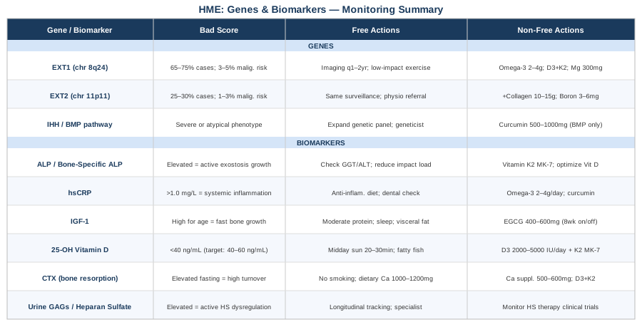
EXT1 (Exostosin Glycosyltransferase 1), located on chromosome 8q24.11, encodes the larger subunit of the EXT1/EXT2 heterodimeric enzyme complex. This complex is responsible for polymerizing HS chains in the Golgi apparatus. Loss-of-function mutations in EXT1 account for approximately 65 to 75 percent of genetically confirmed HME cases. Mutation types include frameshift insertions and deletions, nonsense mutations, splice-site variants, and large intragenic deletions — the majority resulting in a truncated or absent protein product. The full gene entry, including known pathogenic variants, is at EXT1 on NCBI Gene.
Evidence from several genotype-phenotype cohort studies suggests that EXT1 mutations are associated with a higher average exostosis burden and a modestly elevated lifetime risk of malignant transformation to secondary chondrosarcoma — estimated at 3 to 5 percent in EXT1 carriers compared to 1 to 3 percent in EXT2 carriers. The evidence is not fully consistent across cohorts, and individual variability remains large, but this signal justifies heightened vigilance in EXT1-mutated individuals, particularly for lesions in the pelvis, axial skeleton, and proximal limb girdles — locations associated with a higher proportion of malignant transformation events. You can explore the research landscape via PubMed: HME malignant transformation and EXT1.
If EXT1 is mutated — the plan without supplements
No lifestyle or dietary intervention can restore a mutated gene to function. The foundational approach is surveillance and smart systemic management:
- Structured imaging schedule: Radiographic surveillance at minimum every 12 to 24 months during childhood and adolescence, when exostoses are most actively growing. In adulthood, any known lesion that changes in size, pain pattern, or character warrants prompt MRI evaluation. Continued growth after skeletal maturity is a key clinical signal for potential malignant transformation and should never be dismissed as incidental. - Low-impact exercise preference during active growth phases: Repetitive high-impact loading near active lesion sites — particularly the distal femur, proximal tibia, and proximal humerus — can generate local mechanical stress and inflammatory signaling. Shifting to swimming, cycling, or elliptical during rapid growth phases is a rational adjustment. This is not about avoiding exercise; skeletal loading remains broadly beneficial for bone density. It is about being selective regarding loading type and anatomic location. - Sleep duration and quality: Growth hormone is secreted in pulses during slow-wave sleep. Chronic sleep deprivation disrupts GH pulsatility, but adequate deep sleep sustains it — which in growing individuals can translate to increased IGF-1 and accelerated bone growth, including at exostosis sites. Prioritizing 8 to 10 hours of quality sleep in children and adolescents with HME is a meaningful, often underappreciated, management lever. - Patient registry participation: Enrolling in the MHE Research Foundation patient registry is a zero-risk action with genuine benefit — connecting individuals to emerging clinical trials and contributing longitudinal data that advances understanding of phenotypic variability.
If EXT1 is mutated — the plan with supplements or targeted approaches
Several adjuncts have biological plausibility in HME, though none have been validated in dedicated clinical trials in this specific population:
- Omega-3 fatty acids (EPA + DHA): 2 to 4 grams per day of combined EPA and DHA from high-quality fish oil or algal oil. The anti-inflammatory evidence is strong across multiple conditions, and the biological rationale in HME relates to the inflammatory microenvironment around growing exostoses. Assess hsCRP response at 3 months. Side effects: mild blood-thinning at higher doses; pause one week before any elective surgery. Ongoing use; no cycling required. - Vitamin D3 with K2: Target serum 25-OH Vitamin D at 40 to 60 ng/mL. Vitamin D receptors are expressed in growth plate chondrocytes, and Vitamin D modulates both BMP and Hedgehog pathway signaling — the same pathways disrupted in HME. Typical starting dose: 2000 to 4000 IU D3 daily, paired with 100 to 200 mcg Vitamin K2 (MK-7 form, which directs calcium toward bone rather than soft tissue). Recheck serum Vitamin D at 3 months and adjust. Side effects: negligible at monitored doses. - Magnesium glycinate: 300 to 400 mg per day. Magnesium is required for hundreds of enzymatic reactions, including those involved in bone mineralization and chondrocyte function. Many people run suboptimal. The glycinate form is better tolerated gastrointestinally than oxide or sulfate. Side effects: loose stools at higher doses; reduce dose if this occurs. - Anti-inflammatory dietary pattern as a foundation: Mediterranean-style eating — abundant fatty fish, olive oil, vegetables, legumes, minimal refined carbohydrates and industrial seed oils — provides a sustained anti-inflammatory signal that complements any specific supplementation. Effects accumulate over months, not days.
EXT2: The Second Major Gene and Its Distinct Considerations
EXT2 (Exostosin Glycosyltransferase 2), located on chromosome 11p11-12, encodes the smaller subunit of the same EXT1/EXT2 heterodimer. Neither enzyme functions effectively without the other. EXT2 mutations account for approximately 25 to 30 percent of HME cases. Some published genotype-phenotype cohort studies report a somewhat milder average phenotype in EXT2-mutated individuals — fewer exostoses, lower average deformity severity, and modestly lower malignant transformation risk. The full gene entry is available at EXT2 on NCBI Gene.
This "milder on average" finding should be treated with appropriate skepticism at the individual level. Within EXT2-mutated families, there is still wide phenotypic variability, and some individuals with EXT2 mutations carry a significant lesion burden. The genotype does not reliably determine the phenotype, and reduced surveillance based on mutation type alone is not justified.
If EXT2 is mutated — the plan without supplements
- Maintain the same surveillance framework: Annual orthopedic review, growth-phase imaging, and adult monitoring for any lesion that changes. The modestly lower average risk associated with EXT2 does not support reducing vigilance. - Early physiotherapy referral for deformity prevention: Forearm deformity arising from distal ulnar lesions at the distal radioulnar joint is particularly associated with HME and can progress significantly if not monitored. Angular limb deformity and leg length discrepancy are additional orthopedic complications that respond better to conservative management when identified early. A physiotherapist experienced in musculoskeletal conditions can provide targeted joint mobility work and compensatory strengthening that preserves function between surgical interventions. - Functional movement mapping every 2 to 3 years: For individuals with multiple forearm or lower limb lesions, detailed functional movement assessment allows early detection of emerging restriction patterns before they become fixed deformities. Comparing results across time points gives both the patient and the treating team a more nuanced picture than imaging alone.
If EXT2 is mutated — the plan with supplements or targeted approaches
The same foundational supplement approach as for EXT1 applies (omega-3, Vitamin D3 with K2, magnesium glycinate, anti-inflammatory diet). Two additional options are worth noting:
- Hydrolyzed collagen peptides: 10 to 15 grams per day taken alongside 500 mg of Vitamin C. Emerging evidence supports collagen peptide supplementation for cartilage and joint matrix maintenance. No HME-specific trials exist, but the biological plausibility is reasonable given the importance of cartilage cap integrity in HME lesions, and the safety profile is excellent. Ongoing use; no cycling required. Side effects: rare gastrointestinal discomfort in some individuals. - Boron: 3 to 6 mg per day from diet or supplement. Boron supports BMP-2 expression and bone mineralization processes. Dietary sources include avocado, raisins, almonds, and chickpeas. At these doses, side effects are minimal.
Downstream Signaling Pathway Genes as Disease Modifiers
Two individuals with the same EXT1 mutation can experience dramatically different disease severity — different numbers of lesions, different rates of growth, different patterns of deformity. Part of this variability is unexplained, but part may be attributable to variants in genes downstream of the HS biosynthesis pathway. These are not HME-causative mutations, but they may modulate how severely impaired HS signaling manifests at the cellular level.
The Indian Hedgehog Pathway
Indian Hedgehog (IHH) is among the most HS-dependent morphogens in the growth plate. HS chains on cell surfaces and in the extracellular matrix are required for proper IHH gradient formation, receptor binding efficiency, and signaling range. When HS is reduced, IHH signaling is compressed and distorted, leading to abnormal chondrocyte differentiation. Variants in IHH, its receptor PTCH1, or the pathway effector SMO may modulate how severely a given reduction in HS affects Hedgehog signaling output. This interaction is under active investigation: PubMed: HME and Hedgehog signaling.
BMP Signaling Variants
Bone Morphogenetic Proteins drive chondrocyte and osteoblast differentiation, and HS chains regulate BMP gradient formation and receptor binding. Variants in BMP2, BMP4, or BMPR1A may influence how aggressively exostoses grow and how rapidly they mineralize. For individuals with an unusually severe or atypical HME presentation, a comprehensive next-generation sequencing panel covering HS biosynthesis and related signaling pathway genes — beyond targeted EXT1/EXT2 sequencing alone — may yield interpretive value that informs surveillance planning.
If downstream pathway variants are identified — the plan
- Request comprehensive genetic panel coverage if your testing was limited to targeted EXT1/EXT2 analysis. - Bring the full genomic report to a clinical geneticist or specialist in rare bone diseases for contextual interpretation. - Consider enrollment in a patient registry to contribute to collective genotype-phenotype understanding. - For BMP pathway variants: bioavailable curcumin (phospholipid complex or nanoparticle formulation, 500 to 1000 mg/day with food) has demonstrated BMP signaling modulation in preclinical studies. Cycling: 8 weeks on, 2 weeks off. Side effects: GI sensitivity in some individuals; caution with anticoagulants. This is a biologically plausible low-risk adjunct, not a validated treatment.
The Epigenetic Layer: What Can Be Partially Shifted Without Changing the Gene
The germline mutation in EXT1 or EXT2 cannot be reversed by lifestyle or supplementation. But the epigenetic layer — which controls gene expression without altering the DNA sequence — is partially modifiable, and it is relevant in HME in specific ways. Research on sporadic (non-hereditary) osteochondromas has documented abnormal CpG methylation at EXT1 and EXT2 promoters in tumor tissue, suggesting that epigenetic silencing of the functional copy contributes to individual lesion formation. MicroRNAs including miR-21, miR-140, and miR-146a regulate chondrocyte behavior and are influenced by diet, exercise, and metabolic status.
Practical epigenetic levers:
- Methyl donor nutrition: Folate (from leafy greens, legumes, or methylfolate supplement at 400 to 800 mcg/day), methylcobalamin B12 (500 to 1000 mcg/day), betaine, and choline support the DNA methylation machinery that regulates gene expression. A standard active B-complex covering these methyl donors is low-risk and broadly supported by nutritional biochemistry. - Adapted resistance training: Moderate resistance training has documented epigenetic effects, including changes in miRNA expression relevant to inflammation and bone metabolism. Work with a physiotherapist familiar with your specific lesion map to design a program that provides skeletal loading signals without concentrating stress at known exostosis sites. - Endocrine disruptor reduction: Synthetic estrogens and endocrine-disrupting chemicals found in plastics, certain personal care products, and pesticide residues interfere with growth plate signaling pathways that interact with HS function. Minimizing plastic food contact (particularly for hot or fatty foods), choosing lower-pesticide produce where accessible, and using personal care products without parabens or phthalates are practical, if difficult to individually quantify, protective measures.
6 Biomarkers to Track the Biology of HME Year Over Year
Imaging shows you where exostoses are. Biomarkers reveal what is happening metabolically — whether bone turnover is elevated, whether systemic inflammation is driving activity, whether key nutrient status is suboptimal, or whether the body is in a biochemical state that may favor lesion growth. The following six markers form a practical, largely affordable monitoring panel that complements standard orthopedic care. Used longitudinally, they give you the kind of early, actionable information that waiting for symptoms does not.
Alkaline Phosphatase (ALP) and Bone-Specific ALP
Why it matters: Alkaline phosphatase is produced by osteoblasts during active bone formation. In HME, elevated ALP can signal periods when exostoses are growing more actively. Standard ALP is included in a comprehensive metabolic panel (CMP) but reflects both bone and liver sources. Bone-specific alkaline phosphatase (BSAP) isolates the skeletal fraction and gives a cleaner signal.
How to measure it: Standard ALP via CMP: approximately $10 to $30 when bundled. Bone-specific ALP as standalone: approximately $50 to $80. Interpret longitudinally — trends over 6 to 12 months matter more than any single measurement.
If ALP is elevated — the plan without supplements: Rule out hepatic causes first by checking GGT and ALT alongside ALP. If bone-derived, schedule a clinical review and consider updated imaging of known lesions. Reduce high-impact mechanical loading at active sites during this period. Document result, date, and clinical context systematically.
If ALP is elevated — the plan with supplements or equipment: Vitamin K2 (MK-7 form, 100 to 200 mcg/day) supports carboxylation of osteocalcin and helps direct bone-forming activity more efficiently toward the bone matrix. Ensure Vitamin D is in the optimal range (40 to 60 ng/mL) before drawing firm conclusions from ALP in isolation. Side effects of K2: minimal; caution in individuals taking warfarin.
High-Sensitivity C-Reactive Protein (hsCRP)
Why it matters: hsCRP is the most accessible marker of systemic low-grade inflammation. Exostosis development and growth involves a local inflammatory microenvironment, and systemic inflammation may amplify this. Chronically elevated hsCRP — above 1.0 mg/L, with 0.5 mg/L as a tighter optimal threshold used by evidence-based clinicians — indicates a pro-inflammatory state that may accelerate bone and cartilage remodeling processes relevant to HME progression.
How to measure it: Standard blood test. Cost: approximately $20 to $40. Request specifically high-sensitivity CRP, not standard CRP, for clinical relevance at low-grade inflammatory levels.
If hsCRP is elevated — the plan without supplements: Anti-inflammatory dietary pattern as the primary lever. Address identifiable drivers: subclinical dental disease (a major and frequently overlooked source of systemic inflammation), metabolic dysfunction, sleep deprivation, and visceral adiposity.
If hsCRP is elevated — the plan with supplements or equipment: Omega-3 (EPA + DHA, 2 to 4 grams/day) has the strongest clinical evidence for reducing hsCRP across conditions. Recheck at 3 months. Bioavailable curcumin (500 to 1000 mg/day, cycling 8 weeks on and 2 weeks off) is a useful secondary option. Side effects: omega-3 at higher doses may mildly impair platelet aggregation — pause one week before any elective procedure.
IGF-1 (Insulin-Like Growth Factor 1)
Why it matters: IGF-1 is the primary mediator of growth hormone's effects on skeletal tissue. It drives chondrocyte proliferation and bone formation — including at exostosis sites. Elevated IGF-1 for age may correlate with faster exostosis growth in children and adolescents. In adults, persistently elevated IGF-1 driven by excess protein intake, visceral fat, or other factors may sustain a pro-growth signal relevant to anyone monitoring known lesions.
How to measure it: Standard blood draw. Cost: approximately $50 to $100. Always interpret using age-specific reference ranges — IGF-1 peaks in mid-adolescence and declines substantially with age.
If IGF-1 is high — the plan without supplements: Moderate excessive protein intake if present. Time high-protein meals away from the late-evening period when GH pulsatility is highest. Reduce visceral fat — the most potent modifiable driver of elevated IGF-1 in adults. Optimize sleep architecture. Avoid GH-promoting supplements (arginine, ornithine, high-dose GABA before bed) if IGF-1 is already elevated.
If IGF-1 is high — the plan with supplements or equipment: Green tea extract (EGCG, 400 to 600 mg/day) has shown modest IGF-1 lowering in some clinical studies. Cycling: 8 weeks on, 2 weeks off. Side effects: GI sensitivity on an empty stomach; may reduce iron absorption — separate from iron-rich meals by at least 2 hours. This is a modest adjunct, not a primary intervention.
25-OH Vitamin D
Why it matters: Vitamin D deficiency is highly prevalent and carries specific relevance in HME: Vitamin D receptors are expressed in growth plate chondrocytes, and Vitamin D directly modulates both BMP and Hedgehog pathway signaling — the same pathways dysregulated by EXT mutations. Suboptimal Vitamin D status may compound the signaling environment already disrupted by impaired HS synthesis.
How to measure it: Serum 25-OH Vitamin D (25-hydroxyvitamin D). Cost: approximately $30 to $60. Target range for musculoskeletal health: 40 to 60 ng/mL, consistent with evidence-based guidance from clinicians like Peter Attia who emphasize the upper half of normal for bone and inflammatory conditions.
If Vitamin D is suboptimal — the plan without supplements: Regular midday sun exposure (20 to 30 minutes with arms and legs exposed, without sunscreen). Reliable only in lower latitudes and appropriate seasons. Increase dietary sources: fatty fish (salmon, mackerel, sardines), egg yolks, and fortified foods.
If Vitamin D is suboptimal — the plan with supplements or equipment: Vitamin D3, 2000 to 5000 IU/day based on baseline level. Always pair with Vitamin K2 (MK-7, 100 to 200 mcg/day). Recheck 25-OH Vitamin D at 3 months and adjust dose to reach target. Side effects: negligible at monitored doses; prolonged high-dose use without testing is not advisable if there is a personal or family history of hypercalcemia or nephrolithiasis.
CTX (C-Terminal Telopeptide of Type I Collagen)
Why it matters: CTX is a bone resorption marker — it rises when bone matrix is being actively broken down. Paired with ALP (a bone formation marker), it provides a picture of overall bone turnover dynamics. In HME, elevated CTX during periods of active exostosis development indicates that the remodeling process is running at high intensity. High ALP combined with high CTX is a high-turnover signal that may correlate with active disease phases and warrants clinical attention.
How to measure it: Serum CTX, fasting, ideally drawn in the morning before 10 am (CTX exhibits strong diurnal variation). Cost: approximately $50 to $100. Use age-appropriate reference ranges — CTX is naturally higher during adolescence and differs between pre- and postmenopausal women.
If CTX is elevated — the plan without supplements: Eliminate smoking, which is among the most potent modifiable drivers of accelerated bone resorption. Ensure adequate dietary calcium (1000 to 1200 mg/day from food — dairy, sardines, fortified plant milks, leafy greens). Prioritize weight-bearing and resistance exercise, which shifts the bone remodeling balance toward formation. Maintain adequate protein intake (1.2 to 1.6 g/kg body weight) to support bone matrix synthesis.
If CTX is elevated — the plan with supplements or equipment: Calcium supplementation as an adjunct to dietary intake only — 500 to 600 mg per dose (the intestinal absorption limit), never more than 600 mg at once. Vitamin D3 with K2 as described above. Strontium ranelate has bone resorption-reducing evidence but carries cardiovascular risk signals in certain populations — relevant only in specialist context and not for general self-administration.
Urine Glycosaminoglycans and Heparan Sulfate
Why it matters: This is the most mechanistically direct biomarker for HME. Since EXT1 and EXT2 mutations impair heparan sulfate synthesis, measuring urinary excretion of glycosaminoglycans — the broader class that includes heparan sulfate — reflects the global systemic burden of HS dysregulation. Elevated urinary GAGs have been documented in HME patients, particularly during active growth phases, and may serve as a marker of disease activity that complements rather than duplicates imaging information. See current evidence at PubMed: HME and heparan sulfate urinary markers.
How to measure it: Urine GAG quantification is available through metabolic disease laboratories and academic medical centers. Cost: approximately $100 to $200. Not yet part of standard HME management protocols, but increasingly recognized as the field moves toward molecular monitoring. Ask your specialist about availability through their institutional network.
If GAG levels are elevated — the plan without supplements: Use as a longitudinal tracking signal rather than an isolated actionable finding. Elevated urinary GAGs during a clinical review period may justify accelerated imaging scheduling or closer surveillance of known lesions. Discuss the result explicitly with your treating team — this is not a marker most clinicians will interpret without prompting.
If GAG levels are elevated — the plan with supplements or equipment: Research into HS-directed therapies — including heparanase inhibitors and HS mimetics — is active but currently in early clinical stages. Monitor clinicaltrials.gov for studies specifically targeting HS biosynthesis in HME. The MHE Research Foundation maintains a patient registry that connects individuals to emerging trials and provides updated information as the evidence base develops. No commercially available supplement has been validated for normalizing GAG levels in this condition.
10 Research Insights About HME That Are Changing How Specialists Think
Research on HME has accelerated substantially since the discovery of EXT1 and EXT2 in the mid-1990s. What follows is a synthesis of the most impactful insights from the collective body of molecular and clinical research — things that most patients never hear in a standard outpatient visit, and that even some clinicians have not fully integrated into their approach.
1. Each Exostosis Is Genetically Distinct
Individual exostoses within the same patient are not copies of each other — each arises from an independent somatic second-hit event in a unique progenitor cell. This means the exostoses in your knee and your shoulder are genetically distinct lesions. It also means there is no single systemic treatment target that will simultaneously address all lesions, and it underscores why the field is pursuing strategies that address the upstream HS deficiency rather than targeting individual tumors.
2. The Cartilage Cap Thickness Is the Critical Malignant Transformation Signal
Not all exostoses carry the same risk of malignant transformation. The thickness of the cartilage cap on an osteochondroma is the most reliable imaging predictor. A cap greater than 2 cm on MRI in a skeletally mature individual is a significant warning signal; in children, thicker caps are more common and less concerning since caps thin as skeletal maturity approaches. Annual cap measurement in adults with larger lesions should be standard, not optional. This is a finding well-established in the orthopedic oncology literature and worth ensuring your care team is actively measuring.
3. Heparan Sulfate Deficiency Affects More Than the Skeleton
Because HS chains regulate signaling across virtually every tissue type, EXT mutations can have systemic implications beyond bone. Neurological manifestations — including cognitive differences in some HME patients — have been reported in a subset of studies, likely because HS chains regulate axon guidance and synaptic organization. This is an area of ongoing research, and its clinical implications are not yet fully defined, but it is a reason to take the systemic nature of the condition seriously rather than treating HME as purely a skeletal problem.
4. Mosaic EXT Mutations Are Likely Underdiagnosed
A proportion of individuals who appear to have sporadic (non-familial) osteochondromas may actually carry mosaic EXT mutations — mutations present in only a fraction of cells due to a post-zygotic somatic event rather than a germline mutation. Standard Sanger sequencing can miss mosaic variants at low allele fractions. Next-generation sequencing with sufficient depth is more sensitive and may reveal a diagnosis that changes surveillance recommendations for the affected individual and their children.
5. Approximately 10 to 15 Percent of HME Cases Have No Identified EXT Mutation
Genetic testing in HME does not always identify the causative variant. In 10 to 15 percent of clinically diagnosed HME cases, neither EXT1 nor EXT2 mutations are found by standard testing. This may reflect deep intronic variants, large structural rearrangements that require specialized detection methods, mosaic mutations at low allele fractions, or involvement of additional genes. A negative standard genetic test does not exclude HME, and escalating to comprehensive genomic analysis is appropriate when the clinical picture strongly supports the diagnosis.
6. Malignant Transformation Predominantly Occurs in Specific Anatomic Locations
Secondary chondrosarcoma in HME does not occur randomly across all lesion sites. The pelvis, scapula, and proximal limb girdles account for a disproportionate share of malignant transformation events. Lesions in these locations deserve more aggressive and frequent surveillance — including MRI rather than X-ray alone — regardless of size, because their depth makes physical examination unreliable for detecting change. Distal extremity lesions have a much lower transformation rate.
7. Surgical Removal Does Not Prevent New Exostoses from Forming
A common misconception is that removing exostoses reduces the overall disease burden over time. It does not. Surgical excision removes individual lesions — it does not address the germline mutation or the second-hit mechanism that drives new lesion formation. New exostoses can and do form at sites separate from previous excisions, particularly during childhood and adolescence. Surgery is indicated for specific clinical indications (pain, nerve compression, joint deformity, growth disturbance, suspicion of malignant transformation) — not as a prophylactic or disease-modifying strategy.
8. Animal Studies Have Produced the First Pharmacological Proof of Concept
Mouse models carrying Ext1 or Ext2 conditional knockouts have been used to test pharmacological interventions. Studies using compounds that activate Hedgehog signaling, modulate BMP pathways, or restore partial HS function have reduced exostosis formation in these models. While these findings do not translate directly to clinical treatments, they provide a compelling biological proof-of-concept that pharmacological modulation of HME is achievable in principle. Clinical translation is ongoing, and following the MHE Research Foundation's trial updates is the most direct way to track progress.
9. Pain in HME Is Significantly Undertreated and Underreported
Clinical surveys of HME patients consistently reveal that pain is substantially more prevalent and impactful than typically acknowledged in clinical notes. Many patients report adapting their lives around pain rather than disclosing it because they have been told the condition is "benign." The chronic pain associated with HME — from mechanical impingement, bursa formation over exostosis surfaces, nerve compression, and joint restriction — deserves explicit evaluation and management, not passive acceptance. Validated pain assessment tools and access to pain management specialists should be part of standard HME care.
10. The Psychosocial Burden Is a Distinct and Under-Addressed Clinical Domain
HME affects body image, physical capacity, occupational choices, and social participation — particularly during adolescence. Qualitative research with HME patients documents high rates of anxiety related to malignant transformation risk and frustration at the unpredictability of disease course. Psychological support, access to condition-specific peer communities, and explicit conversations about quality of life are not secondary concerns — they are primary clinical needs that improve adherence to surveillance, willingness to report symptom changes, and overall outcomes.
Complementary Approaches with Evidence Worth Knowing
The following modalities do not modify the underlying genetics of HME. What they can do — when selected and applied carefully — is meaningfully reduce pain burden, improve mobility, and support the kind of sustained engagement with physical activity and mental wellbeing that makes long-term management more realistic. Each was selected for having meaningful human clinical evidence relevant to the experience of living with HME.
Low-Level Laser Therapy and Photobiomodulation
Low-level laser therapy (LLLT), also called photobiomodulation, uses specific wavelengths of red and near-infrared light to stimulate cellular energy production, reduce local inflammation, and accelerate tissue repair. In the context of HME, its relevance lies in managing pain and inflammation around exostosis sites — particularly at the bursae that frequently form over bony prominences, and at sites of soft tissue compression or nerve irritation.
A 2014 meta-analysis published in The Lancet identified LLLT as a modestly effective intervention for chronic musculoskeletal pain, including conditions involving joint and periarticular tissue. Specific parameters matter: wavelengths of 810 to 980 nm and energy densities of 4 to 8 J/cm² per session have the most supporting evidence. Multiple studies on musculoskeletal applications are indexed via PubMed: LLLT and musculoskeletal pain.
For practical application in HME: seek a physiotherapist or rehabilitation medicine specialist with a clinical-grade photobiomodulation device. Consumer-grade handheld devices vary widely in output and reliability. A typical protocol involves 6 to 12 sessions over 3 to 6 weeks, targeting specific painful exostosis sites. No significant side effects at appropriate doses. Results should be assessed at completion of an initial course before committing to ongoing treatment.
Mindfulness-Based Stress Reduction (MBSR) for Chronic Pain
Chronic pain associated with HME — from mechanical impingement, pressure over exostosis surfaces, and joint restriction — often outlasts the immediate tissue event and becomes centrally maintained. MBSR, an 8-week structured program combining mindfulness meditation, body scanning, and gentle movement originally developed by Jon Kabat-Zinn, has a substantial evidence base for reducing pain catastrophizing, improving pain tolerance, and enhancing functional capacity in chronic musculoskeletal conditions.
A randomized controlled trial published in JAMA Internal Medicine (Cherkin et al., 2016) demonstrated that MBSR produced significant improvements in chronic back pain — a condition with strong mechanistic parallels to the central sensitization aspects of chronic HME-related pain. The evidence for MBSR across chronic pain conditions is reviewed extensively in PubMed: MBSR and chronic pain.
For realistic application: MBSR programs are available in-person through hospitals and community centers, and evidence-based digital programs have demonstrated comparable outcomes to in-person delivery in some studies. The time commitment — approximately 2.5 hours per week over 8 weeks plus daily home practice — is substantial, but the evidence justifies it for individuals whose HME-related pain has a significant chronic or centrally maintained component. This is not a pain relief technique; it is a training in how the nervous system relates to persistent pain signals.
Massage Therapy for Musculoskeletal Function and Pain
Soft tissue restriction, compensatory muscle tension, and myofascial dysfunction around exostosis sites — particularly in the shoulder girdle, forearm, and knee regions — are common and underappreciated contributors to HME-related functional limitation. Massage therapy addresses these perilesional tissue changes without interacting with the bone directly. It can reduce the secondary muscle splinting and guarding that develops as the body protects around painful or mechanically awkward exostoses.
A systematic review covering evidence for massage in musculoskeletal conditions, indexed at PubMed: massage therapy and chronic musculoskeletal pain, supports its use for pain reduction and short-term functional improvement in a range of soft tissue and joint-adjacent conditions. Evidence specifically in HME does not exist, but the mechanical rationale is direct.
For safe application in HME: work with a massage therapist who has experience with musculoskeletal conditions and bony abnormalities. The therapist must be clearly briefed on the location and size of known exostoses — direct pressure over a prominent exostosis is contraindicated, particularly at sites with possible bursae or thin overlying tissue. Sessions focusing on adjacent muscle groups, fascial restrictions, and proximal or distal segments relative to affected sites are the most appropriate starting framework.
Conclusion
Hereditary multiple exostoses is a condition where the underlying cause is fixed but the trajectory is not. Understanding which gene is mutated and what that means for your specific risk profile, tracking the biomarkers that reveal what is happening metabolically year over year, and applying the most evidence-supported complementary strategies to manage pain and preserve function — these are the levers that are actually within reach.
The next smart step is to assess where you are: Do you know your specific mutation type and what it predicts? Do you have a baseline measurement for the six biomarkers described here? Are there aspects of pain, functional restriction, or psychological burden that are not currently addressed in your care plan? Bringing precise questions to your next clinical appointment — about genetic panel completeness, about monitoring biomarkers, about evidence-based adjuncts — is a more productive form of advocacy than frustration at the limitations of current options. Better conversations, with better information, lead to better outcomes over time.
Cancer & Oncology Mental Health Endocrine & Metabolic
Musculoskeletal: Bone Conditions Joint Conditions
Neurological: Nerve Conditions
Autoimmune: Inflammatory Conditions