This article contains AI generated content.
· UpdatedHereditary Spastic Paraplegia Genes and Biomarkers — 7 Genes and 6 Biomarkers to Track
Introduction
Living with hereditary spastic paraplegia, or watching a family member navigate its progression, brings a particular kind of uncertainty. The condition moves slowly enough that it can feel manageable one year, then noticeably different the next. What most people receive from a standard neurology appointment is a diagnosis, a referral to physical therapy, and very little else — because for most forms of HSP, the mainstream approach is still largely symptomatic management. That gap between what is known in research and what reaches patients is real, and it matters.
HSP is not one disease. It is a family of over 80 genetically distinct conditions, all sharing the hallmark of progressive spasticity in the lower limbs but differing substantially in mechanism, severity, and, critically, how they might be addressed. A one-size-fits-all strategy built around baclofen and stretching does not account for whether your HSP is driven by mitochondrial dysfunction, toxic oxysterol accumulation, impaired axon transport, or defective endoplasmic reticulum shaping. Each of those mechanisms suggests a different set of levers worth pulling.
What is changing now is the availability of information. Genetic panels have become far more accessible and affordable. Certain blood-based biomarkers that once required research-grade equipment are entering clinical use. The field of neurological biomarkers, in particular, has matured dramatically over the past decade, and some of those advances are directly applicable to people managing HSP today. That does not mean there are easy answers, but it does mean there are smarter questions to ask.
This article works through two complementary lenses. The first examines six blood-based biomarkers — from neurofilament light chain to vitamin D — that can reveal what is happening in the nervous system right now and guide targeted action. The second looks at seven key HSP-associated genes and what each one implies for compensation strategies, lifestyle, and targeted supplementation. Together, they offer a framework for moving from a passive stance toward an informed, adaptive one.
Summary
This article covers 6 measurable biomarkers and 7 key genes that matter most in hereditary spastic paraplegia. The biomarker section explains what neurofilament light chain, GFAP, homocysteine, oxysterols, CoQ10, and vitamin D each reveal about neurological health — including cost, how to measure each, and specific action plans when results come back abnormal. The genetics section covers the most clinically relevant HSP genes including SPAST, CYP7B1, and SPG7, with practical compensation strategies for each. A dedicated section then summarizes Dr. Terry Wahls' mitochondrial protocol, which has direct implications for the subset of HSP cases driven by energy metabolism dysfunction. The article closes with five evidence-supported complementary modalities — yoga, biofeedback, rhythmic music therapy, photobiomodulation, and breathing-based approaches — with specific protocols and supporting research for each.
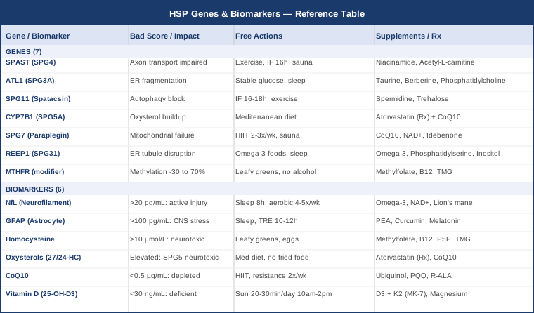
6 Biomarkers to Track in Hereditary Spastic Paraplegia
Biomarkers do not replace a genetic diagnosis or a neurologist's assessment. What they do is provide a running picture of what is happening biologically — and in a condition that progresses over years and decades, having regular data points is far more useful than a single clinical snapshot. The six markers below were selected for their relevance to the mechanisms that drive HSP, their practical accessibility, and the degree to which they can actually inform action.
Biomarker 1: Neurofilament Light Chain (NfL)
Why it matters. Neurofilament light chain is a structural protein found inside nerve axons. When axons are damaged or die, NfL leaks into the cerebrospinal fluid and, to a lesser extent, into the blood. In HSP, where the primary pathology is axon degeneration in the corticospinal tract, elevated NfL signals active neuronal injury. It is one of the few markers that reflects the rate of damage — not just the accumulated deficit — making it genuinely useful for tracking disease activity over time. Several research groups have now validated its use in hereditary spastic paraplegias, and it consistently tracks with disease burden and functional decline. See the current research landscape at PubMed: NfL in HSP.
How to measure it. Blood NfL is measured via the Simoa (Single Molecule Array) platform, now available through specialty neurology labs and a growing number of commercial diagnostics providers. In the United States, some labs are beginning to offer it through standard requisition. Cost range: $200–$500 USD. It is not yet universally covered by insurance for HSP specifically, though coverage is expanding. Optimal reference ranges in blood for adults under 40 are generally below 10 pg/mL; above 20 pg/mL in someone under 50 suggests active neurological injury and warrants investigation.
If the score is high — the plan without supplements. Reducing all modifiable sources of neural inflammation is the priority: optimize sleep to 7.5–9 hours per night (slow-wave sleep is the nervous system's primary repair window), eliminate chronic alcohol use entirely, reduce glycemic variability by spacing meals and lowering refined carbohydrate intake, and engage in moderate aerobic exercise 4–5 times per week — 30–40 minutes at a moderate intensity (zone 2 heart rate). Avoid prolonged sitting, which has been shown to increase inflammatory markers relevant to neurological tissue. Cold water immersion (10–15 minutes, 3–4 times per week) may support mitochondrial efficiency and reduce systemic inflammation, though direct HSP evidence is limited.
If the score is high — the plan with supplements. Omega-3 fatty acids (EPA + DHA, 2–4g/day from high-quality fish oil or algae-based sources) have the most consistent anti-neuroinflammatory evidence and are a reasonable first-line addition. NAD+ precursors (NMN 500mg/day or NR 300mg/day) support axonal energy metabolism and have shown neuroprotective effects in animal models of axonopathy. Phosphatidylserine (100–300mg/day) supports neuronal membrane integrity. Lion's mane mushroom (Hericium erinaceus extract, 500–1000mg/day) promotes nerve growth factor production and may support axonal maintenance — evidence in humans is preliminary but biologically plausible. Cycle NMN/NR with one week off per month to avoid potential habituation. Side effects are generally mild (GI sensitivity with omega-3; mild headache early with NAD precursors). Retest NfL every 6–12 months to track trajectory.
Biomarker 2: GFAP (Glial Fibrillary Acidic Protein)
Why it matters. GFAP is the structural protein of astrocytes, the support cells of the central nervous system. When the CNS is under stress or experiencing injury, astrocytes become activated and GFAP is released into blood and CSF. In the context of HSP, elevated GFAP signals reactive astrogliosis — a sign that the environment surrounding degenerating motor neurons is under active stress. GFAP is often measured alongside NfL, and the two together give a more complete picture than either alone: NfL reflects axonal damage, GFAP reflects glial activation and CNS inflammatory status.
How to measure it. Measured through the same Simoa blood testing platforms used for NfL. Often available as a bundled panel with NfL, reducing per-test cost. Cost range: $200–$400 USD standalone; $350–$600 combined with NfL. Normal blood GFAP in adults under 50 is generally below 100 pg/mL, though reference ranges vary by lab and age.
If the score is high — the plan without supplements. Astrocyte activation is strongly driven by systemic inflammation, blood-brain barrier disruption, and metabolic stress. Correcting sleep architecture is the single most impactful non-supplement intervention: poor sleep dramatically increases CNS inflammatory markers including GFAP. Adopt a time-restricted eating window (10–12 hours), reduce ultra-processed food, and prioritize anti-inflammatory whole foods (olive oil, fatty fish, berries, cruciferous vegetables, leafy greens). Heat therapy — sauna 3–4 times per week, 15–20 minutes at 80°C — has demonstrated reductions in systemic inflammatory markers and may support glial health over time.
If the score is high — the plan with supplements. Palmitoylethanolamide (PEA), 600–1200mg/day, is a naturally occurring fatty acid amide with well-documented anti-neuroinflammatory properties and a strong safety profile. It modulates astrocyte reactivity and has been studied in several chronic neurological conditions. Curcumin in bioavailable form (theracurmin or phosphatidylcholine-bound, 400–800mg/day) reduces NF-κB-driven astrocyte inflammation in animal models; human CNS penetration with standard curcumin is poor, so formulation matters. Melatonin (0.5–3mg at night) has specific antioxidant activity in glial cells. Cycle PEA continuously; take curcumin 5 days on, 2 days off to reduce GI burden. Retest GFAP every 6 months alongside NfL.
Biomarker 3: Homocysteine
Why it matters. Homocysteine is a sulfur-containing amino acid that accumulates when the methylation cycle is impaired. Elevated homocysteine is directly neurotoxic — it promotes excitotoxicity, impairs DNA repair, increases oxidative stress in neuronal mitochondria, and accelerates vascular damage. In HSP, where the long axons of the corticospinal tract are already metabolically vulnerable, chronically elevated homocysteine creates an additional toxic load. It is also a key readout of MTHFR status, one of the most common genetic modifiers in the broader HSP population. The test is inexpensive and widely available, making it one of the easiest actionable markers in this list.
How to measure it. Standard fasting blood test, available through virtually any clinical lab. Cost range: $30–$80 USD. Optimal homocysteine is below 7–8 μmol/L; values above 10 μmol/L are considered elevated; above 15 μmol/L is classified as hyperhomocysteinemia. Request fasting homocysteine specifically — some panels only include it if specifically ordered.
If the score is high — the plan without supplements. Diet-first interventions are highly effective for homocysteine. Increase folate from whole food sources: dark leafy greens (spinach, arugula, romaine), legumes, asparagus, broccoli. Moderate methionine intake by reducing red meat and processed meat frequency. Avoid alcohol entirely when homocysteine is elevated — alcohol strongly impairs folate metabolism. Increase choline-rich foods (eggs, liver, fish) which support the betaine pathway for homocysteine clearance independently of the methylation cycle.
If the score is high — the plan with supplements. The methylation B vitamins are the cornerstone: methylfolate (L-5-MTHF, 400–1000mcg/day — especially important if MTHFR variants are present, as folic acid is ineffective in this case), methylcobalamin (B12, 1000–2000mcg/day sublingually or injected), and pyridoxal-5-phosphate (B6 as P5P, 25–50mg/day). Trimethylglycine (TMG, 500–1000mg/day) directly remethylates homocysteine via the betaine pathway and is a powerful adjunct. Monitor with retesting every 3 months until stable. Note: in rare cases, high-dose B6 can cause peripheral neuropathy above 200mg/day; stay well below this threshold. Once homocysteine is optimized, maintenance doses can be reduced.
Biomarker 4: Oxysterols (24S-Hydroxycholesterol and 27-Hydroxycholesterol)
Why it matters. This biomarker is of particular relevance to individuals with CYP7B1 mutations (SPG5A). The enzyme encoded by CYP7B1 normally metabolizes oxysterols — oxidized cholesterol derivatives including 27-hydroxycholesterol and 25-hydroxycholesterol. When this enzyme is defective, these oxysterols accumulate to toxic levels in the nervous system, where they are directly cytotoxic to motor neurons, impair mitochondrial function, and trigger apoptotic pathways. Even beyond SPG5, oxysterol balance is relevant to broader neurological health: 24S-hydroxycholesterol is the brain's main cholesterol excretion product and serves as a marker of neuronal activity and integrity. Tracking these markers can guide treatment decisions and monitor treatment response. See PubMed: SPG5 oxysterol treatment research.
How to measure it. Oxysterol panels are specialized tests not available at standard clinical labs. They require referral to research-affiliated metabolic labs or neurological reference laboratories. Cost range: $300–$800+ USD. In the US and Europe, a handful of academic centers (including those affiliated with NIH Undiagnosed Diseases Network sites) can process these. For SPG5 patients, this test should be a priority; for other HSP subtypes, it remains informative but less critical.
If the score is high (SPG5 context) — the plan without supplements. Reduce dietary sources that drive endogenous oxysterol production: limit oxidized fats (fried foods, rancid vegetable oils, processed snack foods), reduce dietary cholesterol from red meat and processed meats, and adopt a Mediterranean-style diet emphasizing olive oil, fish, and vegetables. Antioxidant-rich foods (berries, pomegranate, green tea) can reduce cholesterol oxidation at the cellular level. Avoid smoking, which dramatically increases oxysterol burden through lipid peroxidation.
If the score is high (SPG5 context) — the plan with supplements or medication. Statins — specifically atorvastatin — have been studied and used in SPG5 patients with demonstrated reductions in oxysterol levels and preliminary evidence of stabilization. This is the only truly disease-modifying pharmacological approach for a specific HSP subtype currently available, and it requires physician prescription and monitoring. If a statin is used, supplement CoQ10 (ubiquinol, 200–300mg/day) to counter statin-related mitochondrial depletion. Vitamin E as mixed tocotrienols (200–400mg/day) can reduce lipid oxidation in neuronal membranes. Monitor oxysterol levels every 6–12 months to assess treatment response.
Biomarker 5: Coenzyme Q10 (CoQ10) and Mitochondrial Function Markers
Why it matters. Coenzyme Q10 is essential for mitochondrial electron transport chain function and serves as a fat-soluble antioxidant in cell membranes. In HSP subtypes involving mitochondrial dysfunction — particularly SPG7 (paraplegin), which encodes a mitochondrial AAA protease critical for protein quality control in the mitochondrial inner membrane — CoQ10 levels are functionally relevant. Beyond SPG7, the extraordinarily high metabolic demands of long corticospinal tract axons (stretching from cortex to sacral spinal cord) make them especially sensitive to any mitochondrial inefficiency. Low CoQ10 accelerates axonal degeneration and worsens the energy deficit that drives HSP pathology in multiple subtypes.
How to measure it. Plasma CoQ10 (total ubiquinone + ubiquinol) is available through most standard labs and many specialty labs. Cost range: $50–$150 USD. Optimal plasma CoQ10 is generally considered above 0.8–1.0 μg/mL; below 0.5 μg/mL suggests meaningful depletion. For a more complete picture of mitochondrial function, add a fasting lactate and pyruvate ratio — available through specialty metabolic labs ($100–$200 USD).
If the score is low — the plan without supplements. Mitochondria respond strongly to exercise — specifically high-intensity interval training (HIIT) and resistance training stimulate mitochondrial biogenesis via PGC-1α activation. A practical protocol for HSP: 2 sessions per week of resistance training (upper body and core focus to minimize fall risk), plus 2 sessions of cycle ergometer or recumbent bike intervals (20–30 seconds hard, 90 seconds easy, 8–10 rounds). Ketogenic metabolic cycling — 3–4 days per week of reduced carbohydrate intake (under 50g/day) — can upregulate mitochondrial efficiency without requiring permanent restriction. Cold exposure (cold shower, 3–5 minutes daily) activates brown adipose tissue and mitochondrial uncoupling proteins.
If the score is low — the plan with supplements. Ubiquinol CoQ10 (the reduced, active form) at 200–400mg/day is the most direct intervention; take with a fat-containing meal for optimal absorption. PQQ (pyrroloquinoline quinone) at 20mg/day stimulates mitochondrial biogenesis independently of CoQ10 and works synergistically with it. R-alpha lipoic acid (200–300mg/day) regenerates CoQ10 and other antioxidants within the mitochondrial membrane. Ribose (D-ribose, 5g/day) supports ATP regeneration in energy-depleted cells. Cycle on for 3 months, off for 2–3 weeks, then retest. Side effects are minimal; high-dose R-ALA can cause hypoglycemia in diabetics.
Biomarker 6: Vitamin D (25-OH-D3)
Why it matters. Vitamin D functions more as a steroid hormone than a simple vitamin, regulating hundreds of genes involved in inflammation, immune function, calcium signaling, and neuronal survival. In the neurological context, Vitamin D receptors are expressed throughout the brain and spinal cord; deficiency has been associated with accelerated neurodegeneration across multiple conditions. For HSP specifically, chronic low Vitamin D contributes to neuroinflammation, impairs remyelination capacity, reduces motor neuron survival signaling, and worsens secondary complications (osteoporosis from reduced mobility, immune vulnerability). Many people with HSP, due to reduced outdoor activity, are at significantly elevated risk of deficiency.
How to measure it. Standard blood test: 25-hydroxyvitamin D. Available at any clinical lab. Cost range: $30–$60 USD. Optimal levels for neurological health, according to researchers like Peter Attia and neurological literature, are 40–70 ng/mL (100–175 nmol/L). Levels below 30 ng/mL represent frank deficiency; levels below 20 ng/mL represent severe deficiency.
If the score is low — the plan without supplements. Sun exposure on the arms and legs (not through glass) for 20–30 minutes between 10 AM and 2 PM, at least 4 times per week, produces approximately 10,000–20,000 IU of Vitamin D in lighter-skinned individuals. This is highly variable by skin tone, latitude, and season — and for those with limited mobility, consistent sun exposure may be impractical. Dietary sources (fatty fish, egg yolks, liver) contribute modestly. For most HSP patients with deficiency, supplementation is typically necessary.
If the score is low — the plan with supplements. Vitamin D3 (cholecalciferol) at 2000–5000 IU/day is the standard starting point; always combine with Vitamin K2 (MK-7 form, 100–200mcg/day) to direct calcium into bones rather than soft tissues. Magnesium glycinate (300–400mg/day) is a co-factor for vitamin D activation and is commonly depleted alongside it. For severe deficiency, loading doses under physician supervision (50,000 IU weekly for 8 weeks) followed by maintenance dosing bring levels up faster. Retest 25-OH-D every 3–4 months until stable in the optimal range, then annually. Toxicity is unlikely below 10,000 IU/day in adults but monitoring remains prudent.
The six biomarkers above, taken together, form a practical dashboard for neurological health in HSP. None of them replaces genetic testing or specialist care, but they provide something those cannot: a dynamic, updatable picture of what is happening right now — and where targeted intervention may produce measurable benefit.
The Genetic Landscape of HSP: 7 Key Genes and What They Mean
Understanding the genetic underpinning of a given HSP case is not just academic. Different HSP genes imply different biological mechanisms — and increasingly, different therapeutic opportunities. What follows covers the seven most clinically relevant genes, their functional impact, and the most evidence-informed compensation strategies.
Gene 1: SPAST (SPG4) — The Most Common HSP Gene
What it does. SPAST encodes spastin, a microtubule-severing enzyme critical for regulating axon transport, vesicle trafficking, and cell division. Mutations in SPAST are haploinsufficient — one defective copy is enough to disrupt spastin activity. The result is defective microtubule dynamics in long corticospinal axons, impairing the transport of mitochondria, vesicles, and proteins over the meter-long distances involved. This is the most common cause of autosomal dominant HSP, accounting for roughly 40% of familial cases. See PubMed: SPAST/spastin HSP research.
Plan without supplements. Axon transport is strongly supported by consistent moderate exercise — walking, cycling, and resistance training promote tubulin expression and improve the efficiency of motor proteins. Reducing protein aggregation stress through intermittent fasting (14–16 hours daily) and caloric moderation activates autophagy, clearing damaged transport machinery. Heat exposure (sauna) enhances heat shock protein expression, which stabilizes misfolded proteins including those in the transport complex.
Plan with supplements. HDAC6 inhibition is a research-informed target: tubastatin A and related compounds inhibit HDAC6, which stabilizes microtubules — directly compensating for spastin loss. Clinical compounds are not yet approved, but some researchers note that niacinamide (vitamin B3, 500mg twice daily) has partial HDAC inhibitory activity. Acetyl-L-carnitine (1000–2000mg/day) supports mitochondrial transport along axons. Frequency: continuous. Monitor liver enzymes if using high-dose niacinamide.
Gene 2: ATL1 (SPG3A) — Early-Onset Dominant HSP
What it does. ATL1 encodes atlastin-1, a GTPase responsible for ER membrane fusion and the formation of the three-way junctions that give the endoplasmic reticulum its tubular network architecture. Mutations cause fragmentation and dysfunction of the axonal ER, impairing calcium homeostasis, lipid metabolism, and protein folding in long motor axons. SPG3A typically presents in early childhood and progresses slowly, often with a more benign course than SPG4.
Plan without supplements. ER stress is mitigated by avoiding prolonged unfolded protein response triggers: maintain stable blood glucose (avoid glycemic spikes), limit alcohol, and prioritize adequate sleep. Regular physical activity maintains calcium handling in muscle and neurons. A diet rich in phospholipids (eggs, liver, fish roe) supports ER membrane quality.
Plan with supplements. Taurine (1–3g/day) supports ER calcium homeostasis. Berberine (500mg twice daily with meals) activates AMPK and ER stress resolution pathways. Phosphatidylcholine (2–4g/day from lecithin) supports ER membrane integrity. Cycle berberine 5 days on, 2 days off to avoid GI tolerance. Side effects: GI discomfort with berberine; taurine is exceptionally well-tolerated.
Gene 3: SPG11 (SPATACSIN) — The Most Common Recessive HSP
What it does. SPG11 is the most common cause of autosomal recessive HSP, and it typically causes a more complex form — often including cognitive impairment, thin corpus callosum, and peripheral neuropathy in addition to spasticity. SPATACSIN is involved in lysosomal tubulation and autophagy — the cellular recycling system. When spatacsin is absent, autophagosomes fail to properly resolve, and toxic protein and lipid aggregates accumulate in neurons, eventually triggering cell death.
Plan without supplements. Activating autophagy is central: consistent intermittent fasting (16–18 hours daily), moderate-intensity exercise (which strongly induces autophagy), and caloric moderation all stimulate lysosomal function. Reducing saturated fat and eliminating ultra-processed foods reduces lysosomal lipid burden. Cognitive engagement (learning, reading, complex motor tasks) may support neuroplasticity reserve.
Plan with supplements. Spermidine (1–5mg/day from wheat germ extract) is the most studied autophagy activator in dietary supplement form and has shown neuroprotective potential in early studies. Trehalose (a disaccharide, 10–20g/day in divided doses) activates TFEB-mediated lysosomal biogenesis in animal models of protein aggregation diseases — human evidence is limited but mechanistically compelling. Rapamycin (low-dose, prescribed off-label) is the most potent mTOR inhibitor/autophagy inducer but requires physician oversight. Cycle spermidine 5 days on, 2 days off. Monitor blood glucose with trehalose supplementation.
Gene 4: CYP7B1 (SPG5A) — The Treatable HSP
What it does. CYP7B1 encodes a cytochrome P450 enzyme that hydroxylates oxysterols (particularly 25-hydroxycholesterol and 27-hydroxycholesterol) into bile acids, clearing them from the body. Loss of function leads to progressive accumulation of toxic oxysterols in spinal motor neurons. This is critical because SPG5A represents one of the very few forms of HSP with a documented disease-modifying pharmacological intervention: statin therapy reduces oxysterol production by inhibiting the HMG-CoA reductase pathway. This gene should be identified promptly — treatment may slow progression in a meaningful way. See PubMed: CYP7B1 SPG5 statin research.
Plan without supplements. Reduce dietary cholesterol and oxidized fat intake aggressively: eliminate fried foods, oxidized cooking oils, and processed meat. Mediterranean diet is the strongest evidence-based dietary model here. Regular exercise (which reduces hepatic cholesterol production and increases LDL receptor activity) serves as a biological statin-lite approach.
Plan with supplements or medication. Atorvastatin (physician-prescribed, typical dose 10–40mg/day) is the priority intervention — this is the only genotype-specific pharmacological treatment currently available in HSP. Always combine with CoQ10 ubiquinol (200–400mg/day) to counter statin-mediated CoQ10 depletion. Cholestyramine (bile acid sequestrant, prescribed) may augment oxysterol clearance. Monitor liver enzymes and CoQ10 levels every 3–6 months. Track oxysterol panels to assess treatment effectiveness.
Gene 5: SPG7 (Paraplegin) — Mitochondrial HSP
What it does. SPG7 encodes paraplegin, a subunit of the mitochondrial AAA protease complex (m-AAA protease) located in the inner mitochondrial membrane. This protease performs quality control on mitochondrial proteins, including components of the respiratory chain. Mutations cause progressive mitochondrial dysfunction in neurons — impaired ATP production, increased reactive oxygen species, reduced mitochondrial membrane potential, and aberrant mitochondrial dynamics (fusion/fission imbalance). The phenotype often includes optic atrophy, cerebellar signs, and a complex HSP picture in addition to spasticity.
Plan without supplements. Mitochondrial biogenesis is the target: HIIT-style exercise 2–3 times per week is the most potent non-pharmacological stimulator of PGC-1α, the master regulator of mitochondrial synthesis. Sauna exposure post-exercise compounds the effect. Minimize mitochondrial toxins: alcohol, certain antibiotics (fluoroquinolones, macrolides), statins without CoQ10 support, and excessive acetaminophen.
Plan with supplements. Ubiquinol CoQ10 (400mg/day), MitoQ (a mitochondria-targeted CoQ10 derivative, 10–20mg/day) — if available — and NAD+ precursors (NMN 500mg/day or NR 300mg/day) form the core mitochondrial support stack. Idebenone (150–900mg/day, divided doses) has demonstrated efficacy specifically in mitochondrial disease and is a short-chain CoQ10 analogue with superior CNS penetration; it is prescription in some countries, over-the-counter in others. Cycle NAD+ precursors with 1 week off per month. Side effects: mild GI sensitivity with high-dose ubiquinol; idebenone can cause elevated liver enzymes at high doses — monitor.
Gene 6: REEP1 (SPG31) — ER-Shaping Protein Pathway
What it does. REEP1 encodes receptor expression-enhancing protein 1, a hairpin-containing integral membrane protein that shapes ER tubules. It interacts directly with spastin and atlastin-1, making SPG31 part of the same ER-shaping network affected in SPG4 and SPG3A. Loss of REEP1 disrupts the tubular ER network specifically in long axons, impairing lipid transport, calcium signaling, and ER-mitochondria contact sites. The clinical presentation is typically pure or complicated spastic paraplegia with onset in childhood or early adulthood.
Plan without supplements. Given the ER-shaping overlap with SPG4, the same lifestyle principles apply: stable glucose, adequate sleep for ER stress resolution, and phospholipid-rich diet. Focus additionally on omega-3 intake (EPA and DHA), which incorporates into ER membranes and improves membrane fluidity and ER function.
Plan with supplements. Omega-3 fatty acids (3–4g/day EPA+DHA) directly incorporate into ER and mitochondrial membranes, improving their physical properties. Phosphatidylserine (300mg/day) supports ER-mitochondria contact site integrity. Inositol (2–4g/day) is a key phospholipid precursor particularly important for ER membrane function. Continuous use is appropriate; side effects are minimal.
Gene 7: MTHFR — The Methylation Modifier
What it does. While MTHFR is not a direct HSP gene, it is the most common genetic modifier of neurological conditions in the general population. The C677T and A1298C variants of methylenetetrahydrofolate reductase reduce the enzyme's efficiency by 30–70%, impairing the conversion of folate to its active form (5-MTHF) needed for methylation reactions, neurotransmitter synthesis, DNA repair, and myelin maintenance. In an already metabolically stressed HSP nervous system, impaired methylation compounds axonal vulnerability and accelerates homocysteine accumulation. MTHFR status is typically included in standard genetic panels and direct-to-consumer genetic tests.
Plan without supplements. Prioritize whole food folate (dark leafy greens, legumes, liver) over folic acid fortified foods. Folic acid (the synthetic form) actually competes with natural folate for absorption and is metabolically problematic for MTHFR variant carriers. Limit alcohol and caffeine (both impair folate metabolism). Adequate hydration is important for methylation cycle efficiency.
Plan with supplements. Methylfolate (L-5-MTHF, 400–800mcg/day) bypasses the impaired MTHFR enzyme entirely and directly supports methylation. Methylcobalamin (1000mcg/day, sublingual) and P5P (Pyridoxal-5-phosphate, active B6, 25mg/day) complete the methylation cofactor set. Trimethylglycine (TMG, 1000–2000mg/day) provides an alternative methylation route via the betaine pathway. Start with lower doses of methylfolate (100–200mcg) and increase gradually — some MTHFR variant carriers experience overmethylation symptoms (anxiety, irritability) at higher doses. Side effects are otherwise mild. Retest homocysteine every 3 months to assess response.
The genetic picture matters because it shifts the question from "how do I manage symptoms" to "what is the underlying mechanism and how might it be addressed at the source." For several of these genes — particularly SPG5A — that distinction is the difference between symptomatic management and genuine disease modification.
What the Mitochondrial Protocol Teaches Us About Progressive Neurological Conditions
Dr. Terry Wahls is a clinical professor of medicine at the University of Iowa and a person living with secondary progressive multiple sclerosis. After her condition deteriorated despite standard immunomodulatory treatment, she reviewed the basic science of neuronal energy metabolism and developed a structured dietary and lifestyle protocol that she eventually trialed on herself — with significant and documented functional improvement. Her book The Wahls Protocol (originally published 2014, updated 2020) and the subsequent research she conducted represent a serious, physician-led investigation into what nutrition and lifestyle can do for progressive neurological conditions. The principles translate directly to the subset of HSP cases driven by mitochondrial and axonal metabolic dysfunction.
Key Insight 1: Long Axons Have Extraordinarily High Energy Demands
Motor neurons projecting from the cortex to the sacral spinal cord are among the longest cells in the human body. Maintaining the electrical potential gradient across a meter or more of axon requires continuous ATP production. When mitochondrial efficiency falls — as it does in SPG7, in vitamin deficiency, in oxidative stress — these axons are the first to fail. The clinical implication: any intervention that improves mitochondrial energy output preferentially protects the cells most affected in HSP.
Key Insight 2: The Wahls Protocol Targets Mitochondrial Inputs Directly
The protocol structures nutritional intake around the specific molecules that mitochondria require: thiamine, riboflavin, niacinamide, pantothenic acid, coenzyme Q10, alpha-lipoic acid, carnitine, and sulfur amino acids. Rather than supplementing these piecemeal, Wahls organizes them into a food-first framework: 3 cups of leafy greens (B vitamins, folate), 3 cups of sulfur-rich vegetables (cabbage, onion, garlic, mushrooms — CoQ10 precursors), and 3 cups of deeply pigmented vegetables and fruits (antioxidants) — daily. This is not theoretical; it directly addresses the input side of the mitochondrial equation.
Key Insight 3: Myelin Requires Specific Fats That Most Diets Lack
Myelin — the insulating sheath of axons — is 70% lipid by dry weight, with a specific composition of sphingomyelin, cerebroside, and sulfatide. Synthesis requires long-chain omega-3 fatty acids (DHA specifically), cholesterol, and sulfur compounds. The protocol emphasizes regular consumption of fatty fish (wild salmon, mackerel, sardines), organ meats (particularly brain and liver), and coconut oil as a medium-chain triglyceride source that bypasses impaired mitochondrial transport in damaged neurons. The specific relevance for HSP: axons that are already structurally stressed from SPG mutations have less margin to tolerate myelin degradation driven by dietary insufficiency.
Key Insight 4: The Gut-Brain Axis is Not Peripheral to Neurological Disease
Wahls incorporates a prebiotic and fermented food approach, based on evidence that gut microbiome composition affects systemic inflammation, short-chain fatty acid production (which directly fuels enterocytes and indirectly supports the blood-brain barrier), and neurotransmitter precursor availability. For HSP patients, systemic inflammation is a modifiable accelerant of neurodegeneration. The practical protocol: daily fermented foods (sauerkraut, kimchi, kefir if tolerated), 8+ cups of vegetables and fruit for prebiotic fiber, and elimination of processed seed oils that dysregulate gut microbiota.
Key Insight 5: E-Stim (Electrical Muscle Stimulation) Was Part of Her Recovery
Dr. Wahls combined her nutritional protocol with neuromuscular electrical stimulation (NMES) applied to weakened muscle groups, working with physical therapists familiar with e-stim protocols for paralysis recovery. For HSP, NMES applied to the tibialis anterior and hip flexors — muscles frequently weakened by spasticity — can maintain muscle mass and support motor neuron-to-muscle signaling pathways even when voluntary activation is reduced. This is an underused tool in HSP management.
Key Insight 6: Heat Shock Proteins Are a Neurological Defense Mechanism
Sauna use, 3–4 times per week at 70–80°C for 15–20 minutes, is a consistent recommendation in Wahls' updated protocols. Heat stress induces HSP70 and HSP90 expression — heat shock proteins that stabilize misfolded proteins, reduce protein aggregation, and support mitochondrial quality control. In SPG4 (spastin) and SPG11 (spatacsin), where protein aggregation and quality control are central to pathology, repeated heat exposure is mechanistically relevant.
Key Insight 7: Removing Gluten and Dairy Reduced Her CNS Inflammation
Wahls eliminated gluten and dairy in the advanced form of her protocol, based on their ability to trigger systemic immune activation and intestinal permeability in susceptible individuals. For neurological conditions, this matters because lipopolysaccharide (LPS) from gram-negative gut bacteria that crosses a leaky gut drives microglial activation in the CNS — the same process that elevates GFAP. This is not universally necessary, but for individuals with HSP who have concurrent GI symptoms or who show elevated GFAP without an obvious explanation, a 90-day elimination trial is a low-risk experiment.
Key Insight 8: CoQ10 Is a Food-First Priority, Not Only a Supplement
Wahls emphasizes that CoQ10 is found in meaningful quantities in heart muscle (beef heart, chicken heart), organ meats, fatty fish, and to a lesser extent in dark leafy greens. Relying on supplementation alone without dietary reform misses the broader context: CoQ10 from food comes in a biological matrix that includes fat-soluble cofactors that enhance its absorption and utilization. Incorporating organ meat once or twice per week is one of the most nutrient-dense dietary changes available to someone managing neurological disease.
Key Insight 9: Movement is Medicine for Axonal Maintenance
The Wahls protocol is not sedentary. Even with significant motor disability, daily movement — whatever form is feasible — is central to the approach. Movement increases BDNF (brain-derived neurotrophic factor), supports mitochondrial biogenesis, reduces neuroinflammation, and maintains the neuromuscular junction health that determines functional status. For HSP, aquatic therapy, recumbent cycling, and seated resistance training are practical modalities that allow significant cardiovascular and neuromuscular work with reduced spasticity and fall risk.
Key Insight 10: The Protocol Was Formally Trialed in MS — With Documented Results
Dr. Wahls conducted an open-label feasibility study (published in peer-reviewed literature) demonstrating significant reduction in fatigue and improvement in quality of life in secondary progressive MS patients following the Wahls Elimination Diet. While HSP and MS are mechanistically distinct, both involve corticospinal axon dysfunction, mitochondrial stress, and neuroinflammation — making the metabolic framework broadly applicable. The evidence grade for HSP specifically remains low (no dedicated trials), but the biological rationale is coherent and the risk-to-benefit profile of the dietary and lifestyle components is favorable.
Complementary Approaches for Spasticity and Neurological Function
None of the approaches below replaces medical care or the biomarker-informed strategies above. Each has a specific body of human evidence that makes it worth considering as a structured adjunct — particularly for managing spasticity, improving gait, and supporting neurological resilience.
Yoga
Yoga combines sustained muscle lengthening with conscious motor control, making it directly relevant to the spasticity, reduced range of motion, and gait disturbance that characterize HSP. Regular yoga practice can reduce spastic tone through reciprocal inhibition mechanisms, improve proprioceptive feedback (often impaired in HSP), and maintain functional range of motion in hip flexors, hamstrings, and ankle dorsiflexors — the muscle groups most commonly compromised.
A 2012 randomized study published by Garrett et al. examined yoga in multiple sclerosis, a condition sharing corticospinal spasticity with HSP, and demonstrated significant improvements in balance, fatigue, and spasticity scores after 6 weeks of twice-weekly practice. While direct HSP trials are limited, the mechanism is shared. See general spasticity and yoga evidence at PubMed: yoga spasticity neurological.
For HSP specifically, a chair yoga or wall-supported yoga format is most practical. Focus on hip flexor lengthening (low lunge, pigeon modifications), hamstring stretching (seated forward fold), and ankle mobility. Practice 30–40 minutes, 3–4 times per week. Inform the yoga instructor of HSP-related balance limitations before beginning; avoid unsupported standing poses if balance is compromised. Progression should be gradual over 8–12 weeks.
Biofeedback
Biofeedback involves real-time monitoring of physiological signals — muscle activation (EMG biofeedback), movement patterns (kinematic biofeedback), or pressure (balance platform biofeedback) — with the goal of training conscious motor control. For HSP patients, EMG biofeedback applied to the lower limb allows patients to visualize spastic co-contraction patterns and learn to modulate them — a form of motor relearning that is often impossible without this direct feedback.
A randomized controlled trial by Armagan et al. (2003) demonstrated significant gait speed improvement and spasticity reduction in stroke patients using EMG biofeedback on ankle dorsiflexors — a protocol directly applicable to HSP, where foot drop and spastic plantar flexion are common. Spasticity mechanisms in stroke-related upper motor neuron syndrome and HSP are closely related.
In practice, seek a physical therapist or rehabilitation specialist trained in biofeedback-assisted gait training. EMG biofeedback sessions of 30–40 minutes, 2–3 times per week, with a focus on tibialis anterior activation during the swing phase of gait, represent the most evidence-aligned approach. Home biofeedback devices are now available (Myo, Delsys) for more frequent practice. Results are typically visible within 4–8 weeks of consistent training.
Music Therapy and Rhythmic Auditory Stimulation
Rhythmic Auditory Stimulation (RAS) is a specific music therapy technique in which a rhythmic auditory cue — usually a metronome or rhythmic music — is used to entrain and regularize gait cadence. It exploits the brain's strong tendency to synchronize movement timing to external auditory rhythm, bypassing the impaired internal timing circuits in motor neuron disease. For HSP, where gait dysrhythmia and shuffling contribute significantly to fall risk and functional limitation, RAS is a logical and evidence-grounded tool.
A systematic review by Thaut et al. on RAS in neurological rehabilitation (including Parkinson's disease, stroke, and traumatic brain injury) demonstrates consistent improvements in gait speed, cadence, stride length, and symmetry. See PubMed: rhythmic auditory stimulation gait. The mechanism — auditory-motor coupling via premotor cortex and basal ganglia circuits — remains partially functional in HSP and is worth recruiting.
The practical protocol: select a metronome app set to 10–15% above your natural cadence. Walk in time with the beat for 20–30 minutes, 4–5 days per week. Music with a strong, steady beat (typically 100–120 BPM for most HSP patients) can replace a metronome for more enjoyable sessions. Gradually increase beat tempo as gait improves. Work with a music therapist or neurological physiotherapist trained in RAS for the most structured results.
Low-Level Laser Therapy and Photobiomodulation
Photobiomodulation (PBM) involves the application of near-infrared or red light (typically 600–1100nm) to tissue, where it is absorbed by cytochrome c oxidase in mitochondria, improving electron transport chain function, increasing ATP production, and reducing oxidative stress. In the neurological context, transcranial PBM has been studied for neurodegenerative conditions, with preliminary evidence of neuroprotective and anti-inflammatory effects. For HSP, spinal cord PBM (applied to the dorsal spine) is the most anatomically rational target.
Animal studies of PBM applied to injured spinal cord demonstrate reduced neuroinflammation, improved axonal preservation, and better motor recovery outcomes. Human evidence in HSP specifically does not yet exist, but small clinical trials in multiple sclerosis (a shared corticospinal pathology) and ALS have shown safety and preliminary functional signals. See PubMed: photobiomodulation spinal cord.
A practical protocol involves a Class 2 or 3B near-infrared device (810nm or 850nm wavelength) applied to the thoracic and lumbar spine for 10–15 minutes per session, 4–5 times per week. Power density matters: target 20–50 mW/cm². Commercial PBM panels (Joovv, Mito Red, BioMax) offer home-use options. This is a low-risk intervention with a favorable safety profile; evidence strength is currently low to moderate for neurological conditions specifically. Use cautiously and monitor for any changes (improvement or worsening) over 8–12 weeks.
Breathing-Based Therapies
Diaphragmatic and controlled breathing practices have direct neurological relevance: the vagus nerve — the primary conduit for parasympathetic signaling — is activated by slow, deep expiration. Reduced vagal tone increases systemic inflammation, worsens neurological stress responses, and contributes to the autonomic dysregulation sometimes seen in complex HSP forms. Additionally, respiratory muscle weakness can develop in some HSP subtypes — particularly those with cerebellar or bulbar involvement — making respiratory training a prophylactic priority.
Evidence from clinical trials of inspiratory muscle training (IMT) in neurological conditions including ALS and MS demonstrates meaningful improvements in respiratory muscle strength, exercise tolerance, and quality of life. Slow-paced breathing at 4–6 breaths per minute (approximately 5 seconds inhale, 5–7 seconds exhale) activates the baroreflex and dramatically increases heart rate variability — a key marker of healthy autonomic function and a measurable indicator of nervous system resilience.
For HSP, a practical protocol is 10–15 minutes of diaphragmatic breathing (lying supine, placing one hand on the abdomen, inhaling to 6 seconds and exhaling to 7 seconds) twice daily. Physiological Sigh (two short inhalations through the nose followed by a long, complete exhale through the mouth, as studied by Stanford researchers including Dr. Andrew Huberman) can be used in acute spasticity episodes to reduce cortisol and spastic tone within seconds. For those with measurable respiratory compromise, an Inspiratory Muscle Trainer (POWERbreathe or Threshold IMT device) used 20–30 breaths per session, once daily, 5 days per week, is the structured version of this protocol.
Conclusion
Hereditary spastic paraplegia is not one condition, and it does not have one solution. What the evidence — from biomarkers to gene-specific mechanisms to metabolic interventions — consistently shows is that the nervous system is far more responsive to informed support than a purely symptomatic management framework would suggest. Tracking NfL and GFAP reveals whether active neurodegeneration is happening and whether interventions are working. Knowing whether your HSP is SPG4, SPG5A, or SPG7 changes what actions are most relevant. Optimizing homocysteine, mitochondrial function, and vitamin D removes modifiable accelerants that compound genetic vulnerability.
None of this is a cure. But there is a meaningful difference between managing symptoms passively and actively supporting the biological systems under stress. The next smart step is to identify your genetic subtype if you have not already, request the two or three biomarkers most relevant to that subtype, and bring those results to a neurologist or metabolic medicine physician willing to work with them. That combination — specific knowledge, measurable markers, targeted intervention — is where the most useful progress happens.