This article contains AI generated content.
· UpdatedHip Labral Tear - 4 Genes and 6 Biomarkers to Track
Living with a hip labral tear often means navigating a persistent cycle of physical restriction and frustration. The sharp, deep pinch in the groin during a sudden pivot, the aching stiffness that sets in after sitting, and the constant apprehension about certain movements are deeply disruptive. This is not just a structural issue; it is a daily challenge that impacts sleep, activity, and emotional well-being.
Too often, standard medical advice for this condition is generic and unsatisfying. Patients are frequently told to rest, take over-the-counter anti-inflammatories, or perform basic rehabilitation exercises like clamshells. While these recommendations may offer temporary relief, they fail to address why one person's labrum tears under normal mechanical loads while another's remains intact, or why some individuals experience progressive joint degeneration while others heal.
To truly understand and manage a hip labral tear, we must look deeper than simple structural wear and tear. A successful recovery strategy requires a thorough investigation of both the mechanical stresses acting on the joint and the internal biochemical environment. By analyzing how our body builds connective tissue, regulates local inflammation, and processes mechanical load, we can transition from passive symptom management to targeted joint support.
Grounded hope lies in precise, actionable data. This article outlines two advanced pathways to help you take control of your joint health. First, we examine a primary biochemical strategy focused on tracking key blood and urine biomarkers that reflect joint-tissue turnover and systemic inflammation. Second, we explore a genetic pathway that identifies structural variations in collagen and joint morphogenesis, offering specific compensation strategies. Together with targeted exercise science and evidence-based complementary modalities, these tools provide a comprehensive framework to support your hip joint.
Summary
This article provides a science-backed framework for managing and reversing the degenerative cycle associated with hip labral tears. By focusing on biochemical markers, readers will discover how tracking specific inflammatory and cartilage-turnover indicators can reveal the rate of joint degradation before permanent damage occurs. We investigate crucial biomarkers, including high-sensitivity C-reactive protein (hs-CRP) and matrix metalloproteinase-3 (MMP-3), alongside concrete lifestyle and supplemental protocols to optimize their levels. Additionally, we analyze how genetic variations in collagen synthesis (such as the COL5A1 and COL1A1 genes) and joint morphology (the GDF5 gene) predispose individuals to joint laxity and structural impingement, offering practical ways to compensate for these genetic vulnerabilities.
Furthermore, we translate complex exercise science and physical recovery methodologies into actionable daily habits. This includes a detailed summary of evidence-based joint repair protocols—covering progressive mechanical loading, local microcirculation enhancement, and tissue-healing peptides—designed to stimulate avascular cartilage repair. Finally, we explore complementary modalities such as modified yoga, photobiomodulation, electromyographic biofeedback, and mindfulness-based stress reduction, each supported by human clinical trials. This holistic approach empowers readers to move beyond temporary relief and build a resilient, structurally stable hip joint.
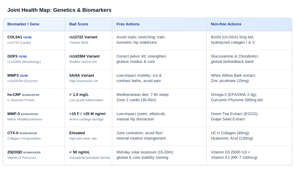
Biochemical Tracking: 6 Joint and Inflammatory Biomarkers to Measure
Managing a hip labral tear requires looking beyond structural imaging like MRIs. While an MRI shows the physical tear, it cannot measure the active biochemical environment of the joint. The rate at which cartilage degrades, the intensity of local synovial inflammation, and the body's capacity to synthesize new connective tissue are all driven by cellular pathways. By tracking specific biomarkers in the blood and urine, you can obtain a real-time assessment of your joint's metabolic state.
This biochemical strategy focuses on six critical biomarkers. Tracking these markers allows you to determine whether your joint is in a catabolic (tissue-breaking) or anabolic (tissue-building) phase. Armed with this data, you can tailor your physical therapy, nutrition, and lifestyle habits to suppress cartilage degradation and promote structural stability.
Biomarker 1: High-Sensitivity C-Reactive Protein (hs-CRP)
High-sensitivity C-reactive protein (hs-CRP) is an acute-phase reactant synthesized by the liver in response to inflammatory cytokines. In joint health, hs-CRP serves as a highly sensitive marker of systemic, low-grade inflammation. While classic osteoarthritis was once considered a purely mechanical "wear-and-tear" disease, modern research shows that low-grade systemic inflammation accelerates joint degeneration. A study linking systemic hs-CRP to local joint inflammation demonstrated that elevated hs-CRP levels correlate directly with the presence of synovitis—inflammation of the joint lining—in patients with joint pathology. When the synovium is inflamed, it releases catabolic enzymes that degrade the labrum and surrounding articular cartilage, turning a minor structural tear into a progressive degenerative issue.
How to measure it
The hs-CRP test is a standard, widely available blood test. It must not be confused with a standard CRP test, which is not sensitive enough to detect the low-grade systemic inflammation associated with joint degeneration. The cost ranges from $20 to $50 when ordered through online laboratory services or standard clinical practitioners. Optimal levels for joint preservation and overall health are below 1.0 mg/L, while levels between 1.0 and 3.0 mg/L indicate moderate systemic inflammation, and levels above 3.0 mg/L signal high inflammatory activity.
If the score is bad, the plan without supplements
If your hs-CRP is elevated (above 1.0 mg/L), the primary non-supplemental focus must be on dietary and lifestyle modification to lower systemic inflammatory cytokines. Transition to a strict Mediterranean diet rich in extra virgin olive oil, wild-caught fish, leafy greens, and cruciferous vegetables, while completely eliminating refined sugars, ultra-processed foods, and industrial seed oils. Prioritize sleep quality, aiming for 7 to 9 hours of restorative sleep nightly, as sleep deprivation is a powerful driver of systemic inflammation. Additionally, incorporate Zone 2 cardiovascular exercise (moderate-intensity aerobic activity where you can maintain a conversation) for 30 to 45 minutes, 3 to 4 times per week. Zone 2 training stimulates systemic mitochondrial function and decreases circulating inflammatory markers without overloading the hip joint.
If the score is bad, the plan with supplements or equipment
To actively drive down elevated hs-CRP, implement a targeted supplement regimen. - Omega-3 Fatty Acids (EPA/DHA): Take 2,000 to 3,000 mg of combined EPA and DHA daily. This dose is required to displace pro-inflammatory arachidonic acid in cell membranes. Run this protocol continuously; side effects are rare but may include mild gastrointestinal upset or minor blood-thinning effects at very high doses. - Curcumin Phytome (highly bioavailable formulation): Take 500 mg twice daily. Curcumin downregulates inflammatory pathways by inhibiting NF-kB. Cycle this supplement by taking it for 12 weeks, followed by a 2-week break. Avoid curcumin if you have gallbladder disease or are taking prescription blood thinners.
Biomarker 2: Matrix Metalloproteinase-3 (MMP-3)
Matrix metalloproteinase-3 (MMP-3), also known as stromelysin-1, is a zinc-dependent enzyme produced by chondrocytes and synovial fibroblasts. MMP-3 plays a major role in joint degradation because it directly breaks down the extracellular matrix of cartilage and the labrum, specifically targeting proteoglycans, fibronectin, and type II, IV, and IX collagens. A study on matrix metalloproteinases highlighted that pathologically elevated MMP-3 levels in the joint fluid and serum are strong indicators of active cartilage degradation and joint space narrowing. When a labral tear occurs, the mechanical instability triggers local cells to overproduce MMP-3, which then eats away at the surrounding healthy tissue, preventing the tear from stabilizing and accelerating the onset of osteoarthritis.
How to measure it
MMP-3 is measured via a serum blood test using an ELISA (enzyme-linked immunosorbent assay) method. It is typically offered by specialized diagnostic laboratories and costs between $70 and $150. Reference ranges vary by sex, but optimal values for active joint protection are under 15 ng/mL for females and under 25 ng/mL for males.
If the score is bad, the plan without supplements
When MMP-3 levels are high, the joint is in an active catabolic state. The non-supplemental plan must focus on reducing structural shear stress on the hip joint while maintaining joint lubrication. Avoid repetitive high-impact activities (such as long-distance road running or jumping exercises) that create high peak force on the torn labrum. Instead, utilize low-impact, closed-kinetic-chain exercises like swimming, aqua jogging, or elliptical training. Work with a physical therapist to perform manual hip distraction techniques, which temporarily open the joint space, reduce mechanical friction, and help downregulate the mechanical triggers that stimulate chondrocytes to secrete MMP-3.
If the score is bad, the plan with supplements or equipment
Supplements for high MMP-3 must focus on natural enzyme inhibition and collagen protection. - Green Tea Extract (standardized to 45% EGCG): Take 400 mg daily. Epigallocatechin gallate (EGCG) has been shown to inhibit the expression of MMP-3 in human chondrocytes. Take this supplement for 12 weeks, then take a 4-week break. Side effects can include mild nausea if taken on an empty stomach; do not exceed recommended doses due to potential liver strain. - Grape Seed Extract (standardized to 95% oligomeric proanthocyanidins): Take 200 mg daily. Proanthocyanidins bind to collagen fibers, protecting them from enzymatic cleavage by MMPs. This can be taken continuously without cycling. Side effects are rare but may include mild headaches or dizziness.
Biomarker 3: C-Terminal Cross-Linked Telopeptide of Type II Collagen (CTX-II)
C-terminal cross-linked telopeptide of type II collagen (CTX-II) is a specific degradation product of type II collagen, which is the primary collagen type found in articular cartilage and the hip labrum. When cartilage is degraded by catabolic enzymes, CTX-II fragments are released into the synovial fluid, eventually entering the bloodstream and being excreted in the urine. A biomarker investigation on CTX-II confirmed that urinary CTX-II is one of the most reliable and validated indicators of active cartilage destruction and is highly predictive of progressive joint space narrowing in hip and knee osteoarthritis. Elevated CTX-II indicates that the mechanical irritation from your labral tear is actively wearing down the protective cartilage of the hip socket.
How to measure it
CTX-II is most accurately measured via a urine test (usually corrected for creatinine levels) or a specialized serum ELISA test. Because it is primarily a research-grade biomarker, it may require ordering through functional medicine laboratories or specialized orthopedic centers, with costs ranging from $100 to $250. Optimal levels are in the lowest quartile of the reference range, indicating minimal cartilage breakdown.
If the score is bad, the plan without supplements
If your CTX-II score is elevated, the immediate mechanical priority is to restore optimal joint centration—ensuring the head of the femur sits perfectly within the acetabulum during movement. Focus on neuromuscular training of the deep hip rotators (gemelli, obturators, and piriformis) and the gluteus medius to prevent the femoral head from sliding forward and rubbing against the labrum. Avoid exercises that require end-range hip flexion combined with internal rotation (the impingement position), as this mechanically grinds the cartilage and drives up CTX-II. Additionally, utilize a supportive hip compression sleeve or brace during daily activities to improve proprioception and joint stability.
If the score is bad, the plan with supplements or equipment
To combat active cartilage degradation reflected by high CTX-II, utilize supplements that stimulate cartilage matrix repair and protect type II collagen. - Undenatured Type II Collagen (UC-II): Take 40 mg daily on an empty stomach before bed. UC-II works through oral tolerance, modulating the immune system to prevent the autoimmune-mediated degradation of joint collagen. This must be taken continuously; it has an excellent safety profile with virtually no reported side effects. - Oral Hyaluronic Acid (high molecular weight): Take 120 mg daily. This helps restore the viscoelastic properties of the synovial fluid, reducing mechanical friction. Take for 6 months, then cycle off for 1 month. No known side effects.
Biomarker 4: Interleukin-6 (IL-6)
Interleukin-6 (IL-6) is a multifunctional cytokine that acts as a major mediator of joint inflammation and pain. In the hip joint, IL-6 is secreted by osteoblasts, chondrocytes, and inflamed synovial cells. It plays a dual role: it acts as a systemic inflammatory signal and directly stimulates the production of matrix-degrading enzymes while promoting osteoclast activity, which leads to subchondral bone resorption and joint space narrowing. A clinical trial on pain sensitivity demonstrated that elevated systemic IL-6 levels are strongly associated with increased pain sensitivity and decreased functional scores in patients with joint degeneration. Elevated IL-6 not only damages the physical structure of the hip but also sensitizes local nociceptive pathways, making the pain of a labral tear feel more intense.
How to measure it
IL-6 is measured via a standard serum blood test. It is highly sensitive to recent physical activity and stress, so the test must be performed in a fasted, rested state. The cost of the test ranges from $50 to $100. The optimal clinical range for joint preservation is under 1.5 pg/mL, with lower levels indicating a highly controlled inflammatory state.
If the score is bad, the plan without supplements
Elevated IL-6 is heavily influenced by systemic stress and overtraining. The non-supplemental plan must focus on stress reduction and balancing physical activity. Avoid exhaustive, prolonged workouts, which cause a massive surge in systemic IL-6. Instead, keep resistance training sessions under 45 minutes and prioritize recovery. Implement daily vagal nerve stimulation techniques, such as deep diaphragmatic breathing or structural meditation, for 10 to 15 minutes. Ensure you maintain a consistent circadian rhythm by exposing your eyes to morning sunlight and avoiding blue light before bed, as disrupted sleep patterns directly elevate morning IL-6 levels.
If the score is bad, the plan with supplements or equipment
Targeted supplementation can help lower IL-6 by modulating the cellular pathways that produce this cytokine. - Resveratrol (trans-resveratrol): Take 250 mg daily. Resveratrol is a potent polyphenol that inhibits the activation of the inflammatory cytokine cascade, specifically reducing IL-6 expression. Cycle this supplement: take it for 8 weeks, followed by a 2-week break. Side effects at this dose are rare but can include mild digestive upset. - Ashwagandha Extract (standardized to high withanolides, e.g., KSM-66): Take 600 mg daily. Ashwagandha helps reduce systemic cortisol, which in turn helps stabilize IL-6 levels. Cycle this by taking it for 8 weeks, then taking 2 weeks off. Avoid ashwagandha if you have autoimmune thyroid conditions.
Biomarker 5: 25-Hydroxyvitamin D [25(OH)D]
Vitamin D is a steroid hormone precursor essential for calcium absorption, bone mineralization, and skeletal integrity. In the context of a hip labral tear, Vitamin D is crucial for maintaining the structural density of the acetabular cup and the femoral head. Low Vitamin D levels compromise subchondral bone remodeling, leading to micro-instability at the joint interface. This instability increases the mechanical shear stress transferred directly to the soft labrum. Furthermore, Vitamin D is a potent immunomodulator; deficiency is associated with increased production of pro-inflammatory cytokines and a reduced capacity for tissue repair.
How to measure it
Vitamin D status is assessed by measuring serum 25-hydroxyvitamin D [25(OH)D] through a standard blood draw. The test is inexpensive, costing between $30 and $60. While conventional laboratory reference ranges define sufficiency as anything above 30 ng/mL, optimal levels for orthopedic health, tissue repair, and immune regulation are between 50 and 80 ng/mL.
If the score is bad, the plan without supplements
To naturally improve Vitamin D status, maximize safe solar exposure. Expose large areas of skin (arms, legs, back) to mid-day sunlight for 15 to 20 minutes daily, depending on your skin tone and geographic location, without sunscreen (avoiding any sunburn). Increase your intake of Vitamin D-rich foods, such as wild-caught sockeye salmon, mackerel, sardines, and pastured egg yolks. However, because food sources contain relatively low amounts of Vitamin D, mechanical and lifestyle adjustments must also focus on maximizing joint stability through progressive strength training of the gluteals and core to protect the bone-cartilage interface.
If the score is bad, the plan with supplements or equipment
Supplementation is highly effective for correcting a Vitamin D deficiency. - Vitamin D3 + Vitamin K2 (MK-7): Take 5,000 IU of D3 combined with 100 mcg of K2 daily. Vitamin K2 is critical because it ensures that the calcium absorbed via Vitamin D is directed into the bones and teeth, rather than soft tissues or arterial walls. Run this continuously, but re-test your blood levels every 3 months to ensure you do not exceed 100 ng/mL. Side effects are virtually non-existent at this dose when taken with K2.
Biomarker 6: Tumor Necrosis Factor-Alpha (TNF-Alpha)
Tumor Necrosis Factor-alpha (TNF-alpha) is a master pro-inflammatory cytokine that plays a primary role in joint destruction. Secreted by macrophages and T-cells, TNF-alpha binds to receptors on chondrocytes and synovial cells, triggering a cascade that upregulates the production of other destructive cytokines (like IL-6) and catabolic enzymes (like MMP-3). TNF-alpha actively suppresses the synthesis of type II collagen and proteoglycans, halting the body's natural joint repair mechanisms. In patients with hip labral tears, elevated TNF-alpha levels indicate an aggressive inflammatory environment that prevents tissue healing and accelerates the breakdown of the surrounding articular cartilage.
How to measure it
TNF-alpha is measured via a serum blood test using specialized high-sensitivity assays. The cost typically ranges from $60 to $120. An optimal score for joint preservation is under 1.5 pg/mL, with lower values indicating that the master inflammatory switch is turned off, allowing tissue recovery.
If the score is bad, the plan without supplements
To lower TNF-alpha without supplements, implement dietary and physiological stressors that trigger cellular autophagy and anti-inflammatory pathways. Incorporate time-restricted eating (e.g., a 16:8 fasting schedule where you fast for 16 hours and eat within an 8-hour window) 3 to 4 days per week, as fasting has been shown to downregulate macrophage activation and reduce TNF-alpha secretion. Additionally, utilize cold thermogenesis (cold showers or cold plunges at 50-55°F for 2 to 3 minutes, 3 times per week). The acute cold exposure triggers a systemic norepinephrine release that suppresses systemic TNF-alpha production.
If the score is bad, the plan with supplements or equipment
To suppress TNF-alpha pharmacologically through natural agents, focus on targeted anti-inflammatory compounds. - Boswellia Serrata Extract (standardized to 30% AKBA, e.g., 5-Loxin): Take 100 mg daily. AKBA is a potent inhibitor of 5-lipoxygenase (5-LOX) and has been shown to significantly reduce TNF-alpha levels and joint pain. Cycle this: take for 12 weeks, followed by a 3-week break. Side effects may include mild heartburn or acid reflux. - Ginger Root Extract (standardized to 5% gingerols): Take 500 mg daily. Gingerols act as natural dual inhibitors of cyclooxygenase (COX) and lipoxygenase (LOX), reducing TNF-alpha. This can be taken continuously. Side effects are rare but can include mild stomach warmth.
Between biochemical tracking and genetic analysis lies a critical connection: while biomarkers tell us what is happening in the body right now, our genes define the underlying structural blueprint. By understanding this blueprint, we can better interpret our biomarkers and customize our long-term recovery strategy.
Genetic Compensation: 4 Genes and Epigenetic Factors Influencing Hip Laxity
To understand why a hip labral tear occurs, we must look at our genetic blueprint. The structural integrity of the hip joint depends not only on muscle strength but also on the molecular composition of the ligaments, joint capsule, and cartilage. Genetic variations can influence the diameter of collagen fibrils, the mechanical elasticity of connective tissues, and even the developmental shape of the hip socket.
This section covers four key genes: COL5A1, COL1A1, GDF5, and MMP3. By identifying variations in these genes, you can implement specific lifestyle, physical therapy, and supplemental protocols to compensate for these structural vulnerabilities, helping to protect your joints and prevent further damage.
Gene 1: COL5A1 (The Collagen Alpha-1 V Chain / Joint Laxity Gene)
The COL5A1 gene provides instructions for making the alpha-1 chain of type V collagen. Type V collagen is a regulatory collagen; it forms long, thin fibrils that act as a structural template, controlling the diameter and organization of the much larger type I collagen fibers in ligaments, tendons, and the joint capsule. Variations in the COL5A1 gene—most notably the BstUI polymorphism (rs12722)—are associated with altered collagen fibril structure. A study on COL5A1 polymorphisms and flexibility demonstrated that specific genotypes correlate with significant differences in joint range of motion, muscle-tendon flexibility, and susceptibility to soft-tissue injuries. Individuals with the "laxity-associated" alleles have thinner, more elastic collagen fibrils, leading to generalized joint hypermobility and hip instability. In the hip, this laxity allows the femoral head to slide excessively within the socket, placing high shear stress on the labrum and leading to micro-instability and tears.
If the gene is bad, the plan without supplements
If you carry the COL5A1 laxity variant, your primary goal is to build structural stability through muscle activation, compensating for the lack of passive ligamentous support. Avoid all passive, end-range static stretching of the hip (such as deep lunges or yoga pigeon pose), as this further stretches the already loose joint capsule. Instead, focus entirely on high-tension, isometric strengthening of the hip stabilizers. Perform isometric gluteal bridges, lateral band walks, and single-leg balance exercises with a focus on pelvic alignment. Emphasize eccentric muscle contractions, which train the nervous system to control the hip joint throughout its entire range of motion, preventing the femoral head from sliding forward.
If the score is bad, the plan with supplements or equipment
To support collagen synthesis and strengthen loose connective tissues, utilize supplements that stimulate endogenous collagen production. - Choline-Stabilized Orthosilicic Acid (ch-OSA, e.g., BioSil): Take 5 mg twice daily. Silicon is a key co-factor for the enzymes prolyl hydroxylase and lysyl hydroxylase, which are required for cross-linking collagen fibers to give them structural strength. Cycle this by taking it for 3 months, followed by a 1-month break. There are no known side effects at this dose. - Hydrolyzed Type I and V Collagen Peptides: Take 15 grams daily, consumed 30 to 60 minutes before your rehabilitation exercises. Consuming collagen peptides before exercise delivers key amino acids (like glycine and proline) to the joint when blood flow is elevated, helping to strengthen the connective tissue matrix. Run this protocol continuously. Side effects are rare but may include mild bloating.
Gene 2: COL1A1 (The Type I Collagen Structural Gene)
The COL1A1 gene provides instructions for producing the pro-alpha-1 chain of type I collagen, the most abundant collagen type in the human body. Type I collagen forms the dense, structural fibers that give the labrum its tensile strength, allowing it to act as a seal and shock absorber for the hip joint. A common polymorphism in the promoter region of the COL1A1 gene (Sp1 binding site, rs1800012) can alter the ratio of collagen chains produced, resulting in collagen fibers with reduced structural integrity. Individuals with this variant have a weaker labral matrix that is more susceptible to tearing under normal mechanical loads.
If the gene is bad, the plan without supplements
For individuals with structural collagen weakness, progressive mechanical loading is essential to stimulate the body to produce stronger tissue. However, this loading must be applied carefully. Avoid sudden, high-velocity movements, explosive plyometrics, and heavy lifting at end-ranges of motion, which can shear the fragile labrum. Implement a program of slow, controlled resistance training (using a 3-second concentric and 3-second eccentric tempo) within a pain-free range of motion. Emphasize exercises like squat variations to parallel, step-ups, and cable hip extensions. These movements apply axial compression to the joint, stimulating chondrocytes and fibroblasts to remodel the collagen matrix without causing micro-trauma to the labrum.
If the score is bad, the plan with supplements or equipment
To compensate for a genetic weakness in type I collagen synthesis, prioritize the raw materials and co-factors required for collagen assembly. - Vitamin C (as ascorbic acid): Take 500 mg twice daily with meals. Vitamin C is an essential co-factor for collagen synthesis; without it, the proline and lysine residues in collagen cannot be hydroxylated, resulting in structurally weak fibers. Take this continuously; high doses can cause mild loose stools, so adjust the dosage if necessary. - Copper (as copper bisglycinate): Take 1 mg daily. Copper is a vital cofactor for lysyl oxidase, the enzyme responsible for covalent cross-linking of collagen and elastin fibers. Run this for 8 weeks, then take a 4-week break to prevent imbalances with zinc. Avoid taking copper on an empty stomach to prevent nausea.
Gene 3: GDF5 (The Growth Differentiation Factor 5 / Hip Morphology Gene)
Growth Differentiation Factor 5 (GDF5) is a member of the transforming growth factor-beta (TGF-beta) superfamily and is critical for joint development, cartilage maintenance, and bone remodeling. A well-studied single nucleotide polymorphism in the 5′-untranslated region of the GDF5 gene, **rs143384**, leads to reduced transcriptional activity and lower levels of GDF5 expression in joint tissues. A study on GDF5 variants and osteoarthritis demonstrated that this reduced expression is a robust genetic risk factor for developmental dysplasia of the hip (DDH) and early-onset osteoarthritis. When GDF5 expression is low, the hip socket may develop with a slightly shallower angle (mild dysplasia), or the cartilage may be thinner and less resilient. This structural variation alters the contact area of the joint, concentrating mechanical forces onto the labrum and making it highly vulnerable to tearing.
If the gene is bad, the plan without supplements
When dealing with a genetic predisposition to altered joint morphology (such as a shallow socket), the primary strategy is to optimize joint biomechanics to distribute load evenly. You must work with a physical therapist to identify and correct any anterior pelvic tilt, which shifts the pelvis forward and reduces the contact area of the acetabulum over the femoral head, increasing labral wear. Focus on strengthening the hamstrings, gluteals, and deep lower abdominal muscles (transversus abdominis) to keep the pelvis in a neutral position. Additionally, focus on building strength in the gluteus medius and minimus, which help keep the femoral head securely centered within the socket during single-leg movements.
If the score is bad, the plan with supplements or equipment
For individuals with low GDF5 expression, the goal is to supply the joint with compounds that stimulate cartilage matrix repair and counteract the catabolic state. - Glucosamine Sulfate (1,500 mg) and Chondroitin Sulfate (1,200 mg): Take this combined dose daily. These compounds provide the building blocks (glucosaminoglycans) for proteoglycan synthesis, helping to keep cartilage hydrated and resilient despite structural impingement. Take this continuously; it may take 4 to 8 weeks to see benefits. Side effects are generally mild and limited to mild digestive discomfort. - Gluteal Biofeedback Band (using a resistance band with integrated pressure sensors): Use this device during rehabilitation sessions to ensure proper gluteal activation and prevent compensatory quad or hip flexor dominance during exercises.
Gene 4: MMP3 (The Matrix Metalloproteinase-3 Gene)
The MMP3 gene encodes the matrix metalloproteinase-3 enzyme, which degrades the extracellular matrix of joint tissues. A common promoter polymorphism in this gene, known as the 5A/6A polymorphism (rs3025058), directly affects transcription levels. The 5A allele has been shown to drive significantly higher transcription of MMP3 compared to the 6A allele. Individuals carrying the 5A allele produce higher levels of the MMP-3 enzyme, particularly when the joint is stressed or inflamed. This genetic variation results in an overactive enzymatic environment, where collagen and proteoglycans in the labrum and cartilage are degraded more rapidly in response to minor mechanical irritation or inflammation.
If the gene is bad, the plan without supplements
If you carry the high-expressing MMP3 variant, your joint is genetically prone to rapid tissue breakdown when inflamed. The non-supplemental plan must focus on managing inflammation. Never push through joint pain; any sharp pinch or ache is a sign of mechanical irritation that will trigger a release of MMP-3. Use a physical therapy approach that prioritizes low-impact mobility and joint distraction over heavy loading during flare-ups. Utilize ice or contrast baths (alternating warm and cold water) around the hip joint to reduce local tissue temperature and downregulate inflammatory activity.
If the score is bad, the plan with supplements or equipment
To suppress the overproduction of the MMP-3 enzyme, use natural compounds that inhibit its activity. - White Willow Bark Extract (standardized to 15% salicin): Take 240 mg daily. Salicin acts as a natural inhibitor of pro-inflammatory pathways that trigger MMP-3 secretion. Cycle this supplement: take for 6 weeks, followed by a 2-week break. Avoid if you are sensitive to aspirin or taking blood thinners. - Zinc (as zinc picolinate): Take 15 mg daily. Since MMPs are zinc-dependent enzymes, maintaining balanced zinc levels is crucial. Do not exceed this dose, as excessive zinc can disrupt copper levels. Take continuously, preferably with food to avoid stomach irritation.
Understanding your genetics helps you identify your joint's structural vulnerabilities. To bridge the gap between these genetic tendencies and real-world healing, we can look to modern recovery protocols, such as those discussed in detail on the Andrew Huberman Podcast, which focus on stimulating tissue repair and optimizing recovery.
Podcast Analysis: 10 Core Insights from the Andrew Huberman Joint Health Protocol
In his podcast, neurobiologist Dr. Andrew Huberman frequently explores the science of tissue repair, joint health, and physical recovery. While many medical approaches focus on structural interventions, Huberman emphasizes the physiological levers we can control to stimulate healing in connective tissues. Connective tissues like the hip labrum have low vascularity (blood flow), making their recovery slow and dependent on specific cellular signals.
This section translates ten core insights from the Andrew Huberman podcast into practical protocols. By understanding these mechanisms, you can optimize your recovery and support the healing of your hip joint.
1. The Role of Synergistic Loading in Collagen Synthesis
One of Huberman’s key insights is that complete immobilization is often the worst approach for a soft-tissue injury. Connective tissues require mechanical loading to signal the cells (fibroblasts) to synthesize new collagen fibers. Without this mechanical stimulus, the body will not rebuild the tissue, and the surrounding structures will weaken. The protocol is to perform progressive, pain-free loading. For a hip labral tear, this means engaging in controlled, closed-chain exercises (such as isometric wall sits or partial squats) that compress the joint without causing pain. This loading helps align new collagen fibers along the lines of mechanical stress, improving the structural strength of the healing tissue.
2. Blood Flow and Microcirculation Enhancement
Because the hip labrum is largely avascular, it receives very little direct blood supply. It relies on the diffusion of nutrients from the surrounding synovial fluid to heal. To facilitate this process, you must increase local blood flow and joint movement. Huberman recommends high-repetition, low-load movement. For the hip, this can be achieved by riding a stationary bike with zero resistance for 15 to 20 minutes daily. This gentle, repetitive motion acts as a pump, circulating synovial fluid through the joint capsule and delivering essential nutrients to the avascular labrum.
3. Temperature Cycling and Local Microcirculation
Contrast therapy—alternating exposure to heat and cold—is a powerful tool to increase local circulation and support tissue healing. Heat dilates blood vessels, bringing nutrient-rich blood to the area, while cold constricts them, helping to flush out waste products. Huberman recommends a protocol of 15 to 20 minutes in a hot sauna (or using a hot pack around the hip) followed immediately by 2 to 3 minutes in a cold plunge or cold shower. Repeating this cycle 3 to 4 times helps stimulate local circulation and can trigger a systemic release of growth hormone, which supports tissue repair.
4. Glucosamine and Chondroitin Sulfate Synergy
Huberman has discussed the clinical evidence supporting the use of glucosamine and chondroitin sulfate for joint health. These compounds are structural components of joint cartilage and help maintain the hydration and shock-absorbing capacity of the joint matrix. To get the most benefit, they must be taken consistently over several months. The recommended protocol is to take 1,500 mg of glucosamine sulfate and 1,200 mg of chondroitin sulfate daily. This helps maintain the thickness and hydration of the joint cartilage, reducing the mechanical friction on the torn labrum.
5. Undenatured Type II Collagen (UC-II) and Oral Tolerance
UC-II works through a unique mechanism called oral tolerance. Unlike hydrolyzed collagen, which is broken down into amino acids, UC-II is ingested in its intact form. It interacts with specialized immune cells in the gut, training the immune system to stop attacking and degrading the body's own type II collagen in the joints. Huberman highlights this as an effective way to reduce joint inflammation and support cartilage preservation. The protocol is to take 40 mg of UC-II daily on an empty stomach before bed, allowing it to interact with gut-associated lymphoid tissue without interference from other foods.
6. Sleep Architecture and Growth Hormone Release
Deep sleep is the primary driver of physical recovery. During slow-wave sleep, the brain releases a surge of growth hormone, which is essential for cell regeneration and connective tissue repair. Huberman emphasizes that maximizing sleep quality is crucial for healing any injury. To optimize your sleep architecture, maintain a consistent sleep schedule, sleep in a cool room (65°F or lower), and avoid alcohol and heavy meals close to bedtime. This helps maximize your time in deep, restorative sleep, allowing your body to repair joint tissues.
7. The Impact of Systemic Cortisol on Joint Matrix Integrity
Chronic stress elevates systemic cortisol levels. While acute cortisol is anti-inflammatory, chronically elevated cortisol is catabolic, meaning it breaks down tissues and inhibits the synthesis of new collagen and proteoglycans. This can weaken the joint matrix and delay healing. To manage cortisol, Huberman recommends tools like Non-Sleep Deep Rest (NSDR) protocols or short meditation sessions during the day. Reducing systemic stress helps lower cortisol levels, creating a more favorable environment for tissue repair.
8. Hydration Dynamics and Proteoglycan Swelling Pressure
Cartilage is composed of water, collagen, and proteoglycans. Proteoglycans act like molecular sponges, binding water to create a pressurized cushion that absorbs shock and protects the joint. When you are dehydrated, this cushion loses pressure, increasing mechanical friction on the labrum and cartilage. Huberman emphasizes the importance of proper hydration for joint health. The protocol is to drink water consistently throughout the day, ensuring you consume adequate electrolytes (sodium, potassium, magnesium) to help your cells retain water and maintain joint hydration.
9. Amino Acid Precursors and Cellular Timing
To build new collagen, the body needs a steady supply of specific amino acids, particularly glycine, proline, and hydroxyproline. Huberman discusses the benefit of consuming these precursors shortly before training. The protocol is to take 10 to 15 grams of hydrolyzed collagen peptides or gelatin, combined with 500 mg of Vitamin C, 30 to 60 minutes before your physical therapy exercises. The combination of targeted nutrients and increased blood flow from exercise helps deliver these amino acids directly to the hip joint, supporting collagen synthesis.
10. Peptides and Tissue Healing Regimens
In discussions on advanced recovery, Huberman has noted the emerging research on tissue-healing peptides, such as BPC-157 and TB-500. These compounds are being studied for their potential to accelerate angiogenesis (the formation of new blood vessels) and soft-tissue repair, particularly in avascular regions like tendons and ligaments. While these therapies are still in the research phase and require medical supervision, they represent a promising avenue for accelerating repair in stubborn joint injuries.
While these systemic protocols help optimize the body's internal environment for healing, incorporating targeted, evidence-based alternative modalities can provide direct, localized support for the hip joint.
Evidence-Based Alternative Modalities for Hip Support
In addition to systemic lifestyle modifications and biochemical tracking, several targeted alternative modalities can support recovery from a hip labral tear. To ensure safety and efficacy, these approaches must be backed by clinical evidence. The following four modalities offer complementary pathways to manage pain, improve neuromuscular activation, and support tissue healing.
Modified Yoga
Yoga is a mind-body practice that combines physical postures, breathing exercises, and meditation to improve flexibility, strength, and pain management. For a hip labral tear, yoga can help strengthen the supporting muscles of the hip joint and improve pelvic alignment. However, standard yoga poses that involve extreme ranges of motion must be modified to avoid aggravating the tear. A meta-analysis on yoga for joint inflammation demonstrated that consistent, modified yoga practice significantly reduces pain and stiffness while improving physical function in individuals with joint degenerative conditions.
To apply this modality safely, follow a gentle Hatha or Iyengar yoga protocol that avoids deep hip flexion, internal rotation, and extreme abduction. Focus on postures that strengthen the gluteals and core, such as modified Bridge Pose (Setu Bandhasana) with a block between the thighs to engage the adductors, and Warrior I (Virabhadrasana I) with a shortened stance to protect the hip flexors. Perform this modified practice for 20 to 30 minutes, 3 times per week, ensuring you use blocks and straps to avoid pushing the joint past its comfortable, pain-free range of motion.
Low-Level Laser Therapy / Photobiomodulation (PBM)
Photobiomodulation (PBM), also known as low-level laser therapy (LLLT), uses specific wavelengths of red and near-infrared light to stimulate cellular function. When applied to the hip joint, these wavelengths penetrate deep into the tissue, where they are absorbed by mitochondria. This process increases adenosine triphosphate (ATP) production, reduces oxidative stress, and downregulates pro-inflammatory cytokines, helping to manage joint pain. A clinical study on low-level laser therapy showed that PBM is effective at reducing pain and improving range of motion in patients with joint pain, and serves as a helpful drug-free addition to physical therapy.
To apply this therapy, seek out a physical therapist or clinic equipped with a Class 3B or Class 4 cold laser device. The protocol involves applying near-infrared light (wavelengths of 808 to 830 nm) directly to the anterior and lateral aspects of the hip joint, delivering a dose of 6 to 10 Joules per point. Treatments are typically administered 2 to 3 times per week for a duration of 4 to 6 weeks. It is important to ensure the laser is applied directly to the skin to achieve the necessary tissue penetration, and protective eyewear must always be worn during treatment.
Electromyographic (EMG) Biofeedback
Electromyographic (EMG) biofeedback is a therapeutic technique that uses surface electrodes to detect muscle activity and provide real-time visual or auditory feedback to the patient. For individuals with a hip labral tear, the pain and instability often lead to altered movement patterns, such as gluteal amnesia and compensatory dominance of the TFL (tensor fasciae latae) or hip flexors. EMG biofeedback helps patients identify and re-activate the gluteus medius and minimus, restoring pelvic stability and reducing shear stress on the labrum. An electromyographic biofeedback clinical trial demonstrated that EMG biofeedback is highly effective at correcting abnormal gait patterns (such as Trendelenburg gait) by retraining the gluteus medius during functional movement.
To use this modality, work with a physical therapist who utilizes surface EMG biofeedback. Electrodes are placed over the belly of the gluteus medius and the TFL. During exercises like single-leg balances, side-lying clamshells, or step-downs, you monitor the biofeedback screen to ensure the gluteus medius is actively firing while the TFL remains quiet. Perform this biofeedback-guided training for 15 to 20 minutes, 2 to 3 times per week, until the correct neuromuscular activation patterns become automatic during daily activities.
Mindfulness-Based Stress Reduction (MBSR)
Mindfulness-Based Stress Reduction (MBSR) is a structured program that uses mindfulness meditation, body awareness, and gentle yoga to help individuals manage chronic pain and stress. Chronic joint pain can sensitize the central nervous system, amplifying pain signals and leading to fear of movement. MBSR helps break this cycle by training the mind to observe pain sensations without judgment, reducing the emotional reactivity and muscle tension that can worsen joint pain. A meta-analysis on mindfulness and chronic pain confirmed that MBSR leads to significant improvements in pain control, physical function, and psychological well-being in individuals living with chronic musculoskeletal pain.
To implement this approach, participate in a structured 8-week MBSR course or practice daily mindfulness meditation using guided audio sessions. Dedicate 20 to 30 minutes daily to a "body scan" meditation, focusing on releasing tension in the muscles surrounding the pelvis, lower back, and hip joint. Sit in a comfortable, supported position, close your eyes, and direct your breath to the hip area, consciously relaxing the surrounding tissues. Consistent daily practice is key to retraining the nervous system and reducing pain sensitivity.
Conclusion
Recovering from a hip labral tear requires a comprehensive approach that addresses both the structural and biochemical factors affecting the joint. While physical therapy helps restore mechanical alignment, tracking key biomarkers like hs-CRP and MMP-3 and understanding genetic factors like COL5A1 can provide valuable insights into your joint's healing process. By combining biochemical tracking, genetic compensation strategies, and evidence-based alternative modalities, you can move beyond simple symptom management and build a resilient hip joint. The next step is to discuss these biomarker and genetic testing options with a qualified healthcare professional, allowing you to design a personalized recovery plan tailored to your body's unique needs.
Musculoskeletal: Tendon & Ligament Conditions
Autoimmune: Inflammatory Conditions Connective Tissue Conditions