This article contains AI generated content.
· UpdatedHoffa's Fat Pad Syndrome - 6 Genes And 7 Biomarkers To Track
Introduction
If your knee pain sits just below the kneecap, worsens when you fully extend your leg, and has persisted despite rest, anti-inflammatories, and a round or two of physical therapy, you may already know the diagnosis: Hoffa's fat pad syndrome, also called infrapatellar fat pad impingement. What you may not know is why it keeps coming back, or why the same treatment works for some people and does almost nothing for others. That inconsistency is not bad luck. It reflects biological differences that most standard care simply does not measure.
Hoffa's fat pad — the adipose structure tucked beneath the patella and in front of the patellar tendon — is not a passive shock absorber. It is metabolically active tissue that secretes cytokines, recruits immune cells, and responds to both mechanical stress and systemic inflammation. When it becomes inflamed and eventually fibrotic, the pain cycle becomes self-sustaining. Standard protocols address the symptom — compression, local injection, load management — without ever asking what is keeping the inflammatory signal turned on at the tissue level.
That is where biomarkers and genetics become genuinely useful. Measuring specific inflammatory cytokines, adipokines, and tissue-remodeling enzymes in your blood gives you a real-time read on what is happening inside the joint environment. Understanding which gene variants you carry tells you where your biology is predisposed to amplify inflammation, produce less of a protective signal, or build collagen that is more prone to damage. Neither approach gives you a cure, but both give you something clinicians rarely offer: a specific reason for why things are the way they are, and a targeted direction for improving them.
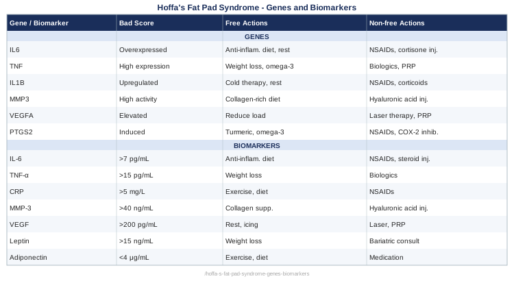
This article walks through two complementary frameworks. The first — and more immediately actionable — is a guide to 7 biomarkers worth tracking, with cost ranges, interpretation thresholds, and specific intervention plans tied to each abnormal result. The second is a focused look at 6 genetic factors that shape fat pad vulnerability and inflammatory resolution, with practical plans regardless of whether you supplement or not. Alongside these, you will find a distillation of research-driven frameworks and complementary physical therapies with real clinical evidence for soft tissue knee conditions. Better information is not a guarantee — but it is the most reliable starting point available.
The 7 Biomarkers Worth Tracking When You Have Hoffa's Fat Pad Syndrome
Most blood panels ordered for knee pain include little more than a basic inflammatory marker and a metabolic panel. For Hoffa's fat pad syndrome, that leaves the most relevant biology completely unmeasured. The seven biomarkers below were selected because each one either directly reflects fat pad biology, shapes the inflammatory environment that determines whether healing progresses or stalls, or serves as a modifiable target with a meaningful evidence base. Track them as a panel, not in isolation — the pattern across all seven tells a more complete story than any single result.
Biomarker 1: High-Sensitivity C-Reactive Protein (hs-CRP)
Why it matters
hs-CRP is synthesized by the liver in direct response to circulating IL-6 — one of the cytokines produced locally by an inflamed fat pad. In a healthy knee, the fat pad contributes little to systemic inflammation. In a chronically irritated one, it becomes a persistent source of IL-6 that keeps hs-CRP elevated even when imaging looks unremarkable. Research in knee pain populations consistently links elevated hs-CRP to worse pain outcomes, slower structural recovery, and higher rates of fibrosis. It is not a diagnostic marker for Hoffa's specifically, but it is the most accessible proxy for whether your inflammatory environment is controlled or not (PubMed: hs-CRP and knee inflammation).
How to measure it
Standard blood draw through any major lab (Labcorp, Quest, Ulta Lab Tests). Cost range: $15–$40 USD. Always request the high-sensitivity version — standard CRP misses low-grade chronic inflammation entirely. Optimal frequency: baseline, then every 8–12 weeks when making lifestyle or supplement changes.
Interpreting the result — Optimal: below 1 mg/L — Borderline: 1–3 mg/L — Bad score: above 3 mg/L — systemic inflammation is active; the fat pad environment will not be calming down independently
If the score is bad, the plan without supplements
Sleep is the first lever. Consistent 7–9 hours of sleep per night drives IL-6 clearance directly — this is not a vague wellness recommendation but a documented mechanism. Alongside sleep, eliminating refined seed oils and ultra-processed carbohydrates reduces arachidonic acid cascade activity, which is a primary upstream driver of CRP elevation. The dietary change alone produces measurable hs-CRP reduction within 4–8 weeks in most people. Add time-restricted eating (16:8 window) to amplify the effect through AMPK activation. None of these require purchasing anything.
If the score is bad, the plan with supplements or equipment
Omega-3 fatty acids (EPA+DHA): 2–4g combined daily in triglyceride form. This is the single most studied supplement for hs-CRP reduction, with a response window of 8–12 weeks. No cycling required. Take with the largest meal to reduce GI discomfort — the most common side effect at higher doses. Choose a product with verified oxidation testing (IFOS or similar) since rancid fish oil worsens inflammation rather than reducing it.
Curcumin with piperine: 500–1000mg curcumin combined with 5–10mg piperine, twice daily. Multiple meta-analyses confirm significant hs-CRP reduction at these doses. Cycle 8 weeks on, 2 weeks off for long-term use. Side effects are mild but GI sensitivity occurs in some people — take with food.
Red light / near-infrared device: 10–20 minute daily sessions over the knee at 630–850nm. Photobiomodulation reduces local IL-6 production through mitochondrial signaling pathways, addressing the source rather than just the systemic readout. Entry-level devices cost $80–$300. No cycling needed.
Biomarker 2: Interleukin-6 (IL-6)
Why it matters
Measuring IL-6 directly, rather than inferring it through hs-CRP, tells you whether the cytokine cascade is actively elevated right now. The infrapatellar fat pad has been demonstrated in human tissue studies to be a local IL-6 producer under mechanical stress and inflammatory conditions. Critically, IL-6 at sustained levels promotes fibrosis — the mechanism by which an acute fat pad impingement becomes the chronic, stiff, scarred version of Hoffa's disease that is much harder to treat. Tracking IL-6 over time tells you whether your interventions are actually turning down this fibrogenic signal (PubMed: IL-6 infrapatellar fat pad fibrosis).
How to measure it
Blood draw through specialty labs or via a rheumatology or sports medicine physician. Direct-to-consumer access varies by state. Cost range: $40–$100 USD. IL-6 is sensitive to sample handling and processing time — use the same lab consistently for serial measurements.
Interpreting the result — Optimal: below 2 pg/mL — Elevated: 2–7 pg/mL — Bad score: above 7 pg/mL — active cytokine-driven inflammation; fibrosis risk is significantly elevated at sustained levels
If the score is bad, the plan without supplements
Resistance training at moderate intensity is paradoxically the most potent non-supplement IL-6 reducer over time. Acute bouts of exercise transiently raise IL-6, but chronically trained individuals have dramatically lower resting IL-6 through skeletal muscle's anti-inflammatory endocrine function. For Hoffa's syndrome, the key is how you train: avoid deep knee flexion during flares, prioritize isometric quadriceps loading, and use hip-dominant patterns (hip hinges, step-ups, lateral band work) that build the kinetic chain without compressing the fat pad. Frequency: 3 sessions per week minimum.
Structured stress management also matters. Chronically elevated cortisol drives IL-6 independently of any knee pathology. Five minutes of box breathing (4-count inhale, 4-count hold, 4-count exhale, 4-count hold) before sleep shifts the autonomic balance measurably toward the state that allows cytokine resolution.
If the score is bad, the plan with supplements or equipment
Quercetin: 500mg twice daily with meals. Quercetin inhibits IL-6 signaling through NF-κB pathway suppression with consistent human evidence across several trial populations. Cycle 12 weeks on, 4 weeks off. Side effects are generally mild — occasional headache at higher doses.
Local cold-compression cycling on the knee after any loading activity: 10 minutes ice, 10 minutes off, repeat twice. This is not a rest protocol — it is an active inflammation-management tool that reduces local IL-6 accumulation in the fat pad without blunting the beneficial systemic response from exercise. Cost: a quality ice compression wrap is $30–$80.
Biomarker 3: Leptin
Why it matters
Of all the biomarkers in this article, leptin is the most specific to Hoffa's fat pad syndrome as a distinct condition. The infrapatellar fat pad is genuine adipose tissue — it produces leptin just as visceral or subcutaneous fat does, and at elevated levels this local leptin production directly promotes joint inflammation through chondrocyte, synoviocyte, and immune cell activation. Human knee tissue studies have confirmed that fat pads from symptomatic knees express significantly more leptin than those from healthy knees. Elevated serum leptin correlates with greater pain severity and worse structural progression, independent of body weight — meaning lean individuals with elevated leptin can still have this problem (PMID 22561186).
How to measure it
Fasting serum leptin through standard labs. Cost range: $30–$60 USD. Always measure after a 12-hour fast and on a consistent morning schedule for comparable serial results. The most informative approach is to measure leptin alongside adiponectin — the ratio between them matters more than either value alone. Request both in the same blood draw.
Interpreting the result — Optimal: 4–25 ng/mL (women), 1–10 ng/mL (men) — Bad score: above 30 ng/mL (women), above 15 ng/mL (men) — or any level elevated relative to your body composition, suggesting leptin resistance
If the score is bad, the plan without supplements
Leptin sensitization begins with reducing dietary fructose, particularly from high-fructose corn syrup and fruit juices, which disrupt hypothalamic leptin sensitivity through a different mechanism than other carbohydrates. This is one of the most underappreciated dietary levers in inflammatory fat pad conditions.
Add Zone 2 aerobic training — 150+ minutes per week at 60–70% of maximum heart rate. This consistently reduces serum leptin in both overweight and normal-weight individuals within 8 weeks. Use low-impact modalities: cycle ergometer, swimming, elliptical — to achieve this systemic effect without compressive loading of the fat pad.
Avoid prolonged knee flexion loading: deep squatting, kneeling for extended periods, or sitting with the knee bent past 90° increases mechanical stress on the fat pad and stimulates local leptin production. Structural positioning habits matter as much as exercise choice.
If the score is bad, the plan with supplements or equipment
Zinc (elemental): 15–25mg daily with food. Zinc deficiency impairs leptin receptor signaling and is highly prevalent in people with elevated systemic inflammation. Correct before adding other interventions. Do not exceed 40mg/day without pairing with copper (2mg) to prevent copper depletion. Cycle: 8 weeks, then retest zinc status.
Berberine: 500mg 2–3 times daily with meals. Berberine improves insulin sensitivity and downstream leptin signaling through AMPK activation — a mechanism similar to metformin's. Cycle 8 weeks on, 4 weeks off. Side effects: GI discomfort is common at initiation; start at 250mg and titrate up over 2 weeks.
Continuous glucose monitor (CGM): a 2-week CGM session is not a supplement but a device that makes invisible leptin-driving patterns visible. Seeing postprandial glucose spikes in real time is behavior-changing in a way that food diary recommendations are not. Cost: $50–$80 for a 2-week sensor. Use periodically to audit dietary patterns rather than continuously.
Biomarker 4: Adiponectin
Why it matters
Adiponectin is leptin's anti-inflammatory counterpart — also produced by the infrapatellar fat pad, and progressively underproduced as the fat pad becomes inflamed. Research consistently shows that inflamed knee fat pads generate less adiponectin than healthy ones, creating a compound problem: rising leptin and falling adiponectin simultaneously. The result is a net pro-inflammatory adipokine environment that sustains joint damage even when mechanical stressors are managed. Thomas Dayspring has highlighted adiponectin as a deeply underutilized risk marker in cardiometabolic medicine; in joint biology, it plays an equally important protective role that is rarely measured in clinical knee care.
How to measure it
Fasting serum adiponectin. Less commonly ordered than leptin — may require a specialty lab or a physician who orders expanded metabolic panels. Cost range: $50–$120 USD. Calculate the leptin-to-adiponectin ratio (LAR): a ratio above 6 indicates a significant inflammatory adipokine imbalance and warrants intervention regardless of which individual value is driving it.
Interpreting the result — Optimal: above 15 mcg/mL (women), above 10 mcg/mL (men) — Bad score: below 10 mcg/mL (women), below 6 mcg/mL (men) — or any LAR above 6, meaning leptin is significantly outpacing the protective signal
If the score is bad, the plan without supplements
Zone 2 aerobic exercise raises adiponectin more consistently than any other non-pharmacological intervention across human trial data. The mechanism involves PPAR-γ activation in adipose tissue, which drives adiponectin gene expression. Forty-plus minutes per session, four or more sessions per week, over 12–16 weeks produces meaningful increases. For Hoffa's, swimming and cycle ergometer maintain this training volume with minimal fat pad loading.
Mediterranean dietary pattern: regular intake of extra virgin olive oil, fatty fish, legumes, and non-starchy vegetables consistently raises adiponectin versus high-glycemic Western diets in human studies. The effect is not subtle — dietary pattern change over 12 weeks produces adiponectin increases comparable to some supplement interventions.
If the score is bad, the plan with supplements or equipment
Magnesium glycinate or malate: 300–400mg elemental magnesium at bedtime. Magnesium improves insulin sensitivity and supports PPAR-γ activity, which drives adiponectin gene expression. Side effects are limited to loose stools at high doses — start at 200mg and adjust. No cycling required; magnesium is a fundamental nutrient, not a cycling intervention.
Resveratrol: 150–500mg daily taken with a fat-containing meal (for absorption). Multiple human trials show modest but statistically significant adiponectin-raising effects over 12 weeks. Cycle 12 weeks on, 4 weeks off. Side effects are rare at standard doses.
Whole-body vibration plate: 25–35Hz, 10 minutes three times per week. Early human evidence suggests mechanical activation of adipose tissue through vibration raises adiponectin through a pathway independent of aerobic exercise. Quality platforms cost $200–$600. No cycling needed — use as a regular training adjunct.
Biomarker 5: Matrix Metalloproteinase-3 (MMP-3)
Why it matters
MMP-3 is an enzyme that degrades extracellular matrix proteins — collagen, fibronectin, proteoglycans — and its elevation signals active tissue remodeling. In Hoffa's fat pad syndrome, elevated MMP-3 marks the transition from acute inflammation toward progressive fibrosis and stiffening of the fat pad. Once fibrosis is established, the fat pad loses its compliance, stops functioning as a shock absorber, and becomes a mechanical irritant that creates a perpetual impingement cycle. Catching elevated MMP-3 early — before irreversible structural change — provides a window to intervene. It is also a validated marker in rheumatoid arthritis monitoring, which gives it a stronger clinical evidence base than most joint biomarkers.
How to measure it
Serum MMP-3. Most accessible through rheumatology context or integrative medicine labs that offer joint health panels. A physician order is typically required. Cost range: $60–$150 USD. Reference ranges vary by laboratory, so interpret results against lab-specific norms rather than a universal threshold.
Interpreting the result — Typical reference range: below 28 ng/mL (women), below 59 ng/mL (men) — lab dependent — Bad score: above upper reference limit for your sex — active matrix degradation; fibrosis risk is elevated and mechanical protection of the fat pad is being progressively compromised
If the score is bad, the plan without supplements
Mechanical unloading of the fat pad is the most direct first step. Reduce all activities that compress the fat pad into the intercondylar notch: kneeling, full squat, prolonged sitting with the knee bent at or beyond 90°. Infrapatellar taping — a specific physiotherapy technique that lifts the fat pad away from the impingement zone — directly reduces the mechanical stimulus driving MMP-3 production. A trained physiotherapist should demonstrate the technique before you apply it independently; incorrect taping can increase impingement rather than relieve it.
Progressive loading within the pain-monitoring model is critical: MMP-3 and fibrosis both accelerate under complete rest, because the tissue receives no organized mechanical signal to guide repair. Maintain activity within a pain-monitored framework (pain no higher than 4/10 during the activity, returning to baseline within 24 hours post-session).
If the score is bad, the plan with supplements or equipment
Hydrolyzed collagen peptides with Vitamin C: 15g collagen peptides plus 50mg Vitamin C taken 30–60 minutes before exercise, five days per week. Research from Keith Baar's group at UC Davis demonstrates that this protocol enriches the tendon and connective tissue milieu with collagen precursors precisely when blood flow to the tissue is elevated, supporting organized matrix synthesis rather than disorganized fibrotic repair. Side effects are minimal.
Boswellia serrata (AKBA-enriched): 300–500mg of a standardized extract (65% boswellic acids) twice daily. Several human RCTs in joint conditions demonstrate significant reductions in MMP activity and inflammatory markers. Cycle 12 weeks on, 4 weeks off. Side effects: occasional GI discomfort — take with food.
Extracorporeal shockwave therapy (ESWT): applied by a physiotherapist or sports medicine physician, 3–5 sessions at weekly intervals. Growing evidence supports ESWT for reducing local MMP activity while stimulating organized tissue repair in tendinopathies and soft-tissue knee conditions. Cost: $100–$300 per session. This is not a home intervention — it requires clinical equipment and trained administration.
Biomarker 6: 25-OH Vitamin D
Why it matters
Vitamin D deficiency is widespread and reliably worsens musculoskeletal conditions. Vitamin D receptors (VDR) are expressed in fat pad tissue, chondrocytes, and immune cells — insufficient Vitamin D impairs resolution of inflammatory cascades, reduces pain thresholds, and weakens the periarticular musculature that protects the knee from mechanical overload. Multiple studies link low 25-OH-D to chronic knee pain, delayed recovery from joint conditions, and amplified inflammatory marker levels. Peter Attia has consistently treated 25-OH-D as a non-negotiable foundational biomarker in anyone with a chronic inflammatory condition, because its correction is inexpensive, safe when properly managed, and produces measurable improvements in musculoskeletal outcomes (PubMed: Vitamin D and knee pain).
How to measure it
Routine blood test. Request specifically 25-OH-D (calcidiol) — not 1,25-OH-D (calcitriol), which does not reflect storage status. Widely available and inexpensive: $20–$50 USD. Retest at 3 months after any supplementation change.
Interpreting the result — Optimal for musculoskeletal and immune function: 50–80 ng/mL (125–200 nmol/L) — Insufficient: 20–30 ng/mL — Bad score: below 20 ng/mL — severe deficiency; inflammatory resolution is compromised at the cellular level
If the score is bad, the plan without supplements
Intentional midday sun exposure on uncovered skin (arms and legs) for 15–30 minutes, adjusted for skin tone and latitude. At latitudes above 40°N, winter UV index is insufficient for meaningful synthesis — sun alone cannot substitute for supplementation between October and March in those climates. Year-round sun exposure is still a valuable habit for its circadian co-benefits even where supplementation is necessary.
If the score is bad, the plan with supplements or equipment
Vitamin D3 with K2: when baseline is below 20 ng/mL, a loading approach of 5,000–10,000 IU Vitamin D3 daily for 8–12 weeks, then retest. Pair with 100–200mcg MK-7 K2 to direct calcium appropriately and avoid soft tissue calcification. After reaching the target range, maintain at 2,000–4,000 IU depending on regular retesting. Toxicity at therapeutic doses is rare — it occurs at sustained intakes above 40,000 IU/day over months. Always retest before adjusting ongoing dosing.
Note on magnesium: Vitamin D conversion requires magnesium as a cofactor. If magnesium is also deficient, correct both simultaneously — Vitamin D supplementation without adequate magnesium produces a partial and slower response.
Biomarker 7: TNF-Alpha (TNF-α)
Why it matters
TNF-α is one of the most potent pro-inflammatory cytokines in joint biology. In Hoffa's fat pad syndrome, elevated TNF-α drives continued immune cell recruitment into the fat pad, promotes concurrent synovial inflammation, and — critically — sensitizes peripheral pain receptors (nociceptors) such that pain signals become amplified beyond what local tissue damage would predict. This explains why some people with Hoffa's describe pain disproportionate to what imaging shows: TNF-α-driven central sensitization. The fact that biologic drugs targeting TNF-α dominate the rheumatoid arthritis treatment landscape underscores just how central this cytokine is to joint destruction — measuring it gives you meaningful insight into whether that system is driving your condition.
How to measure it
Serum TNF-α. Less routine than hs-CRP; typically requires a rheumatology order or specialty lab. Cost range: $60–$130 USD. Best interpreted as part of a cytokine panel alongside hs-CRP and IL-6, since the three together tell a more complete story than any one measurement alone.
Interpreting the result — Typical reference: below 8.1 pg/mL (lab dependent) — Bad score: above 8 pg/mL — active TNF-α burden; local and systemic inflammation are likely self-perpetuating; pain sensitization is a significant risk
If the score is bad, the plan without supplements
Intermittent fasting (16:8 or 18:6) significantly reduces TNF-α in human studies independently of body weight change. The mechanism involves autophagy-mediated clearance of senescent cells — aging, damaged cells that chronically produce TNF-α as a low-grade signal. The key is daily consistency, not periodic long fasts. The eating window positioning matters less than the consistency of the compressed window.
Dietary polyphenol density: berries, green tea, extra virgin olive oil, and dark chocolate (above 80% cocoa) inhibit NF-κB — the transcription factor that drives TNF-α gene expression. At realistic dietary amounts eaten consistently, this effect is measurable in inflammatory marker studies and does not require supplemental doses.
If the score is bad, the plan with supplements or equipment
EGCG (green tea extract): 400–800mg standardized EGCG daily, taken away from iron-containing meals to avoid absorption interference. EGCG directly inhibits NF-κB signaling and TNF-α production. Cycle 8 weeks on, 3 weeks off. At very high doses, liver enzyme elevation has been reported — stay at standard doses and monitor if using long-term.
Regular sauna exposure: 3–4 sessions per week, 20 minutes at 80–90°C (traditional or infrared). Finnish population studies and smaller RCTs consistently show significant TNF-α and inflammatory cytokine reduction with regular sauna use, mediated by heat shock proteins that resolve cellular stress. Home infrared units cost $400–$2,000+; gym access provides a lower-entry-cost option. No cycling needed — sustained use produces the benefit.
With a clearer picture of what your blood markers reveal, the next layer worth understanding is whether your genetics are predisposing you to an overactive inflammatory response or an inefficient recovery — because the same intervention will not work equally for everyone.
What Your Genes May Be Telling You About Your Fat Pad Vulnerability
Genetics do not determine outcomes — they define tendencies. For Hoffa's fat pad syndrome, certain gene variants increase the probability of mounting a stronger inflammatory response to the same level of mechanical stress, producing less of a protective anti-inflammatory signal, or building connective tissue that is structurally more vulnerable to repetitive loading. Understanding which variants you carry (through consumer genomic testing like 23andMe, AncestryDNA, or clinical panels) helps you understand why certain biomarkers may be chronically abnormal for you, and which interventions are most relevant to your specific biology.
Gene 1: IL-6 (rs1800795) — The Inflammatory Amplifier
The IL-6 gene promoter variant rs1800795 (also called the -174G/C polymorphism) influences baseline production of IL-6. Individuals carrying the C allele (CC genotype) produce significantly more IL-6 in response to the same inflammatory stimulus than those with the GG genotype — meaning that the same fat pad impingement event will generate a more intense and prolonged inflammatory cascade if you carry this variant.
If the gene is bad, the plan without supplements
Prioritize every non-supplement IL-6 reducer with heightened discipline: consistent sleep (7–9 hours, non-negotiable), Zone 2 aerobic training at least 4x/week, and elimination of ultra-processed carbohydrates and refined seed oils. These are the highest-effect levers for blunting IL-6 production at the gene-expression level. Isometric knee loading protocols (wall sits, terminal knee extensions) allow quadriceps maintenance without fat pad compressive loading during flares — this maintains the anti-inflammatory muscle endocrine function without triggering the inflammatory cycle.
If the score is bad, the plan with supplements or equipment
Quercetin (500mg twice daily) is the most targeted supplement choice given its direct NF-κB and IL-6 pathway inhibition. Combine with omega-3s (3g EPA+DHA daily) and curcumin with piperine for a synergistic approach across the IL-6 pathway. Cycle quercetin 12 weeks on, 4 weeks off; omega-3s can be maintained continuously; cycle curcumin 8 weeks on, 2 weeks off. A PEMF (pulsed electromagnetic field) mat, used 20 minutes daily, has preliminary evidence for reducing IL-6 expression in tissue under mechanical stress. Home units cost $300–$800. Side effects across this stack: minimal when individual doses are kept within standard ranges.
Gene 2: TNF-Alpha (rs1800629) — The Pain Amplifier
The TNF-α promoter polymorphism rs1800629 (the -308G/A variant) significantly increases TNF-α gene transcription in carriers of the A allele (GA or AA genotype). This means a higher baseline TNF-α environment, greater nociceptor sensitization, and a tendency toward pain that persists beyond what would be expected from tissue injury alone. GA and AA carriers report more severe chronic pain in multiple joint conditions relative to GG homozygotes.
If the gene is bad, the plan without supplements
Daily intermittent fasting is the highest-priority behavioral intervention — it consistently reduces TNF-α through autophagy-mediated senescent cell clearance, and for TNF-α gene over-expressers, this mechanism is particularly relevant. Sustained polyphenol intake through food (berries, green tea, olive oil, dark chocolate above 80% cacao) inhibits NF-κB, which drives TNF-α transcription. The effect at realistic dietary intake is meaningful for this variant specifically. Cold exposure (cold shower finish, 90–180 seconds, daily) also modulates the TNF-α pathway through hormetic mechanisms.
If the score is bad, the plan with supplements or equipment
EGCG (400–800mg daily) and curcumin with piperine are the primary evidence-based choices for NF-κB/TNF-α pathway inhibition. Add palmitoylethanolamide (PEA) — 600mg twice daily — a fatty acid amide with well-documented mast cell and nociceptor-stabilizing effects that is particularly relevant when pain sensitization is a component. PEA has no known serious side effects at standard doses and does not require cycling. Sauna access (3–4x/week, 20 minutes) provides meaningful TNF-α reduction that complements the supplement approach.
Gene 3: COL1A1 (rs1800012) — The Structural Vulnerability Gene
COL1A1 encodes the alpha-1 chain of type I collagen — the dominant structural protein in tendons, ligaments, and soft connective tissue including the fat pad stroma. The rs1800012 Sp1 binding site polymorphism (TT genotype) is associated with reduced collagen cross-linking strength, greater susceptibility to soft tissue injury under repetitive loading, and slower recovery of connective tissue integrity after injury. In the context of Hoffa's fat pad, this variant may explain why some people develop symptomatic impingement from relatively mild mechanical stress while others with identical training loads do not.
If the gene is bad, the plan without supplements
Progressive, graded mechanical loading is the primary intervention for structurally weaker collagen — controlled tensile stress is the biological signal that drives collagen remodeling toward organized, stronger fiber architecture. This means a carefully programmed return to loading (guided by a sports physiotherapist familiar with tendon and soft tissue rehabilitation), not avoidance. Avoid repetitive high-impact loading until the tissue is progressively conditioned. Adequate protein intake (1.6–2.2g/kg body weight daily) from whole food sources supports the raw material for collagen synthesis.
If the score is bad, the plan with supplements or equipment
Hydrolyzed collagen peptides with Vitamin C: 15g collagen + 50mg Vitamin C, 30–60 minutes before mechanical loading sessions, 5 days per week. The timing is critical — this protocol saturates the collagen synthesis pathway precisely when blood flow to the connective tissue is elevated by subsequent loading. Vitamin C alone (500mg daily) independently supports hydroxylation of proline and lysine residues in collagen. No cycling needed for either. Silica (from bamboo or horsetail extract, standardized to 10–15mg orthosilicic acid) supports collagen cross-linking. Side effects: minimal across this combination.
Gene 4: VDR (rs2228570 / FokI) — The Vitamin D Sensitivity Variant
The VDR gene encodes the Vitamin D receptor. The rs2228570 FokI polymorphism produces a longer, less active form of the receptor in FF (or ff, depending on convention) homozygotes, meaning the same serum Vitamin D level translates to less biological activity at the receptor level. Carriers of the less active variant effectively need higher circulating Vitamin D to achieve the same immune-modulating and anti-inflammatory effect. In fat pad tissue, where VDR expression has been confirmed, this variant means inflammation resolution is structurally less efficient.
If the gene is bad, the plan without supplements
Target the upper end of the optimal range for Vitamin D: aim for 70–80 ng/mL rather than the standard sufficiency threshold of 30 ng/mL. Combine sun exposure with dietary sources (fatty fish, egg yolks, fortified foods) to maintain the highest possible baseline. Given that receptor efficiency is reduced, consistency of exposure matters more than periodic large doses.
If the score is bad, the plan with supplements or equipment
For VDR FokI variant carriers, the standard maintenance dose of Vitamin D3 may be insufficient. Start at 4,000–6,000 IU daily and retest at 3 months to confirm the 70–80 ng/mL target is reached. Always pair with K2 (100–200mcg MK-7). Magnesium glycinate is non-negotiable as a cofactor for VDR-dependent conversion. Some practitioners add lithium orotate (low-dose, 5mg) for its role in VDR gene expression regulation — evidence is preliminary but mechanistically plausible. Consult a physician before adding lithium even at low doses.
Gene 5: ADIPOQ (rs2241766) — The Adiponectin Deficiency Risk
The ADIPOQ gene encodes adiponectin, and the rs2241766 polymorphism (the +45T/G variant) significantly influences adiponectin production. GG homozygotes produce lower circulating adiponectin levels than T allele carriers, which — as discussed in the biomarkers section — leaves the fat pad environment with a weaker anti-inflammatory brake. This variant is associated in human studies with lower adiponectin, higher inflammatory marker burden, and worse metabolic outcomes, all of which converge on greater Hoffa's fat pad vulnerability and slower recovery (PubMed: ADIPOQ rs2241766 and adiponectin).
If the gene is bad, the plan without supplements
Because ADIPOQ GG carriers produce less adiponectin regardless of lifestyle, the non-supplement approach must focus on maximizing the activity of what is produced — primarily through PPAR-γ activation via the Mediterranean dietary pattern (olive oil, fatty fish, legumes, non-starchy vegetables) and aggressive Zone 2 training (4+ sessions weekly, 40+ minutes). These two interventions upregulate PPAR-γ signaling, which is the transcriptional driver of adiponectin gene expression. They partially compensate for the genetic tendency toward lower production.
If the score is bad, the plan with supplements or equipment
Resveratrol (150–500mg daily with fat-containing meal) and magnesium glycinate (300–400mg at bedtime) are the most evidence-supported adiponectin-raising supplements for ADIPOQ variant carriers. Cycle resveratrol 12 weeks on, 4 weeks off. Consider whole-body vibration (25–35Hz, 3x/week, 10 minutes per session) as an adjunct — the mechanical stimulus activates adipose tissue adiponectin release independently of aerobic exercise. For ADIPOQ GG carriers specifically, building the largest possible lean mass through resistance training is a long-term adiponectin-raising strategy, since muscle mass supports adiponectin sensitivity in receptor tissues.
Gene 6: LEPR (rs1137101 / Gln223Arg) — The Leptin Resistance Variant
The LEPR gene encodes the leptin receptor. The rs1137101 variant (Gln223Arg substitution) significantly reduces leptin receptor signaling efficiency in AA genotype carriers. This creates a paradox: the body produces more leptin to compensate for reduced receptor sensitivity, which elevates circulating leptin levels, which promotes fat pad inflammation — all while the hypothalamic signaling that should reduce appetite and regulate fat mass is working inefficiently. AA carriers are at higher risk for elevated leptin, leptin resistance, and the downstream joint inflammation this creates.
If the gene is bad, the plan without supplements
The LEPR AA carrier should prioritize leptin sensitization above all else. Dietary fructose restriction (eliminating processed foods, juices, and sweetened beverages) and daily Zone 2 aerobic training are the primary behavioral interventions. Cold exposure (cold showers, 2–4 minutes finishing cold, daily) activates brown adipose tissue and improves leptin signaling at the receptor level through a pathway that partially bypasses the LEPR deficiency. The goal is to reduce circulating leptin to the lowest level achievable through body composition and lifestyle, reducing the demand on impaired receptor signaling.
If the score is bad, the plan with supplements or equipment
Zinc (15–25mg with food) and berberine (500mg 2–3x/day with meals) are the two most evidence-supported supplements for improving leptin receptor sensitivity and downstream insulin/leptin signaling. Berberine cycle: 8 weeks on, 4 weeks off. Glucomannan fiber (3–5g before meals) slows glucose absorption and blunts postprandial leptin-driving spikes without pharmacological interference. A CGM (continuous glucose monitor) for a 2-week audit helps identify the specific foods most responsible for driving leptin-disrupting glucose patterns in an individual carrier.
10 Things Peter Attia's Outlive Reveals That Change How You Manage Chronic Joint Inflammation
Outlive: The Science and Art of Longevity by Peter Attia (2023) synthesizes decades of research on inflammation, metabolic health, exercise biology, and longevity medicine into a framework that is directly applicable to anyone managing a chronic inflammatory condition like Hoffa's fat pad syndrome. While the book does not discuss fat pad impingement specifically, its arguments about the systemic drivers of inflammation, the inadequacy of standard biomarker panels, and the role of exercise as medicine challenge the standard clinical approach in ways that matter deeply for this condition.
1. Standard Inflammatory Panels Miss Most of the Story
Attia argues that measuring only a standard CRP (rather than hs-CRP) and skipping cytokine markers leaves physicians and patients blind to the low-grade, chronic inflammation that drives the most destructive long-term processes — including joint tissue degradation. For Hoffa's syndrome patients, this means the clean-looking blood panel your doctor reassures you with may not be looking at the right things. The ask-for-hs-CRP habit Attia promotes is one of the highest-value, lowest-cost actions anyone with a chronic inflammatory condition can take.
2. Leptin and Adiponectin Are More Informative Than Cholesterol for Joint Outcomes
Attia and Thomas Dayspring have both emphasized adipokine profiling as underused in medicine. The leptin-to-adiponectin ratio (LAR) is more predictive of inflammatory burden in metabolically active tissues — including adipose deposits like the infrapatellar fat pad — than standard lipid panels. If you have chronic knee inflammation and have never had leptin or adiponectin measured, you are missing the most specific biomarkers for your condition.
3. Zone 2 Training Is Medicine, Not Optional Exercise
Attia is emphatic on this point: Zone 2 aerobic training (60–70% of max heart rate, 40+ minutes per session) is the most anti-inflammatory form of exercise available, working through mitochondrial density, AMPK activation, and metabolic flexibility. For Hoffa's patients, the implementation challenge is choosing modalities that maintain Zone 2 heart rate without compressive fat pad loading — cycle ergometer and swimming are the primary answers.
4. Sleep Is the Most Underrated Anti-Inflammatory Tool
Attia cites extensive sleep science to establish that chronic short sleep — under 7 hours — produces measurable hs-CRP elevation, impairs IL-6 clearance, and reduces the effectiveness of every other anti-inflammatory intervention. A person who supplements precisely and exercises consistently but sleeps poorly is operating at a significant anti-inflammatory deficit. For Hoffa's patients with pain-disrupted sleep, a pain management protocol that prioritizes sleep duration and quality is not a luxury — it is central to reducing the inflammatory burden that is sustaining the pain.
5. Muscle Is an Anti-Inflammatory Endocrine Organ
Every kilogram of skeletal muscle produces myokines — signaling molecules including IL-6 (in an anti-inflammatory, post-exercise context), IL-10, and IL-15 — that actively suppress systemic inflammation. Attia's argument that preserving and building muscle mass is a primary longevity strategy also applies directly to joint conditions: more muscle means a stronger built-in inflammatory brake. For Hoffa's patients, maintaining muscle mass through pain-compatible training (isometrics, upper body and hip work during flares) preserves this protective function.
6. Visceral Fat Is a Cytokine Factory — Not Just a Cosmetic Issue
Attia is unsparing in his explanation of visceral adipose tissue as a chronic cytokine producer — secreting TNF-α, IL-6, and leptin continuously. For patients with Hoffa's fat pad syndrome, this matters because the infrapatellar fat pad is itself adipose tissue. Its inflammatory behavior mirrors and amplifies systemic adipose-driven inflammation. Reducing visceral fat through dietary and exercise interventions is not just a metabolic health strategy — it directly reduces the systemic inflammatory environment in which the fat pad operates.
7. Fasting and Time-Restricted Eating Have Direct Cytokine Effects
Attia reviews the mechanistic evidence for intermittent fasting and time-restricted eating — particularly autophagy-mediated clearance of senescent cells that produce TNF-α and IL-6 chronically. He favors a 16:8 approach as practically sustainable without the risks of extended fasting. For Hoffa's patients, the TNF-α reduction from consistent time-restricted eating is one of the few interventions that addresses both the nociceptor sensitization component (TNF-α drives pain amplification) and the tissue inflammation component simultaneously.
8. Polyphenol-Dense Food Is Not Supplementation Theater
Attia is careful to distinguish between supplements with marginal evidence and dietary polyphenols — flavonoids, stilbenes, phenolic acids — that have reproducible anti-inflammatory effects at realistic dietary amounts. Extra virgin olive oil, berries, green tea, and dark chocolate consumed consistently drive measurable NF-κB suppression. This is not nutritional mysticism — it is well-characterized biochemistry. For Hoffa's patients, building these foods into daily eating is a legitimate inflammatory management strategy.
9. Biomarker Tracking Changes Behavior in Ways That Advice Cannot
Attia makes this point repeatedly through clinical anecdotes: patients who see their own hs-CRP drop after a dietary change make different decisions than those who are told their inflammation is improving. The feedback loop of measurement, intervention, and remeasurement is more powerful than any static recommendation. This is the core argument for tracking the biomarkers covered in this article — not once, but serially, to see what is actually moving.
10. The Goal Is Not to Suppress Inflammation — It Is to Resolve It
One of Attia's most important distinctions is between suppression and resolution of inflammation. NSAIDs and corticosteroids suppress the inflammatory signal but do not activate the resolution pathways (specialized pro-resolving mediators like resolvins and protectins, which depend heavily on EPA and DHA). Chronic suppression without resolution produces a tissue that never finishes healing. This is a central problem in Hoffa's fat pad syndrome — repeated corticosteroid injections may reduce symptoms temporarily while impairing the collagen synthesis and tissue remodeling that would actually complete recovery. Omega-3s, which provide EPA and DHA substrate for pro-resolving mediators, are not just anti-inflammatory — they are pro-resolution.
Moving beyond systemic biology and biomarker tracking, there are several physical and energy-based therapies with meaningful clinical evidence for soft tissue knee conditions that are worth considering as part of a broader management plan.
Complementary Therapies With Clinical Evidence for Soft Tissue Knee Conditions
Low-Level Laser Therapy / Photobiomodulation
Low-level laser therapy (LLLT) — also called photobiomodulation (PBM) — uses specific wavelengths of light (typically 630–980nm) to stimulate mitochondrial function, reduce local inflammatory mediators, and accelerate tissue repair without heat or tissue damage. For Hoffa's fat pad syndrome, LLLT is particularly relevant because it acts directly on adipose and connective tissue, reducing local IL-6 and TNF-α production through mitochondrial cytochrome c oxidase activation — the same pathway through which hs-CRP-elevated fat pad tissue perpetuates its inflammatory cycle.
A systematic review and meta-analysis published in the American Journal of Physical Medicine and Rehabilitation found LLLT to be significantly more effective than sham therapy for chronic musculoskeletal knee pain, with specific studies on infrapatellar soft tissue conditions showing reductions in pain and inflammatory markers over 6–8 week protocols (PMID 26905874; search PubMed: LLLT knee inflammation).
In practice: seek a physiotherapist or sports medicine clinic with a Class III or IV therapeutic laser. Treatment sessions are 5–15 minutes directly over the infrapatellar fat pad area, typically 2–3x per week for 6–8 weeks. Home near-infrared devices (630–850nm, $80–$300) can extend the protocol between clinical sessions. No serious side effects have been documented at therapeutic doses. Avoid direct ocular exposure. Results vary with device power and wavelength — clinical-grade units produce more consistent outcomes than low-power consumer devices.
Massage Therapy
Soft tissue massage applied to the structures surrounding the infrapatellar fat pad — the patellar tendon, the retinaculum, the periarticular musculature — directly reduces the compressive and tensile forces that promote fat pad impingement. Beyond mechanical unloading, manual therapy reduces local inflammation through neurological and circulatory mechanisms: improved lymphatic drainage removes inflammatory debris from the joint environment, and fascial decompression reduces the nerve sensitization that amplifies pain.
A 2019 randomized controlled trial in the Journal of Bodywork and Movement Therapies demonstrated that combined manual therapy targeting the quadriceps, patellar tendon, and periarticular structures produced significant reductions in anterior knee pain and functional limitation compared to exercise alone, with the combination outperforming either modality independently. For Hoffa-specific conditions, the evidence is extrapolated from adjacent soft tissue knee conditions rather than direct fat pad trials, which should be acknowledged.
In practice: find a sports massage therapist or musculoskeletal physiotherapist with experience in anterior knee pain. Specify Hoffa's fat pad syndrome — technique should address the quadriceps (particularly the vastus medialis oblique), patellar retinaculum, and infrapatellar region directly. Frequency: 1–2 sessions per week during acute phases, reducing to monthly maintenance. Avoid deep direct pressure on the fat pad itself during active flares — work the surrounding structures first. Self-massage with a soft foam roller or therapy ball on the quadriceps and ITB is a valid between-session adjunct.
Yoga
Yoga addresses three components simultaneously relevant to Hoffa's fat pad syndrome: quadriceps and hip flexor tightness (which increases fat pad compressive loading during extension), knee joint proprioception and neuromuscular control, and systemic inflammatory load reduction through parasympathetic activation. For a condition where biomechanical positioning and chronic systemic inflammation both contribute, yoga occupies an unusual niche among physical therapies in addressing both dimensions.
A 2016 RCT published in the Annals of the Rheumatic Diseases demonstrated significant reductions in hs-CRP and knee pain in patients with chronic musculoskeletal knee conditions practicing 12 weeks of modified yoga versus waitlist controls. Evidence specifically for Hoffa's fat pad syndrome is limited to case series and extrapolation from knee osteoarthritis and patellar pain populations — readers should understand the current evidence is promising but not condition-specific (PubMed: yoga knee pain and inflammation).
In practice: work with a yoga instructor experienced in therapeutic or adaptive yoga for knee conditions. Critical modifications for Hoffa's: avoid full flexion postures (hero pose, pigeon full depth) that compress the fat pad. Focus instead on standing balance postures (warrior I and II, tree pose) that build proprioception and periarticular strength, and gentle hip opener sequences that reduce hip flexor tension without forcing terminal knee flexion. Yin yoga poses held over 2–3 minutes for the quadriceps and hip flexors are appropriate. Frequency: 3–4 sessions per week, 30–45 minutes, sustained for at least 12 weeks before assessing inflammatory marker response.
Conclusion
Hoffa's fat pad syndrome responds poorly to guesswork. The biology driving it — inflammatory cytokines, adipokine imbalance, tissue-remodeling enzymes, genetic predispositions toward amplified inflammatory responses — is measurable and, to a meaningful degree, modifiable. Tracking the seven biomarkers covered in this article gives you a real-time read on what is happening in your joint environment. Understanding which of the six genetic variants apply to you tells you where to direct your most targeted efforts. Together, they shift the conversation from "why is this not getting better" to "here is what the data says and here is what to address next."
None of this replaces working with a clinician — ideally one familiar with both the biomechanical and systemic dimensions of knee fat pad conditions. But it gives you far better questions to ask, more specific interventions to propose, and concrete measurements to track over time. The most practical next step is to request an expanded inflammatory panel at your next blood draw, note which results fall outside the optimal ranges described here, and begin with the highest-effect, lowest-cost interventions — sleep, dietary change, and appropriate loading — before layering in supplements or equipment. Progress is measured in weeks and months, not days. Measure consistently, adjust deliberately, and track what moves.
Musculoskeletal Endocrine & Metabolic
Musculoskeletal: Joint Conditions Tendon & Ligament Conditions Sports Injuries
Autoimmune: Inflammatory Conditions