This article contains AI generated content.
· UpdatedHydroxyapatite Deposition Disease — 5 Genes and 7 Biomarkers to Track
Introduction
Hydroxyapatite deposition disease (HADD) has a way of arriving without warning — a sudden, severe episode of joint pain, imaging that confirms calcific deposits in a tendon, and a clinical visit that ends with anti-inflammatories and a recommendation to wait it out. For some people, that is the end of the story. For many others, it is not. Deposits recur. Pain becomes a long-term companion. And the underlying biology that made those crystals form in the first place is never addressed.
What makes HADD particularly difficult to manage is that it sits at the intersection of mineral metabolism, inflammation, and genetics — three dimensions that a standard clinical workup rarely touches. Most people with HADD never have their magnesium checked. Few are asked about phosphate-heavy diets or vitamin K2 status. And the genetic variants that make some individuals dramatically more prone to pathological soft tissue calcification almost never enter the conversation.
This article does not offer a cure. What it offers is a more complete picture. When you understand which numbers in your blood panel matter for crystal formation, and which genetic variants raise your personal risk, you move from guessing to targeting. Better information does not guarantee better outcomes — but it changes the odds significantly.
The following sections cover two complementary strategies: first, a detailed look at the seven biomarkers most worth tracking in HADD, with practical plans for each one; second, a review of five key genes linked to soft tissue calcification and what each variant may mean for your approach. A summary of the book most directly relevant to misplaced calcium follows, along with evidence-supported complementary approaches. Together, these layers form a framework for more intelligent, personalized management of this condition.
7 Biomarkers Worth Tracking
Hydroxyapatite crystals form when the balance between calcification promoters and calcification inhibitors tips in the wrong direction. The process involves calcium, phosphate, pH, pyrophosphate, magnesium, and inflammation — each of which can be partially assessed through blood testing. The seven biomarkers below give you a meaningful window into each part of that biology.
1. Serum Calcium
Why it matters: Calcium is the primary mineral component of hydroxyapatite crystals. While HADD is not caused by elevated blood calcium alone, persistently high-normal or above-range calcium increases the concentration gradient that drives crystal nucleation in soft tissue. Hypercalcemia — whether from excessive supplementation, primary hyperparathyroidism, or dietary overload — is a recognized contributor to multiple calcium crystal deposition disorders.
How to measure it: A standard serum calcium test is included in most basic or comprehensive metabolic panels and costs $10–$30. For greater precision, request ionized calcium rather than total calcium, which reflects only the biologically active fraction and is less influenced by albumin levels. Ionized calcium runs $20–$60 depending on the lab. Optimal range: 8.5–10.2 mg/dL total; 4.6–5.3 mg/dL ionized.
If the score is bad — plan without supplements: Begin by reducing or eliminating supplemental calcium, which many people take at unnecessarily high doses. Increase physical movement, particularly weight-bearing activities like walking and resistance training, which direct calcium toward bone rather than soft tissue. Staying well hydrated supports renal calcium clearance. Reducing high-sodium processed foods also helps, as sodium excess drives urinary calcium loss and compensatory reabsorption that elevates serum levels.
If the score is bad — plan with supplements or equipment: Vitamin K2 in the MK-7 form (100–200 mcg/day) activates Matrix Gla Protein (MGP), the most potent soft tissue calcification inhibitor in the body. It must be taken alongside vitamin D3 for synergistic effect. Magnesium glycinate (300–400 mg/day) competes with calcium for tissue binding sites and helps regulate parathyroid hormone. Both can be taken continuously. If calcium remains elevated on two consecutive tests, specialist evaluation for primary hyperparathyroidism is warranted.
2. Serum Phosphate
Why it matters: Hydroxyapatite is a calcium phosphate compound — Ca₁₀(PO₄)₆(OH)₂. Elevated serum phosphate raises the calcium-phosphate ion product in tissue and directly lowers the threshold for crystal nucleation. Diet is a major and modifiable driver: phosphate additives in processed foods are far more bioavailable (nearly 100%) than organic phosphate in whole foods (40–60%), and they are largely invisible on standard nutrition labels.
How to measure it: Serum phosphate is included in most comprehensive metabolic panels for under $30. Optimal adult range: 2.5–4.0 mg/dL. Values consistently in the upper range (3.5–4.5 mg/dL), even when labeled normal, warrant dietary attention in someone with recurrent HADD.
If the score is bad — plan without supplements: Eliminating or significantly reducing phosphate-additive foods is the single most impactful dietary change for this biomarker. This includes processed meats, fast food, canned goods, commercial baked goods, and any beverage containing phosphoric acid. Cooking from whole food ingredients reduces phosphate intake by several hundred milligrams per day with measurable effects within weeks.
If the score is bad — plan with supplements or equipment: No supplement is recommended to reduce serum phosphate without medical supervision. If phosphate remains above 4.0 mg/dL despite dietary changes, assessment of kidney function and FGF-23 is appropriate, as these regulate phosphate excretion. In nephrology, phosphate binders are sometimes used — this is a clinical decision, not a self-directed one.
3. 25-OH Vitamin D
Why it matters: Vitamin D regulates both calcium and phosphate absorption from the gut and plays a modulatory role in matrix vesicle-mediated calcification. Both deficiency and excess vitamin D have been associated with abnormal mineral deposition. Low vitamin D elevates PTH, which increases bone resorption and raises circulating calcium and phosphate; high-dose supplementation without K2 co-administration may promote ectopic calcification by increasing calcium availability without activating the proteins that direct it appropriately.
How to measure it: A standard 25-OH vitamin D test costs $30–$80. The most useful range for HADD management is 40–60 ng/mL (100–150 nmol/L). Levels below 30 ng/mL or above 100 ng/mL both warrant action. Testing every 6 months is appropriate when actively supplementing.
If the score is bad — plan without supplements: For deficiency, daily midday sun exposure on bare skin (arms, legs, face) for 15–25 minutes is the most natural correction. Fatty fish (salmon, mackerel, sardines) eaten 3–4 times per week provides meaningful dietary vitamin D. For excess, discontinue high-dose supplementation and recheck in 8 weeks.
If the score is bad — plan with supplements or equipment: For levels below 30 ng/mL, vitamin D3 at 2000–5000 IU daily alongside vitamin K2 (100–200 mcg MK-7) is the standard starting point. The K2 co-administration is not optional here — it prevents the calcium routing dysregulation that can occur with D3 supplementation alone. Retest at 90 days. Doses above 5000 IU should not be self-directed without lab monitoring.
4. Parathyroid Hormone (PTH)
Why it matters: PTH is the primary hormonal regulator of calcium homeostasis. Chronically elevated PTH increases bone resorption, raises circulating calcium and phosphate, and in severe or long-standing cases has been linked to soft tissue and periarticular calcification. Primary hyperparathyroidism — an autonomous parathyroid adenoma driving excess PTH production — should be ruled out in any recurrent or atypical HADD presentation, as it is a correctable cause of crystal deposition disorders.
How to measure it: Serum intact PTH runs $30–$90. Normal range: approximately 15–65 pg/mL. Elevated PTH combined with high-normal or elevated calcium on two occasions is a red flag requiring specialist evaluation. Low PTH combined with high calcium points to a different etiology (often excess vitamin D or malignancy).
If the score is bad — plan without supplements: If PTH is elevated due to vitamin D deficiency, correcting vitamin D status is the primary intervention. If elevated due to low dietary calcium, increase whole-food calcium sources — dairy, canned fish with bones, leafy greens — rather than supplements, which provide calcium in less physiological forms. Adequate hydration and reducing processed food sodium load also support PTH normalization.
If the score is bad — plan with supplements or equipment: Vitamin D3 combined with K2 addresses the most common cause of elevated PTH (vitamin D deficiency). Magnesium glycinate (300–400 mg/day) is essential: magnesium deficiency impairs both PTH secretion and receptor sensitivity, and is often the missing variable in persistent PTH dysregulation. If PTH remains elevated despite 90 days of optimization, request a parathyroid scan (MIBI scintigraphy) to rule out adenoma.
5. Alkaline Phosphatase (ALP)
Why it matters: Tissue-nonspecific alkaline phosphatase (TNAP), encoded by the ALPL gene, hydrolyzes inorganic pyrophosphate (PPi) — the body's primary endogenous inhibitor of hydroxyapatite crystal formation. When TNAP activity is elevated in soft tissue, PPi is depleted and the natural brake on calcification is released. Elevated total ALP in the absence of liver disease is therefore a relevant indirect signal for HADD risk, particularly in people with recurrent calcifications.
How to measure it: ALP is routinely included in liver function panels for $15–$40. If elevated, ALP isoenzyme fractionation can distinguish bone-derived from liver-derived ALP and adds context. Bone-specific ALP testing runs $60–$120 at specialized laboratories. Optimal total ALP for adults: 40–100 U/L; values above 120 U/L outside of growth phases or pregnancy warrant investigation.
If the score is bad — plan without supplements: Weight-bearing exercise modulates bone turnover and osteoblast-driven ALP activity physiologically. Reducing dietary refined carbohydrates and added sugar is important: hyperglycemia upregulates TNAP expression in vascular and tendon tissue via inflammatory signaling. An anti-inflammatory whole-food diet is the foundational intervention. If ALP is elevated due to liver disease, that pathway requires its own clinical workup.
If the score is bad — plan with supplements or equipment: Magnesium inhibits TNAP activity at physiological concentrations, making it directly relevant here. Magnesium glycinate (300–400 mg/day) addresses this while also supporting the broader calcification-inhibitory pathway. Zinc (15–25 mg/day) plays a modulatory role in alkaline phosphatase activity and can be considered. Cycling note: zinc above 20 mg/day should not be taken continuously for more than 3 months without monitoring, as it can deplete copper — supplement copper at 1–2 mg/day if taking zinc long-term.
6. Serum Magnesium
Why it matters: Magnesium is one of the most clinically underappreciated minerals in the context of calcification. It directly competes with calcium for binding sites in soft tissue, inhibits hydroxyapatite crystal nucleation by substituting for calcium in the crystal lattice, and serves as a cofactor for over 300 enzymatic reactions including those governing the pyrophosphate pathway. Low serum magnesium has been associated with increased crystal deposition in multiple tissues. Standard serum magnesium tests only reflect the extracellular pool — RBC (red blood cell) magnesium testing, as recommended by practitioners like Thomas Dayspring for comprehensive mineral assessment, is significantly more sensitive.
How to measure it: Serum magnesium: $15–$30, included in some metabolic panels. Optimal range: 2.0–2.5 mg/dL. RBC magnesium (intracellular, more accurate): $50–$120 at specialty labs. Optimal RBC magnesium: 5.5–6.5 mg/dL. Deficiency by serum test alone is severely underdetected, as the body maintains serum levels by pulling magnesium from intracellular stores.
If the score is bad — plan without supplements: Increase dietary magnesium through dark leafy greens (cooked spinach provides ~150 mg per cup), pumpkin seeds (~150 mg per ounce), almonds, black beans, and dark chocolate. Reduce alcohol, which significantly increases urinary magnesium excretion. Support gut health: magnesium absorption depends heavily on intestinal mucosal integrity, so anything impairing absorption (chronic stress, dysbiosis, proton pump inhibitors) worsens magnesium status.
If the score is bad — plan with supplements or equipment: Magnesium glycinate (300–400 mg/day) is the best-tolerated form for cellular repletion and least likely to cause GI symptoms. Magnesium malate is preferable when fatigue is a co-symptom. Magnesium threonate is the form with best evidence for CNS penetration if neurological symptoms are present. Avoid magnesium oxide, which has approximately 4% bioavailability. Duration: magnesium is generally safe long-term; recheck RBC levels at 90 days. Transdermal magnesium (flakes in baths) may provide local relief during acute flares.
7. High-Sensitivity CRP (hs-CRP)
Why it matters: Hydroxyapatite crystals are potently pro-inflammatory: when deposited in soft tissue, they activate the NLRP3 inflammasome and trigger IL-1β release, driving the acute and sometimes intensely painful HADD flare. Simultaneously, chronic low-grade systemic inflammation may promote the local tissue environment that enables crystal nucleation. Tracking hs-CRP reveals whether systemic inflammation is active, whether it is driving your symptoms, and whether anti-inflammatory strategies are producing measurable results.
How to measure it: A standard hs-CRP test costs $20–$60. Following Peter Attia's clinical thresholds: below 0.5 mg/L is ideal; 0.5–1.0 mg/L is acceptable; 1–3 mg/L indicates chronic low-grade inflammation; above 3 mg/L represents significant systemic inflammation. Important caveat: hs-CRP spikes dramatically during acute HADD flares — always test at baseline, outside any acute episode, for meaningful interpretation.
If the score is bad — plan without supplements: A whole-food, low-refined-carbohydrate diet with emphasis on fatty fish, extra virgin olive oil, and colorful vegetables produces consistent hs-CRP reductions in clinical trials. Eliminating ultra-processed food, addressing sleep quality (7–9 hours per night), reducing chronic psychological stress, and incorporating resistance training 3 times per week each contribute independently. Visible changes in hs-CRP typically appear within 8–12 weeks of consistent dietary change.
If the score is bad — plan with supplements or equipment: Omega-3 fatty acids (2–4 g combined EPA+DHA per day from fish oil or algae-based oil) are among the most consistently evidence-supported anti-inflammatory supplements, with hs-CRP reductions demonstrated in multiple randomized controlled trials. Curcumin with piperine (500 mg twice daily) has moderate supporting evidence as an adjunct. Omega-3 can be taken continuously; curcumin works well on an 8-week-on, 2-week-off cycle. During acute HADD flares, short-course NSAIDs remain medically appropriate and do not need to be avoided in favor of supplements.
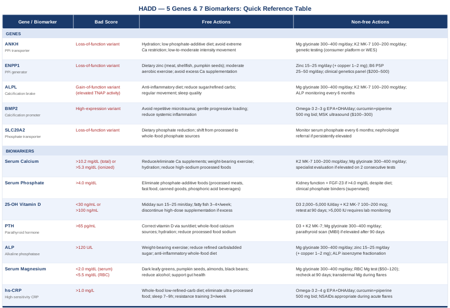
What Your DNA May Reveal: 5 Genes Linked to HADD
Genetics does not predetermine HADD, but it can meaningfully raise your baseline susceptibility to pathological soft tissue calcification. Several genes in the pyrophosphate metabolism and calcification-inhibitory pathways have been identified in research on crystal arthropathies and ectopic mineralization. Understanding your variants — through consumer genetic testing, clinical genetics panels, or whole-exome sequencing — can help you prioritize the most targeted interventions from the biomarker section above.
1. ANKH — The Pyrophosphate Transport Gene
What it does: The ANKH gene encodes a transmembrane protein that transports inorganic pyrophosphate (PPi) out of cells and into the extracellular matrix. PPi is the primary endogenous inhibitor of hydroxyapatite crystal nucleation: it coats nascent crystals and prevents their growth. Loss-of-function ANKH variants reduce extracellular PPi concentration and directly enable soft tissue calcification. In a landmark study published in Science, disruption of the murine homolog of this gene produced generalized tissue calcification and arthritis, establishing ANKH as a central calcification gate.
If the gene is bad — plan without supplements: The most impactful lifestyle intervention is reducing conditions that deplete extracellular PPi. This means maintaining excellent hydration (PPi transport is impaired in acidic environments), following a diet low in phosphate additives, and avoiding extreme dietary calcium restriction, which paradoxically increases PTH and further destabilizes the mineral balance. Regular low-to-moderate intensity movement improves joint fluid turnover, which influences synovial PPi concentrations.
If the gene is bad — plan with supplements or equipment: Magnesium at 300–400 mg/day (glycinate form) partially compensates for reduced PPi by competing with calcium at nucleation sites in soft tissue. Vitamin K2 MK-7 (100–200 mcg/day) activates MGP independently of the PPi pathway, providing a parallel calcification-inhibitory mechanism. ANKH variants can be identified through raw data from consumer genetic testing platforms or clinical whole-exome sequencing. Note that clinical interpretation of specific ANKH SNPs remains an evolving area — results should be reviewed with a genetic counselor.
2. ENPP1 — The PPi Generator
What it does: ENPP1 (ectonucleotide pyrophosphatase/phosphodiesterase 1) is the enzyme responsible for generating extracellular PPi from ATP. It works in tandem with ANKH: ENPP1 produces the PPi, and ANKH transports it to where calcification inhibition is needed. Loss-of-function variants in ENPP1 reduce PPi production at the source. Research by Rutsch et al. in Nature Genetics identified ENPP1 mutations as the cause of severe generalized arterial calcification in infancy, and milder variants are now being studied in the context of broader calcification-prone phenotypes including periarticular crystal deposition.
If the gene is bad — plan without supplements: ENPP1 requires zinc as a structural cofactor for its enzymatic activity. Ensuring adequate dietary zinc through meat, shellfish (especially oysters), and pumpkin seeds supports ENPP1 function without any supplementation. Moderate aerobic exercise supports purine nucleotide cycling (the ATP-to-PPi pathway) and may help maintain ENPP1 substrate availability. Avoid conditions that deplete ENPP1 substrate: excessive calcium supplementation in some contexts may dampen the cellular signaling that drives ENPP1 activity.
If the gene is bad — plan with supplements or equipment: Zinc (15–25 mg/day) directly supports ENPP1 enzymatic function. Vitamin B6 (25–50 mg/day as P5P form) supports phosphate metabolism as a broader cofactor. Cycling note: zinc above 20 mg/day should not exceed 3 months without copper monitoring; supplement copper at 1–2 mg/day alongside long-term zinc. ENPP1 variant analysis is available through clinical metabolic genetics panels ($200–$500) or whole-exome sequencing.
3. ALPL — The Calcification Brake
What it does: The ALPL gene encodes tissue-nonspecific alkaline phosphatase (TNAP), which hydrolyzes PPi — effectively dismantling the PPi-based brake on hydroxyapatite formation. In normal physiology, TNAP activity in bone promotes mineralization by clearing PPi where bone formation should occur. In soft tissue, however, aberrant TNAP activity or gain-of-function variants deplete local PPi and enable inappropriate calcification. This gene is the direct genetic driver of the ALP biomarker discussed above and links the two analytical layers covered in this article.
If the gene is bad — plan without supplements: Reducing chronic inflammation is the most direct lifestyle lever: inflammatory cytokines upregulate TNAP expression in vascular and tendon fibroblast cell lines. An anti-inflammatory whole-food diet, regular movement, and addressing sleep quality all reduce the inflammatory pressure on TNAP expression. High-sugar and high-refined-carbohydrate diets specifically upregulate TNAP through insulin-and-AGE-driven signaling pathways and are worth eliminating independently of any other intervention.
If the gene is bad — plan with supplements or equipment: Magnesium is a physiological inhibitor of TNAP activity and should be a foundational supplement for anyone with high-activity ALPL variants. Vitamin K2 MK-7 provides a downstream compensatory mechanism by activating MGP even when PPi levels are reduced by elevated TNAP. These two supplements work synergistically and can be maintained long-term. Track ALP on standard blood panels every 6 months to monitor TNAP activity trajectory.
4. BMP2 — The Calcification Promoter
What it does: Bone morphogenetic protein 2, encoded by the BMP2 gene, is a potent inducer of osteoblast differentiation and bone formation. While essential for skeletal development, BMP2 signaling in soft tissue drives the transdifferentiation of tendon fibroblasts into osteoblast-like cells — a key step in HADD pathogenesis. Elevated BMP2 expression has been documented directly in calcific tendinopathy tissue specimens. Genetic variants that increase BMP2 expression or receptor sensitivity may therefore predispose tendons to mineralization under conditions of microtrauma or local hypoxia.
If the gene is bad — plan without supplements: Reducing the triggers of BMP2 upregulation is the primary mechanical intervention. Repetitive microtrauma to the affected tendon is a known trigger for fibroblast-to-osteoblast transition via BMP2 — this is the mechanistic reason why conservative tendon load management during active HADD phases matters clinically. Gentle progressive loading (not aggressive loading) is preferable. Reducing chronic systemic inflammation dampens the cytokine environment that amplifies BMP2 signaling in soft tissue.
If the gene is bad — plan with supplements or equipment: Omega-3 fatty acids (2–3 g EPA+DHA daily) reduce the inflammatory cytokine environment that synergizes with BMP2 signaling to drive ectopic calcification. Curcumin with piperine (500 mg twice daily) has been shown in cell studies to inhibit BMP2-induced osteogenic differentiation; clinical translation is still uncertain but the anti-inflammatory rationale is sound. Musculoskeletal ultrasound ($100–$300 per assessment) provides a practical way to track calcification burden over time and evaluate whether interventions are reducing crystal size or density.
5. SLC20A2 — The Phosphate Transporter
What it does: SLC20A2 encodes a type III sodium-dependent phosphate transporter (PiT-2), expressed in various soft tissues including tendons, smooth muscle, and neural tissue. Loss-of-function mutations in SLC20A2 cause primary familial brain calcification, demonstrating that dysregulated phosphate transport directly leads to ectopic mineral deposition. Milder variants and altered expression of phosphate transporter genes are an active area of calcification genetics research, with emerging evidence suggesting they may influence soft tissue calcification risk in non-neurological contexts including periarticular calcification.
If the gene is bad — plan without supplements: The most actionable intervention is dietary phosphate reduction. Since transporter activity determines how much consumed phosphate enters cells, a lower total phosphate load reduces absolute crystal nucleation risk regardless of transporter activity level. Shifting from processed to whole-food sources of phosphate reduces bioavailable phosphate intake by 400–800 mg per day for most people — a meaningful change that takes effect within weeks.
If the gene is bad — plan with supplements or equipment: No targeted supplement addresses SLC20A2 directly. The strategy is keeping serum phosphate in the optimal range (2.5–3.5 mg/dL) through diet and monitoring. If variants in this gene are confirmed through genetic testing and phosphate remains persistently elevated, a nephrologist is the appropriate specialist for further evaluation and management. Serum phosphate monitoring every 6 months is a reasonable surveillance frequency.
The Calcium Paradox: A Book That Reframes Why Calcium Ends Up in the Wrong Places
Kate Rheaume-Bleue's Vitamin K2 and the Calcium Paradox is arguably the most directly relevant popular health book for anyone with HADD. Its core argument — that vitamin K2 determines where calcium is deposited in the body, and that K2 deficiency causes calcium to accumulate in arteries, kidneys, and soft tissue rather than reinforcing bone — maps precisely onto the biology of hydroxyapatite deposition disease.
10 Things Worth Knowing From This Book
1. Matrix Gla Protein is your body's primary soft tissue calcification blocker. MGP is produced in the walls of blood vessels and in soft connective tissue, but it only becomes active after vitamin K2-dependent carboxylation. Without adequate K2, MGP remains undercarboxylated and functionally inert — the brake on soft tissue calcification is simply not engaged.
2. Undercarboxylated MGP (ucMGP) is now a measurable biomarker. Elevated ucMGP in blood is a direct marker of K2 functional deficiency and has been found elevated in patients with soft tissue calcification disorders. Testing is available at specialty labs for $100–$200 and is emerging as a meaningful clinical tool.
3. Most Western diets are severely K2-deficient. The primary dietary sources of MK-7 — natto (fermented soybeans), certain aged cheeses, and grass-fed animal fat — are largely absent from standard Western diets. Vitamin K1 from leafy greens is poorly converted to K2 in humans and does not adequately substitute.
4. Supplemental calcium without K2 may worsen soft tissue calcification. Population data from the Rotterdam Study, published in the Journal of Nutrition (Geleijnse et al., 2004), found that higher dietary K2 intake was associated with significantly lower rates of arterial calcification and cardiovascular mortality. Calcium supplements taken without K2 provide calcium without the routing instructions.
5. Vitamin D3 requires K2 to be used safely. D3 increases intestinal calcium absorption; K2 ensures that absorbed calcium is directed to bone rather than soft tissue via MGP and osteocalcin activation. Taking high-dose D3 without K2 long-term may promote ectopic calcification — this is directly relevant to the vitamin D supplementation guidance in this article.
6. MK-7 is the preferred supplemental form of K2. MK-4 has a half-life of a few hours; MK-7 has a half-life of approximately 3 days, producing more sustained tissue activation of MGP. Effective doses in the research literature range from 45 to 200 mcg/day. Taking K2 with a fat-containing meal improves absorption.
7. Osteocalcin also requires K2-dependent carboxylation. Osteocalcin anchors calcium into the bone matrix; without adequate K2, it remains undercarboxylated and dysfunctional. This means that even people with sufficient bone mineral density can have poor calcium incorporation quality when K2 is deficient.
8. The paradox explains a common clinical puzzle. Patients with the highest vascular and soft tissue calcification burden frequently have osteoporosis simultaneously. They do not have too much calcium; they have a broken routing system. Addressing K2 targets the routing dysfunction rather than the calcium supply.
9. Recurring HADD may be driven by chronic K2 insufficiency. If the proteins responsible for preventing crystal nucleation in soft tissue are chronically inactive, calcific deposits will continue to reform regardless of how individual episodes are treated. K2 optimization is one of the few interventions that addresses a potential root cause of the recurrence pattern.
10. Natto is the most practical dietary source. A modest serving (2–3 tablespoons) of natto 3–4 times per week provides more MK-7 than any other food source by a large margin. Aged Gouda and certain Brie cheeses also contain meaningful amounts. Where natto is impractical, a daily MK-7 supplement at 100–200 mcg is the next best option.
Complementary Approaches With Meaningful Clinical Support
The following three modalities have the best combination of human clinical evidence and relevance to HADD, whether for pain management, tissue mobility, or the inflammatory biology underlying crystal deposition.
Low-Level Laser Therapy (Photobiomodulation)
Low-level laser therapy (LLLT), also called photobiomodulation, uses red and near-infrared light at low intensity to stimulate mitochondrial activity in cells, reduce local pro-inflammatory cytokine production, and promote tissue repair. For HADD, its relevance is twofold: first, it has direct evidence for reducing pain and improving function in calcific shoulder tendinitis; second, it may support the active vascular remodeling phase during which crystals are being reabsorbed, a process that depends on cell-mediated activity within the deposit.
Systematic reviews of LLLT for shoulder tendinopathy have found statistically significant improvements in pain and functional scores at wavelengths of 630–1000 nm and doses of 1–4 J/cm². A Cochrane-adjacent review by Bjordal and colleagues on LLLT for musculoskeletal shoulder disorders found moderate short-term evidence for benefit in rotator cuff-related conditions including calcific forms. Protocols typically involve 6–12 sessions over 3–6 weeks, administered by a physiotherapist or sports medicine practitioner with an appropriate device.
Clinically, LLLT sessions run $50–$150 each. Home devices cleared for musculoskeletal use are available in the $300–$800 range. The approach is well tolerated with minimal adverse effects. For HADD specifically, LLLT is best applied during the subacute or chronic phase — not during a severe acute inflammatory flare. A practical starting protocol is three sessions per week for four weeks, targeting the affected tendon area. It should be positioned as an adjunct to, not a replacement for, standard physiotherapy and medical management.
Mindfulness Meditation / MBSR
Mindfulness-Based Stress Reduction (MBSR) is an 8-week structured program integrating body scan practice, seated meditation, and gentle movement. Its relevance to HADD extends beyond simple relaxation: MBSR has been shown to reduce systemic inflammatory markers including IL-6 and hs-CRP with consistent practice, which connects directly to one of the seven biomarkers tracked above. More practically, recurrent HADD can develop a central sensitization component — where the nervous system amplifies pain signals independently of the physical deposit status — making mind-body training clinically meaningful for a subset of patients.
A landmark randomized controlled trial published in JAMA Internal Medicine demonstrated that MBSR produced significant and durable improvements in pain and function compared to health education controls in patients with chronic musculoskeletal pain. While not specific to HADD, the reduction in pain sensitization and systemic inflammation is directly applicable. The effects on inflammatory biomarkers have been replicated in multiple smaller controlled trials, lending biological plausibility to what might otherwise appear to be a purely psychological intervention.
MBSR is available through hospital-based programs, community health centers, and validated online courses such as the free 8-week curriculum offered by Palouse Mindfulness, based on Jon Kabat-Zinn's original protocol. The time commitment is approximately 45 minutes per day for 8 weeks. Effects accumulate over the 3–6 month range with consistent practice. For HADD patients specifically, the body scan component can help develop a more nuanced and less anxious relationship with chronic joint pain, reducing the muscle guarding and tension that compound local symptoms around the affected tendon.
Massage Therapy
Deep tissue massage and myofascial release address the soft tissue changes that invariably accompany HADD: restricted joint capsule, muscle guarding around the affected tendon, adhesions in periarticular structures, and compensatory movement patterns in surrounding musculature. Massage does not dissolve calcific deposits, but it plays a meaningful role in maintaining tissue mobility, reducing referred pain patterns, and preventing the secondary joint dysfunction that accelerates degeneration. In the shoulder — the most common HADD site — targeted work on the rotator cuff muscles, posterior capsule, and cervical region can substantially improve functional range of motion between flares.
A systematic review on massage therapy for shoulder disorders found moderate evidence for short-term pain reduction and functional improvement in rotator cuff-related conditions. The evidence base for HADD specifically is limited, but the mechanical rationale is sound: periarticular soft tissue restrictions that develop around a calcific deposit create their own pain cycle independent of the deposit itself, and manual therapy is the most direct way to address those restrictions. During acute inflammatory flares, deep tissue work directly over the calcified area is contraindicated and may worsen pain; gentler lymphatic drainage techniques are more appropriate during acute phases.
Practically, a course of 6–10 sessions of 45–60 minutes with a registered massage therapist or physiotherapist trained in deep tissue techniques is a reasonable protocol for HADD-related shoulder or hip restrictions. Sessions run $60–$150 depending on location. Self-massage tools — foam rollers, lacrosse balls for trigger point work — can maintain soft tissue mobility between professional sessions at minimal cost. The therapeutic goal is not to address the deposit itself but to keep the surrounding soft tissue environment from compounding the problem.
Conclusion
Hydroxyapatite deposition disease is a condition where better data genuinely changes the picture. Tracking the seven biomarkers in this article — particularly magnesium, vitamin D, PTH, ALP, and hs-CRP — gives you a personal mineral and inflammatory profile that generic advice cannot account for. Understanding your genetic risk through ANKH, ENPP1, ALPL, BMP2, and SLC20A2 adds a layer of context that helps prioritize which interventions matter most for your individual biology. And for anyone with recurrent calcifications, the K2-MGP axis deserves serious attention as a potential root-cause contributor that most clinicians never address.
The smartest next step is also the most actionable: request a comprehensive mineral panel at your next blood draw — serum calcium, phosphate, magnesium, 25-OH vitamin D, PTH, and hs-CRP — and bring those results to a clinician comfortable interpreting them in the context of crystal deposition disease. That conversation, grounded in numbers rather than symptoms alone, is where more targeted management begins.
Musculoskeletal: Bone Conditions Joint Conditions Tendon & Ligament Conditions
Autoimmune: Inflammatory Conditions