This article contains AI generated content.
· UpdatedHypereosinophilic Syndrome Genes And Biomarkers: 6 Genes And 7 Biomarkers To Track
Introduction
Living with hypereosinophilic syndrome — or watching your eosinophil count creep persistently above 1,500 cells per microliter despite treatment adjustments — is a frustrating experience that most standard care approaches handle incompletely. HES is not a single disease. It is a cluster of overlapping immune disorders that look similar on the surface but have very different drivers underneath, and that distinction matters enormously for how you respond to treatment.
Generic advice about "managing inflammation" or "reducing stress" barely scratches the surface here. HES can silently damage the heart, lungs, skin, and nervous system before symptoms become obvious. By the time someone feels the full weight of the condition, tissue injury is already underway. That gap between what routine testing reveals and what is actually happening at the cellular level is exactly where this article tries to help.
What makes HES particularly difficult to navigate is that its causes span multiple biological systems. In some people, a single genetic fusion event drives runaway eosinophil production. In others, a rogue population of T-lymphocytes secretes cytokines that tell bone marrow to keep making eosinophils at dangerous levels. For many, the mechanism remains genuinely unclear. That heterogeneity is why one-size-fits-all treatment often falls short, and why understanding your own biological picture — through targeted biomarkers and genetic markers — can make a real difference in treatment decisions.
This article takes two practical angles. The first focuses on 7 key biomarkers that give you and your physicians a running picture of eosinophil-driven tissue stress, treatment response, and organ risk. The second explores the 6 most clinically relevant genes and molecular markers implicated in HES subtypes, with practical implications for each. Neither section offers cures. Both offer better questions to ask and clearer signals to track.
Summary
This article covers 7 biomarkers — including some rarely ordered in routine care — that can reveal whether eosinophil activity is causing hidden cardiac damage, mast cell overlap, or treatment resistance. It also covers 6 genetic and molecular markers, including the FIP1L1-PDGFRA fusion that, when present, predicts a near-complete response to a specific targeted drug. You will find a concrete action plan for each marker, whether or not you have access to supplements or advanced testing. Toward the end, you will find a protocol summary that challenges conventional thinking about immune dysregulation management, followed by complementary approaches with real human-study backing. The goal throughout is simple: better information, sharper decisions, fewer surprises.
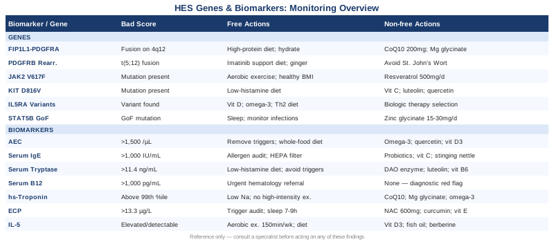
7 Biomarkers to Track in Hypereosinophilic Syndrome
Biomarkers in HES serve two distinct purposes. Some confirm the diagnosis or quantify disease severity. Others detect organ damage early enough to intervene before it becomes irreversible. The seven below cover both functions, ranging from a basic blood count component to specialized cytokine assays. None of them alone tells the full story, but together they create a surveillance framework that puts you ahead of the disease rather than reacting to it.
Biomarker 1: Absolute Eosinophil Count (AEC)
The absolute eosinophil count is the cornerstone of HES diagnosis and monitoring. Unlike the percentage of eosinophils in a white cell differential, the AEC reflects the actual number of eosinophils circulating per microliter of blood. The diagnostic threshold for HES is greater than 1,500 eosinophils/µL persisting for at least one month, though organ damage can occur even at lower sustained counts.
How to Measure It
AEC is derived from a standard complete blood count with differential (CBC-diff). This is one of the most affordable lab panels available, typically costing $20–$50 in the United States, and is covered by most insurance plans. It should be measured serially — at least monthly during active disease or treatment initiation — rather than as a one-time snapshot.
If the Score Is Bad: The Plan Without Supplements
When AEC remains persistently elevated, the first non-pharmacological priorities are identifying and removing reversible causes. Drug-induced hypereosinophilia (triggered by NSAIDs, antibiotics, antiepileptics, or biologics) accounts for a meaningful minority of cases and resolves on discontinuation. Helminth infections, particularly tissue-invasive parasites, must be excluded with stool examination and relevant serology. Reducing systemic inflammation through a whole-food, low-processed-food diet and minimizing toxin exposure (mold, pesticides, heavy metals) provides an anti-inflammatory foundation. During flares, strict rest and avoidance of intense physical exertion reduce the risk of eosinophil degranulation in cardiac tissue.
If the Score Is Bad: The Plan With Supplements or Equipment
Several supplements have anti-eosinophilic or mast-cell-stabilizing properties that may modestly support medical management, though none replace disease-modifying therapy. Omega-3 fatty acids (EPA/DHA, 2–4g/day in 2 divided doses) shift eicosanoid production toward less pro-inflammatory mediators and have shown eosinophil-modulating effects in allergic and eosinophilic disease models. Cycle with a 1-week break every 8–12 weeks; side effects include GI discomfort and mild blood thinning. Quercetin (500–1000mg/day with meals) acts as a mast cell stabilizer and inhibits IL-5 signaling in vitro; cycle 5 days on / 2 days off. Vitamin D3 (2,000–5,000 IU/day with K2) has immunomodulatory effects on Th2 bias; test serum 25-OH-D to target 50–70 ng/mL, adjust dose accordingly. Do not self-supplement without discussing with your physician if you are on corticosteroids or immunosuppressants.
Biomarker 2: Serum IgE
Total serum IgE is elevated in the lymphocytic and allergic subtypes of HES and reflects the degree of Th2 immune skewing. While a high IgE does not diagnose HES on its own, it identifies a subgroup of patients whose eosinophilia is driven by lymphocyte-derived cytokines — particularly IL-4 and IL-13 — rather than intrinsic bone marrow disease.
How to Measure It
Serum IgE is measured via immunoassay (ELISA or chemiluminescence) from a standard venous blood draw. Cost ranges from $30–$80. Normal values in adults are typically below 100 IU/mL, with levels above 1,000 IU/mL suggesting significant allergic or lymphocytic HES contribution. Specific IgE panels (ImmunoCAP) can further identify allergen triggers driving the Th2 response.
If the Score Is Bad: The Plan Without Supplements
Elevated IgE calls for a structured allergen audit. Common environmental triggers include dust mites, mold, pet dander, and certain food proteins. An elimination diet trial (removing the top 8 allergens for 4–6 weeks) followed by systematic reintroduction can reveal dietary contributors without any pharmaceuticals. Reducing indoor air pollution through HEPA filtration (CADR-rated units for bedroom and living areas) addresses inhalant triggers. Gut permeability plays a significant role in IgE-mediated sensitization; a low-lectin, low-gluten dietary approach may reduce antigen translocation.
If the Score Is Bad: The Plan With Supplements or Equipment
Probiotics with Lactobacillus rhamnosus GG or Bifidobacterium longum strains have demonstrated modest IgE-lowering effects in allergic populations in several randomized trials; take daily with a prebiotic fiber source, continuous use for at least 12 weeks before evaluating effect. Vitamin C (1,000mg twice daily with meals) has antihistamine and Th2-modulatory properties; generally well-tolerated, though high doses may cause GI upset. Stinging nettle extract (300–500mg freeze-dried, twice daily) inhibits tryptase and prostaglandins; cycle 6 weeks on / 2 weeks off. Air purifiers with HEPA and activated carbon filters are a practical equipment investment for high-IgE patients.
Biomarker 3: Serum Tryptase
Tryptase is released by activated mast cells and elevated baseline tryptase indicates that mast cell activation — or systemic mastocytosis — is co-occurring alongside eosinophilia. This overlap is more common than clinically recognized in HES and significantly alters treatment strategy. Patients with concurrent mast cell activation syndrome (MCAS) may have blunted responses to corticosteroids and require separate mast cell–directed therapies.
How to Measure It
Serum tryptase is measured via fluorescent enzyme immunoassay. Basal tryptase (drawn outside of acute symptoms, ideally in the morning) costs $80–$150. A normal baseline is below 11.4 ng/mL. Levels above 20 ng/mL, particularly when persistent, warrant bone marrow biopsy to rule out systemic mastocytosis. In patients with HES who are not responding to expected therapies, tryptase should be checked even if never ordered before.
If the Score Is Bad: The Plan Without Supplements
High baseline tryptase requires referral to an allergist-immunologist or hematologist with MCAS expertise. Dietary management follows a low-histamine protocol: avoiding fermented foods, aged cheeses, cured meats, alcohol, leftovers, and high-histamine vegetables (tomato, spinach, eggplant). Cold-storing and consuming fresh food reduces histamine accumulation. Trigger avoidance (heat, cold, pressure, emotional stress, fragrance exposure) is central to mast cell management.
If the Score Is Bad: The Plan With Supplements or Equipment
DAO (Diamine Oxidase) enzyme (taken before histamine-containing meals, 3–6mg per dose) helps degrade dietary histamine; use as needed before high-risk meals, not daily continuously. Luteolin (100–200mg/day), a flavonoid with potent mast cell–stabilizing properties, has shown efficacy in reducing mediator release in cell studies and small clinical observations; cycle 4 weeks on / 1 week off. Vitamin B6 (as P5P, 25–50mg/day) is a cofactor for DAO enzyme activity. Salt-based nasal irrigation may help with upper airway mast cell reactivity. An air purifier and fragrance-free household environment are practical protective measures.
Biomarker 4: Serum Vitamin B12 (Cobalamin)
This marker surprises many patients and physicians. In the context of HES, significantly elevated vitamin B12 — particularly above 1,000 pg/mL in someone not supplementing — is a red flag for myeloproliferative disease. Eosinophilic granulocytes and the neoplastic clones driving myeloproliferative HES release transcobalamin and haptocorrin proteins that elevate circulating B12. In this setting, high B12 is not a sign of nutritional abundance — it is a warning of possible bone marrow pathology.
How to Measure It
Serum B12 is a standard add-on to a metabolic or nutrition panel, costing $20–$50. For full interpretation in HES, it should be measured alongside serum folate, LDH, and uric acid, which together create a profile of bone marrow proliferative activity. If B12 is dramatically elevated with no supplementation history, an urgent hematology referral is warranted rather than any lifestyle intervention.
If the Score Is Bad: The Plan Without Supplements
If B12 is genuinely elevated due to myeloproliferative disease, lifestyle and dietary interventions will not normalize it. The priority is immediate hematological workup: FISH testing for FIP1L1-PDGFRA fusion, bone marrow biopsy, and JAK2 mutation screening. Avoiding B12-rich foods (liver, shellfish, meat concentrates) has negligible impact when the elevation is endogenous. The focus here is on accelerating diagnosis, not dietary adjustment.
If the Score Is Bad: The Plan With Supplements or Equipment
There are no supplements indicated to lower pathologically elevated B12. If B12 is low in an HES patient (which can occur in those on prolonged proton pump inhibitor therapy or with dietary restriction), standard supplementation with methylcobalamin (1,000–2,000 mcg sublingually daily) is appropriate, but this scenario does not suggest myeloproliferative disease — it is an unrelated nutritional correction.
Biomarker 5: High-Sensitivity Cardiac Troponin (hsTnI or hsTnT)
Cardiac involvement is the most feared complication of HES. Eosinophil degranulation in myocardial tissue releases major basic protein and eosinophil peroxidase, which are directly toxic to cardiomyocytes. This produces a staged pathology: first a necrotic phase (acute myocarditis), then a thrombotic phase, then a fibrotic phase with endomyocardial scarring and restrictive cardiomyopathy. High-sensitivity troponin detects subclinical myocardial injury before symptoms develop — arguably making it the most important HES biomarker for preventing irreversible damage.
How to Measure It
hsTnI or hsTnT is measured from a standard blood draw at any accredited laboratory. Cost ranges from $50–$150. Reference ranges vary by assay and laboratory; the 99th percentile upper reference limit is typically below 14–22 ng/L for hsTnI in most commercial assays. In HES patients with any eosinophil count above 5,000/µL, or with any unexplained dyspnea or palpitations, this should be ordered as a baseline and repeated every 3–6 months. Cardiac MRI with gadolinium contrast is the gold standard for detecting endomyocardial fibrosis when troponin trends upward.
If the Score Is Bad: The Plan Without Supplements
Elevated troponin in HES is a cardiology emergency. It demands immediate reduction of eosinophil count (with corticosteroids or directed therapy), cardiology consultation, and echocardiogram. From a lifestyle perspective, avoiding high-intensity exercise during active disease prevents mechanical stress on an already-inflamed myocardium. Moderate activity (walking, gentle cycling at 50–60% of heart rate reserve) may be maintained if the cardiologist approves. Sodium restriction (below 2g/day) reduces volume load on a compromised heart.
If the Score Is Bad: The Plan With Supplements or Equipment
CoQ10 (ubiquinol form, 200–400mg/day with fatty meal) supports mitochondrial energy production in cardiomyocytes and has evidence for improving cardiac function in inflammatory cardiomyopathy; ongoing use without cycling in this context. Magnesium glycinate (300–400mg/day in the evening) reduces arrhythmia risk and supports vascular tone; long-term use is safe at this dose. Omega-3 EPA/DHA (3–4g/day) reduces triglycerides and has anti-inflammatory effects on pericardial and myocardial tissue; cycle as noted above. A pulse oximeter and home blood pressure cuff are practical monitoring tools. Avoid energy drinks, excess caffeine, and stimulant supplements which increase cardiac demand.
Biomarker 6: Eosinophil Cationic Protein (ECP)
While AEC measures how many eosinophils are present, ECP measures what they are doing. ECP is a granular protein released by activated eosinophils that is directly cytotoxic to epithelial, cardiac, and neuronal tissue. Elevated serum ECP indicates active degranulation — meaning tissue damage is ongoing — even when the AEC appears controlled or borderline. In patients who feel symptomatic despite a "normal" eosinophil count, ECP often reveals subclinical disease activity.
How to Measure It
Serum ECP is measured via immunoassay (the Pharmacia UniCAP assay is the most standardized). This is a specialized test not available at all commercial labs, costing approximately $80–$200 out of pocket. Normal adult levels are generally below 13.3 µg/L. Sample handling is critical — blood must be allowed to clot at room temperature for exactly 60 minutes before centrifugation, as temperature and timing affect ECP release from granulocytes during clotting.
If the Score Is Bad: The Plan Without Supplements
High ECP in the absence of a proportionally elevated AEC suggests that each eosinophil is more activated than expected — a pattern seen in allergic triggers, occult infections, and stress-driven immune activation. A systematic environmental and dietary trigger audit is the first step. Sleep optimization (7–9 hours of consistent sleep with circadian alignment) substantially reduces pro-inflammatory cytokine levels including those that prime eosinophil degranulation. Sauna or thermotherapy protocols should be avoided during active flares, as heat can trigger mast cell and eosinophil mediator release.
If the Score Is Bad: The Plan With Supplements or Equipment
N-Acetylcysteine (NAC) (600mg twice daily) is a glutathione precursor that reduces oxidative damage from ECP-associated reactive oxygen species; cycle 8 weeks on / 2 weeks off, as extended high-dose use may downregulate intrinsic antioxidant production. Curcumin with piperine (500–1,000mg curcumin + 5–10mg piperine, twice daily with food) inhibits NF-κB signaling and reduces multiple inflammatory mediators including those governing eosinophil activation; cycle 6 weeks on / 1–2 weeks off. Vitamin E (mixed tocopherols) (400 IU/day) acts as a lipid-phase antioxidant protecting cell membranes from ECP-mediated oxidative assault; take with a fat-containing meal.
Biomarker 7: Interleukin-5 (IL-5)
IL-5 is the master cytokine of eosinophil biology. It drives eosinophil differentiation in bone marrow, prolongs eosinophil survival in tissue, and primes eosinophils for degranulation. In lymphocytic HES, aberrant T-cell clones secrete large quantities of IL-5, sustaining eosinophilia even when no intrinsic bone marrow problem exists. IL-5 measurement directly reveals the upstream driver of eosinophil excess and predicts response to anti-IL-5 therapies (mepolizumab, reslizumab, benralizumab).
How to Measure It
Serum IL-5 is measured via high-sensitivity ELISA in specialized immunology or research-adjacent clinical laboratories. Cost ranges from $150–$300 and availability varies by region. In healthy individuals, IL-5 is typically low or undetectable. In active lymphocytic HES, levels are often meaningfully elevated. Some academic medical centers also offer flow cytometry–based intracellular cytokine staining to identify IL-5-secreting T-cell populations, which is more informative but more expensive and less accessible.
If the Score Is Bad: The Plan Without Supplements
Chronically elevated IL-5 points strongly toward lymphocytic HES and is the most direct rationale for seeking evaluation for biologic anti-IL-5 therapy. Non-pharmacological strategies that modulate Th2 cytokine bias include consistent moderate-intensity aerobic exercise (150 minutes/week), which shifts immune polarization toward Th1 and reduces Th2 cytokine output in multiple human studies. Reduction of dietary advanced glycation end-products (AGEs), found in high-heat processed foods and charred meats, reduces systemic inflammatory tone.
If the Score Is Bad: The Plan With Supplements or Equipment
Vitamin D3 (target 25-OH-D of 60–70 ng/mL, typically requiring 4,000–6,000 IU/day depending on baseline) directly suppresses Th2 cytokine production, including IL-5 and IL-13, in multiple human immune studies; test levels and titrate rather than assuming a standard dose. Fish oil (high EPA) at 3–4g/day shifts eicosanoid production away from prostaglandin E2, which amplifies IL-5 secretion. Berberine (500mg twice daily with meals) modulates gut microbiota and reduces Th2 polarization through AMPK activation; cycle 8 weeks on / 2 weeks off due to potential gut adaptation effects. Note: none of these replace physician-directed evaluation for IL-5–targeted biologics if indicated.
The biomarker picture is only half of the story. Understanding the genetic and molecular architecture underlying your HES subtype can dramatically sharpen treatment selection and predict long-term prognosis. The following section covers the 6 most clinically relevant molecular markers.
6 Genes and Molecular Markers in Hypereosinophilic Syndrome
Genetic testing in HES is not about ancestry or distant risk. It is about determining right now which biological mechanism is driving your eosinophilia, because the answer determines whether you need a targeted kinase inhibitor, a biologic, or a cytotoxic agent — and whether your condition is a true malignancy or a reactive phenomenon. These six markers cover the full diagnostic landscape of HES molecular subtypes.
Gene 1: FIP1L1-PDGFRA Fusion (F/P Fusion)
This is the most clinically important molecular finding in HES. A deletion on chromosome 4q12 fuses the FIP1L1 and PDGFRA genes, creating a constitutively active tyrosine kinase that drives uncontrolled eosinophil production. It is found in approximately 10–20% of HES cases, predominantly in men, and defines a WHO-recognized entity: myeloid/lymphoid neoplasm with PDGFRA rearrangement. The clinical implication is profound: this fusion is exquisitely sensitive to imatinib (Gleevec) at doses as low as 100mg/day, with complete hematologic and molecular remission reported in the vast majority of cases.
If the Gene Is Abnormal: The Plan Without Supplements
Detection of the F/P fusion is an immediate indication for imatinib therapy under hematologist supervision — this is not a lifestyle-manageable condition. However, lifestyle practices that support treatment tolerance include: a high-protein anti-inflammatory diet to offset the fatigue and nausea that sometimes accompany imatinib initiation, ensuring adequate hydration, and avoiding concurrent use of strong CYP3A4 inhibitors (grapefruit juice, certain supplements) which can raise imatinib plasma levels. Cardiac monitoring at baseline and periodically during therapy is standard given the pre-existing cardiac risk in HES.
If the Gene Is Abnormal: The Plan With Supplements or Equipment
While imatinib is the cornerstone, CoQ10 (200–400mg/day ubiquinol) may help offset the mitochondrial suppression and fatigue occasionally associated with tyrosine kinase inhibitor therapy; ongoing use is reasonable. Magnesium supplementation (glycinate, 300mg/day) supports cardiovascular monitoring. A home blood pressure monitor and pulse oximeter enable between-visit surveillance. There are no supplements that substitute for imatinib when the fusion is confirmed — the drug achieves outcomes no natural intervention approaches.
Gene 2: PDGFRB Rearrangements
PDGFRB rearrangements, most commonly the t(5;12) translocation creating the ETV6-PDGFRB fusion, define another WHO-recognized subtype of myeloid/lymphoid neoplasm with eosinophilia. Like PDGFRA rearrangements, these produce constitutively active tyrosine kinases that drive eosinophil excess from the marrow. They are rarer than the F/P fusion but equally important to identify because they also respond well to imatinib. Testing is done via FISH (fluorescence in situ hybridization) or cytogenetic karyotyping.
If the Gene Is Abnormal: The Plan Without and With Supplements
The clinical management parallels FIP1L1-PDGFRA: imatinib under specialist supervision. Lifestyle support focuses on treatment tolerance, cardiovascular monitoring, and managing GI side effects with small frequent meals and ginger tea (500–1,000mg ginger extract for nausea; well-tolerated and compatible with imatinib). Avoid St. John's Wort, which is a strong CYP3A4 inducer that substantially reduces imatinib blood levels.
Gene 3: JAK2 V617F
The JAK2 V617F point mutation is the signature mutation of classic myeloproliferative neoplasms (polycythemia vera, essential thrombocythemia, myelofibrosis) but is occasionally found in HES patients with a myeloproliferative phenotype — particularly those with elevated B12, splenomegaly, or thrombocytosis alongside eosinophilia. When present alongside eosinophilia, it suggests a primary myeloproliferative driver and often correlates with resistance to corticosteroids.
If the Gene Is Abnormal: Plans Without and With Supplements
JAK2-mutant HES with organ involvement typically requires JAK inhibitor therapy (ruxolitinib) or hydroxyurea under hematology supervision. Lifestyle practices supporting JAK2-mutant disease management include: maintaining a healthy BMI (adipose tissue amplifies JAK-STAT inflammatory signaling), avoiding exogenous estrogen (which can worsen thrombotic risk in JAK2-positive states), and consistent low-to-moderate aerobic activity for JAK-STAT pathway modulation. Resveratrol (500mg/day with food) has been studied as a JAK2 signaling modulator in preclinical work, though human evidence in this context remains preliminary; cycle 8 weeks on / 2 weeks off.
Gene 4: KIT D816V
The KIT D816V mutation drives systemic mastocytosis and is present in HES patients who have a concurrent mast cell neoplasm — an underappreciated overlap condition. When eosinophilia co-exists with systemic mastocytosis, elevated baseline tryptase (see biomarker 3 above) is the clinical clue, and KIT D816V is the molecular confirmation. Critically, KIT D816V is resistant to imatinib at standard doses, so identifying it prevents futile treatment and guides use of midostaurin or avapritinib.
If the Gene Is Abnormal: Plans Without and With Supplements
This diagnosis requires specialist management at a center with mastocytosis expertise. Lifestyle management follows the same low-histamine, mast-cell-trigger-avoidance protocol described under biomarker 3. Vitamin C (1g twice daily) is a natural antihistamine and mast cell stabilizer; luteolin and quercetin stacks (as described under biomarkers 1 and 3) are well-tolerated and may reduce mediator burden. Strict avoidance of NSAID use unless previously tested for mast cell tolerance is important.
Gene 5: IL5RA (IL-5 Receptor Alpha Subunit Variants)
IL5RA encodes the alpha subunit of the IL-5 receptor on eosinophil surfaces. Variants in this gene, and particularly in the signaling complex it forms with the common beta chain (IL3RB), influence how sensitive eosinophils are to IL-5 stimulation and, crucially, how well they respond to anti-IL-5 receptor biologics like benralizumab. This pharmacogenomic relevance is clinically underutilized — patients with certain IL5RA configurations may show differential responses to mepolizumab versus benralizumab, which target the ligand versus the receptor, respectively.
If the Gene Is Abnormal: Plans Without and With Supplements
IL5RA variants do not directly cause disease but modulate its severity and treatment responsiveness. If you are on or being considered for biologic therapy, discussing IL5RA variant testing with your immunologist may help predict which anti-IL-5 agent is most likely to achieve complete response. From a lifestyle standpoint, all strategies that reduce upstream IL-5 secretion (vitamin D, EPA-rich omega-3s, Th2-modulating probiotics) remain relevant regardless of receptor variant status.
Gene 6: STAT5B Mutations
STAT5B encodes a transcription factor that sits downstream of multiple cytokine receptors, including those for IL-5, IL-2, and growth hormone. Gain-of-function mutations in STAT5B have been identified in a subset of patients with T-cell lymphocytosis, eosinophilia, and lymphocytic HES. They drive autonomous T-cell–derived cytokine production independent of normal immune regulation. Detection requires next-generation sequencing panels that include STAT5B — it will not appear on standard genetic testing.
If the Gene Is Abnormal: Plans Without and With Supplements
STAT5B gain-of-function mutations are associated with an aggressive clinical phenotype and often require cyclosporine, interferon-alpha, or clinical trial enrollment. There are no dietary or supplement interventions that meaningfully counter constitutive STAT5B activation. The practical lifestyle priority is maximizing treatment adherence, supporting immune resilience through consistent sleep, and closely monitoring for opportunistic infections if on immunosuppressants. Zinc glycinate (15–30mg/day) supports baseline immune function without stimulating Th2 pathways; take with food to reduce GI irritation.
The Huberman Lab Approach to Immune Dysregulation: What a Deep Dive on Inflammation Suggests for HES
Several episodes of the Huberman Lab podcast, hosted by Stanford neuroscientist Andrew Huberman, have addressed the physiological underpinnings of immune regulation, chronic inflammation, and the neuroimmune axis — all directly relevant to HES management. While no single episode covers HES specifically, the mechanistic principles discussed — particularly around cortisol, circadian rhythm, and vagal tone — offer a framework that challenges the conventional "treat the eosinophil count and wait" approach.
1. Morning Cortisol Is Protective, Not Harmful
Huberman emphasizes that the cortisol spike within the first hour of waking is anti-inflammatory and immune-organizing. Suppressing it through late morning wake times or light deprivation disrupts immune regulation. For HES patients, optimizing morning cortisol rhythm — via 10 minutes of outdoor morning light and consistent wake times — may modestly support eosinophil containment through endogenous glucocorticoid signaling.
2. The Vagus Nerve as an Immune Modulator
The cholinergic anti-inflammatory pathway, mediated through vagal efferents, suppresses systemic cytokine production including IL-5 and IL-6. Huberman has detailed protocols for vagal stimulation through physiological sighing (double inhale through the nose, long exhale through the mouth, practiced 1–2 minutes daily) which activates the parasympathetic nervous system. For HES patients with elevated ECP or persistent IL-5, this costs nothing and has a plausible mechanistic basis.
3. Sleep Architecture and Immune Memory
Slow-wave sleep is when the immune system undergoes regulatory consolidation. Disrupted sleep architecture — common in HES patients with pruritus, cough, or cardiac symptoms — creates a feedback loop of worsening immune dysregulation. Huberman's sleep protocol: consistent sleep and wake times, no bright light (especially blue wavelengths) 1 hour before bed, keeping the bedroom below 67°F (19.4°C), and using non-sleep deep rest (NSDR) protocols for midday immune recovery.
4. Cold Exposure and Eosinophil Biology
Cold exposure (cold shower 30–60 seconds or cold immersion at 50–60°F for 1–3 minutes) increases norepinephrine release, which has anti-inflammatory and IL-6-modulating effects. Huberman cites multiple studies on cold-induced catecholamine surges. For HES patients, caution is warranted: cold can be a mast cell trigger in some individuals. Start with cool (not cold) exposure and monitor for urticarial or anaphylactoid reactions.
5. Exercise as an Anti-Cytokine Intervention
Huberman cites evidence that moderate-intensity aerobic exercise (zone 2, sustainable conversation pace, 30–45 minutes, 4–5 times/week) reduces circulating Th2 cytokines including IL-5 and IL-13 while boosting regulatory T-cell populations. High-intensity exercise, by contrast, transiently elevates eosinophil-relevant cytokines and should be approached cautiously in active HES until the condition is controlled.
6. The Gut-Immune Axis
Multiple Huberman episodes emphasize that 80% of immune cells reside in or adjacent to the gut. Fermented foods (1–3 servings/day of kefir, kimchi, yogurt with live cultures) have been shown in a Stanford RCT to increase microbiome diversity and reduce inflammatory markers more effectively than high-fiber diets alone in healthy adults. For HES patients — particularly those with co-existing gut eosinophilia — this is a practically important and low-risk dietary shift.
7. Light and the Immune Clock
Circadian disruption specifically alters the rhythmicity of eosinophil trafficking and degranulation. Eosinophils express circadian clock genes, and their tissue migration peaks at certain circadian phases. Huberman's emphasis on light-based circadian entrainment (bright light in the morning, darkness at night) may be more relevant to HES biology than is generally appreciated.
8. Deliberate Breathing for Mast Cell and Eosinophil Calm
Box breathing (4 counts in, 4 hold, 4 out, 4 hold) practiced for 5–10 minutes prior to known triggers (stress events, allergen exposure, medical procedures) activates the PNS and blunts immediate mast cell and eosinophil degranulation responses. This is a protocol with strong mechanistic logic and essentially no risk.
9. The Role of Foundational Nutrition
Huberman repeatedly highlights that protein sufficiency (at least 1g per pound of ideal body weight daily) is necessary for robust immune function, including maintaining regulatory T-cell populations that keep Th2 inflammation in check. Many HES patients inadvertently undereat protein due to dietary restrictions. Whole food protein sources (eggs, fish, legumes, quality meat) should anchor the diet.
10. Stress Chemistry and Immune Dysregulation
Chronic psychological stress chronically elevates IL-6, reduces regulatory T cells, and amplifies Th2 polarization. Huberman discusses the distinction between acute stress (beneficial, immune-organizing) and chronic low-grade stress (immune-dysregulating). Practical interventions: daily journaling of emotional stress to create cognitive distance from stressors, limiting news exposure, and ensuring at least one daily activity that produces genuine absorption or "flow."
Complementary Approaches With Human Study Support
The strategies below do not replace medical treatment for HES, but they carry meaningful human-study evidence for relevant mechanisms and may reduce disease burden or improve quality of life when integrated thoughtfully.
Mindfulness Meditation and MBSR
Mindfulness-Based Stress Reduction is an 8-week structured program that teaches non-judgmental present-moment awareness through body scans, sitting meditation, and mindful movement. Its relevance to HES lies in its demonstrated capacity to reduce circulating pro-inflammatory cytokines — including IL-6 and TNF-alpha — in chronically stressed populations, and to modulate the neuroimmune axis through vagal and HPA axis normalization.
A randomized controlled trial published in Psychoneuroendocrinology found that MBSR training significantly reduced IL-6 responses to stress compared to active controls in adults with chronic inflammatory conditions. The 8-week standard protocol (available free through Jon Kabat-Zinn's recordings or low-cost programs) involves 45 minutes of formal practice daily.
For HES patients, MBSR is best started during a period of relative disease stability rather than during a severe flare. Begin with 10–15 minutes of daily body scan practice and build over 2–4 weeks before committing to the full 45-minute sessions. The primary caution is that MBSR can surface emotional and somatic awareness that some patients find initially destabilizing — working with a trained MBSR teacher is recommended over self-guided apps for this population.
Breathing-Based Therapies
Structured breathing interventions — specifically slow-paced breathing at 5–6 breaths per minute (resonance frequency breathing) — are among the most evidence-backed non-pharmacological tools for reducing systemic inflammatory tone. Slow breathing increases heart rate variability (HRV), a marker of vagal tone and a surrogate for anti-inflammatory parasympathetic dominance. Several controlled trials have demonstrated that HRV-biofeedback breathing reduces inflammatory markers and sympathetic nervous system hyperactivity in chronic inflammatory conditions.
A 2017 trial published in Frontiers in Human Neuroscience demonstrated that resonance frequency breathing significantly elevated HRV and reduced salivary alpha-amylase (a sympathetic stress marker) versus controls. The protocol: 4.5 seconds in through the nose, 4.5 seconds out through the mouth, for 20 minutes daily, ideally in the morning or during an identified stress period.
HES patients should approach breathing protocols with awareness that some techniques (Wim Hof hyperventilation, for example) can temporarily elevate histamine or trigger vasovagal episodes. Resonance frequency breathing is the safest option for this population. A simple app-based HRV monitor (Polar H10 chest strap or compatible device) can help identify your personal resonance frequency for personalized practice.
The Autoimmune Protocol (AIP) from Sarah Ballantyne
The Autoimmune Protocol, developed by Dr. Sarah Ballantyne (known as The Paleo Mom) and based on her extensive review of peer-reviewed immunology literature, is a phased dietary and lifestyle approach originally designed for autoimmune disease but directly relevant to immune dysregulation including lymphocytic HES. The protocol eliminates pro-inflammatory foods (grains, legumes, dairy, eggs, nightshades, nuts, seeds, alcohol, processed foods) during an elimination phase, then systematically reintroduces them while monitoring immune markers and symptoms.
A pilot study of AIP in inflammatory bowel disease patients, published in Inflammatory Bowel Diseases, demonstrated significant reductions in inflammatory markers and clinical disease scores. The mechanistic basis — reducing gut epithelial permeability, decreasing dietary antigen load, supporting mucosal immune homeostasis — is directly applicable to immune conditions driven by dietary triggers. Ballantyne's book The Paleo Approach remains the most comprehensive implementation resource.
For HES patients, the AIP is best approached with a registered dietitian familiar with elimination protocols. The elimination phase should last at least 30–60 days before evaluation. HES patients with eosinophilic esophagitis or GI eosinophilia are particularly likely to benefit from dietary elimination work, as multiple studies demonstrate food-antigen–driven eosinophil recruitment in the GI tract. The six-food elimination diet (milk, egg, wheat, soy, nuts, seafood) is a validated alternative starting point with strong evidence specifically in eosinophilic esophagitis.
Microbiome-Directed Therapies
The gut microbiome is a principal regulator of systemic immune polarization, and disrupted microbiota diversity — dysbiosis — consistently correlates with Th2 dominance, elevated IgE, and eosinophilic disease. Children and adults with eosinophilic conditions, including eosinophilic esophagitis, consistently show reduced microbiome diversity and altered Firmicutes/Bacteroidetes ratios compared to controls. Microbiome-directed therapy — through fermented foods, targeted probiotics, and prebiotic fiber — addresses this upstream driver.
The Stanford RCT by Wastyk et al., published in Cell (2021), found that 10 weeks of high-fermented food intake (6+ servings/day) significantly increased microbiome diversity and reduced 19 inflammatory proteins, including several Th2-relevant cytokines, compared to a high-fiber control group. This is currently the strongest human-controlled evidence for diet-driven microbiome modulation of systemic inflammation.
For HES patients, fermented food introduction should be gradual — especially in those with mast cell overlap — starting with 1 small serving of live-culture yogurt or kefir daily and increasing over 4–6 weeks as tolerance is confirmed. Prebiotic fiber (partially hydrolyzed guar gum, 5–10g/day in water) supports beneficial bacteria without the histamine risk of fermented foods. Probiotic supplementation with Lactobacillus rhamnosus GG or Bifidobacterium longum provides a targeted approach when fermented food tolerance is limited.
Conclusion
Hypereosinophilic syndrome is not a single diagnosis with a single solution. It is a heterogeneous condition that can look the same on the surface while having completely different drivers underneath — and the distance between knowing your driver and not knowing it is the distance between a highly effective treatment and years of inadequate control.
The 7 biomarkers covered here — from AEC and ECP to cardiac troponin and IL-5 — give you a running picture of where eosinophil activity is currently causing damage, what it is telling you about the underlying mechanism, and what is changing in response to intervention. The 6 molecular markers offer something even more precise: a map of which biological pathway is primarily responsible, and which treatments are therefore most likely to work.
The most actionable next step is to bring this framework to your next specialist appointment and ask which of these markers have never been ordered for you. Cardiac troponin, serum ECP, IL-5, and baseline tryptase are routinely under-ordered in HES yet carry significant clinical implications. FIP1L1-PDGFRA FISH testing, in particular, is a test that — when positive — can change the entire trajectory of care with a single targeted medication.
Work methodically, track serially, and interpret patterns over time rather than reacting to single values. Better biological information, used thoughtfully alongside specialist care, is consistently the most reliable path to improved outcomes in a condition as complex as HES.
Cardiovascular: Heart Conditions
Respiratory: Lung Conditions
Skin: Inflammatory Skin Conditions
Autoimmune: Inflammatory Conditions
Cancer & Oncology: Blood Cancer