This article contains AI generated content.
· UpdatedHypermobility Syndrome Genes And Biomarkers: 6 Genes And 7 Biomarkers To Track
Introduction
Living with hypermobility syndrome means navigating a condition that most practitioners underestimate. Your joints bend further than they should, your pain doesn't follow a predictable pattern, and the advice you receive often amounts to "strengthen your core" and "work with a physio." That advice isn't wrong — but it is incomplete in ways that matter a great deal, particularly when symptoms remain persistent despite following standard protocols.
What makes hypermobility syndrome (HMS), and its more complex presentation hypermobile Ehlers-Danlos syndrome (hEDS), particularly difficult to manage is that it operates across multiple body systems simultaneously. The connective tissue that is too lax in your joints is the same tissue that lines your blood vessels, supports your gut wall, and affects your autonomic nervous system. This systemic nature is why generic rehabilitation programs — designed for people without a collagen disorder — so often fall short after an initial period of improvement.
The emerging science around HMS points toward a more individualized picture. Specific genetic variants influence how your collagen is assembled, how your nervous system calibrates pain signals, and how well your body processes key nutrients for tissue repair. Measurable biomarkers can reveal the degree of inflammation you are carrying, how your autonomic system is coping with daily demands, and whether specific deficiencies are amplifying your symptoms far beyond what the underlying joint laxity alone would explain. Neither piece tells the whole story on its own, but together they offer something more actionable than a one-size-fits-all exercise plan.
This article takes two complementary approaches. The first — and most immediately actionable — covers seven biomarkers you can test today, understand in context, and act on with or without specialist access. The second examines six genetic variants that researchers and functional medicine practitioners are increasingly linking to HMS severity, offering a deeper explanation for why some people respond well to certain interventions and others do not. Beyond these two angles, you will also find a practical look at the connective tissue research that is reshaping rehabilitation thinking, and which complementary modalities have real clinical support for people with HMS specifically. Better information leads to better decisions — and that is the goal here.
7 Biomarkers to Track if You Have Hypermobility Syndrome
Biomarkers give you objective data at a specific moment in time. For HMS, this matters because the condition is not static — inflammation can spike, autonomic tone can shift, and nutrient status can drift without obvious symptoms until they become a significant problem. Tracking the right markers creates a meaningful baseline, reveals patterns over time, and helps you evaluate whether an intervention is actually working rather than simply hoping for improvement.
Biomarker 1: Serum Tenascin-X
Why it matters: Tenascin-X (TNX) is an extracellular matrix protein that regulates collagen fibril spacing and tissue elasticity. Research has found that a significant subset of people with HMS and hEDS have reduced or absent TNX levels in their blood — a finding linked to variants in the TNXB gene. Low TNX is associated with greater joint laxity, skin hyperextensibility, and poor proprioceptive feedback from tendons. It is one of the few HMS-specific biomarkers currently available, and testing it can confirm a biological mechanism behind symptoms that might otherwise be dismissed as functional or psychosomatic.
How to measure it: Serum tenascin-X is measured via a specialized ELISA blood test. It is not available through standard lab panels — you will typically need to request it through a clinical geneticist, a connective tissue clinic, or a functional medicine provider. Cost ranges from approximately $150 to $400 depending on the laboratory and country. Academic medical centers with EDS or connective tissue research programs occasionally include it in diagnostic workups.
If the score is bad — the plan without supplements: No supplement can replace tenascin-X directly. The most effective non-pharmacological approach is structured proprioception training — single-leg balance work, unstable surface exercises, and slow controlled movements that teach the nervous system to compensate for reduced mechanical feedback from lax tendons and fascia. Isometric exercises — holding muscle contractions at fixed angles for 30–45 seconds per set — reduce joint stress while building stabilizer strength effectively. Daily postural habits in both seated and standing positions reduce cumulative micro-injury to connective tissue that is already under mechanical compromise.
If the score is bad — the plan with supplements or equipment: Hydrolyzed collagen peptides (15–20g daily) taken with 500–1000mg of vitamin C approximately 30–60 minutes before exercise have been shown in clinical research to increase collagen synthesis in tendons and ligaments. This does not replace missing TNX but actively supports the quality of the remaining matrix proteins. Glycine (3–5g before sleep) provides additional substrate for collagen production during the overnight repair window. Compression bracing during loaded activities protects hypermobile joints and reduces the cumulative micro-injury that accelerates degeneration. Continuous use of the collagen protocol is standard — no cycling is required, though monitor for digestive tolerance with higher collagen doses.
Biomarker 2: High-Sensitivity CRP
Why it matters: High-sensitivity C-reactive protein (hsCRP) is a widely validated marker of systemic low-grade inflammation. While HMS is not classically described as an inflammatory condition, a growing body of research documents elevated inflammatory markers in a meaningful proportion of patients — particularly those with more severe pain and fatigue profiles. Low-grade neuroinflammation amplifies pain signaling, worsens cognitive symptoms often described as "brain fog," and contributes to the fatigue that is so frequently disproportionate to apparent disease activity. Elevated hsCRP above 1.0 mg/L is clinically significant; above 3.0 mg/L indicates high cardiovascular and systemic inflammatory risk. For HMS patients, this marker helps explain symptom variability across weeks and months.
How to measure it: Standard blood test available through most primary care providers and direct-to-consumer labs. Cost: $20–$50. Request specifically high-sensitivity CRP rather than standard CRP for greater sensitivity at lower inflammation levels. Test while fasting if possible; acute illness or injury will artificially elevate the result, so avoid testing during known flares or infections. Establishing a baseline value and re-testing at 8–12 week intervals gives the most useful data.
If the score is bad — the plan without supplements: The most powerful anti-inflammatory interventions without supplementation are dietary and lifestyle-based. Eliminating ultra-processed foods, refined seed oils, and high-glycemic carbohydrates removes the primary substrate for inflammatory signaling. Zone 2 cardio — 30–45 minutes at a conversational pace, 4–5 days per week — is the best-studied exercise stimulus for reducing hsCRP over an 8–12 week period. Sleep optimization (7–9 hours with consistent timing) has a measurable effect on inflammatory markers within 2–3 weeks when implemented systematically. Cold water exposure (3–5 minutes in water under 15°C, 3–4 times per week) activates anti-inflammatory vagal pathways and reduces inflammatory gene expression.
If the score is bad — the plan with supplements or equipment: Omega-3 fatty acids (2–4g EPA+DHA daily) are among the most robust anti-inflammatory supplements and are consistently recommended by practitioners like Peter Attia as a foundational intervention for elevated hsCRP. Curcumin formulated with piperine (500–1000mg daily with a fatty meal) has randomized controlled trial evidence for measurable CRP reduction. Resveratrol (100–250mg with food) and mixed tocopherol vitamin E are secondary options. Re-test hsCRP at 12 weeks to confirm efficacy before adjusting. At higher omega-3 doses, mild blood-thinning effects are possible — discuss with your physician if you are on anticoagulants.
Biomarker 3: 25-Hydroxyvitamin D
Why it matters: Vitamin D insufficiency is alarmingly common in HMS patients and carries consequences well beyond bone density. Vitamin D receptors are present in connective tissue cells, immune cells, and neurons involved in pain modulation. Suboptimal vitamin D levels — below 40 ng/mL by functional medicine standards, though conventional labs often only flag deficiency below 20 ng/mL — are associated with greater musculoskeletal pain, higher soft tissue injury susceptibility, and impaired immune regulation. Vitamin D also influences the expression of matrix metalloproteinases (MMPs), enzymes that degrade extracellular matrix proteins including collagen. For HMS patients, chronic vitamin D insufficiency may be silently accelerating connective tissue degradation.
How to measure it: The 25-hydroxyvitamin D blood test is standard and available at nearly every laboratory. Cost: $30–$60 direct-to-consumer. It is best interpreted alongside parathyroid hormone (PTH) and serum calcium for a complete picture of vitamin D metabolism. Functional target for HMS: 50–80 ng/mL. Testing at the start of winter and again in late spring reveals the seasonal fluctuation that drives symptom variability in some patients.
If the score is bad — the plan without supplements: Daily midday sun exposure — 15–30 minutes to arms, legs, and face without sunscreen — substantially increases vitamin D synthesis in lighter-skinned individuals, though less effectively in darker skin tones or northern latitudes above 35 degrees. Include oily fish (salmon, mackerel, sardines), egg yolks, and liver regularly. Critically, adequate dietary magnesium is essential for vitamin D hydroxylation and activation — without it, supplementing vitamin D alone may not reliably raise blood levels.
If the score is bad — the plan with supplements or equipment: Vitamin D3 (not D2) at 2000–5000 IU daily, taken with the largest meal of the day for fat-dependent absorption. Always pair with vitamin K2 in the MK-7 form (100–200mcg daily) to direct calcium appropriately to bone rather than arterial tissue. Add magnesium glycinate (200–400mg at night) as a critical cofactor. Re-test at 3 months to adjust the dose. Individuals with VDR gene variants (discussed in the genetics section below) may require higher doses of up to 8000–10000 IU to reach target levels — never supplement at this level without monitoring serum calcium to exclude hypercalcemia.
Biomarker 4: Red Blood Cell Magnesium
Why it matters: Standard serum magnesium is a poor indicator of true magnesium status — the body tightly regulates serum levels by pulling from cells and bone, meaning serum can appear normal while tissues are substantially depleted. Red blood cell (RBC) magnesium reflects actual cellular stores more accurately. For HMS, magnesium deficiency is particularly consequential: it impairs smooth muscle function (worsening dysautonomia and gut dysmotility), lowers pain threshold, increases musculoskeletal spasm frequency, interferes with collagen cross-linking enzymes, and disrupts sleep architecture — all of which compound existing HMS symptoms. Studies suggest that up to 75% of people with chronic musculoskeletal conditions are functionally magnesium-insufficient despite normal serum readings.
How to measure it: Request "RBC magnesium" or "erythrocyte magnesium" explicitly — standard panels report serum magnesium, which is a different and less informative measurement. Available through specialty and functional medicine labs. Cost: $50–$100. Optimal RBC magnesium is generally considered 5.2–6.8 mg/dL; values below 5.0 mg/dL are associated with symptomatic deficiency in sensitive individuals.
If the score is bad — the plan without supplements: Dark leafy greens (spinach, Swiss chard), pumpkin seeds, almonds, and dark chocolate at 85% cacao or above provide the highest dietary magnesium per serving. Mineral water with high magnesium content (Gerolsteiner, Contrex) is a practical daily source. Critically, reducing sugar and refined carbohydrates lowers urinary magnesium excretion significantly, as insulin-driven glucose metabolism increases renal magnesium losses. Excessive caffeine and alcohol both deplete magnesium through the kidneys and should be minimized.
If the score is bad — the plan with supplements or equipment: Magnesium glycinate (200–400mg before bed) is the best-tolerated and most bioavailable form for musculoskeletal and anxiety-related symptoms. Magnesium threonate (providing 145–200mg elemental magnesium) is preferred when cognitive symptoms and brain fog are prominent, as it crosses the blood-brain barrier more effectively. Magnesium malate suits patients with fibromyalgia-type fatigue patterns. Avoid magnesium oxide, which has poor intestinal absorption despite appearing high-dose. Continuous use is safe and appropriate; the main dose-dependent side effect is loose stools — titrate up slowly to identify your individual tolerance threshold.
Biomarker 5: Collagen Turnover — CTX and P1NP
Why it matters: CTX (C-terminal telopeptide of type I collagen) reflects the rate at which collagen is being broken down; P1NP (procollagen type I N-terminal propeptide) reflects the rate at which new collagen is being synthesized. Together, these markers describe the balance of your collagen metabolism. In HMS, accelerated collagen turnover can occur even without obvious injury activity — joints under repetitive mechanical stress from inadequate stabilization shed more connective tissue matrix than healthy joints. An imbalance where CTX is elevated relative to P1NP suggests that synthesis is insufficient to match breakdown, progressively weakening connective tissue over time. While these markers are more commonly used in osteoporosis management, they offer unique mechanistic insight for HMS patients — particularly those who experience worsening joint instability despite consistent rehabilitation.
How to measure it: Both are blood tests, most accurately drawn while fasting in the morning — CTX specifically is diurnally variable and artificially elevated later in the day. Available at most hospital laboratories and specialty clinical labs. CTX: $80–$150; P1NP: $80–$180; some panels offer both together. The goal is a ratio suggesting balanced or net-positive synthesis rather than net degradation.
If the score is bad — the plan without supplements: Weight-bearing exercise is the most potent stimulus for P1NP (collagen synthesis signaling) — walking, gentle resistance training, and body weight loading all promote collagen production when applied consistently and progressively. Adequate dietary protein — at minimum 1.5–2g per kilogram of body weight daily — provides the amino acid substrate necessary for collagen assembly. Sleep quality is foundational: the majority of tissue anabolism occurs during deep slow-wave sleep stages, and less than 7 hours measurably reduces overnight tissue repair regardless of nutritional status.
If the score is bad — the plan with supplements or equipment: The combination of hydrolyzed collagen peptides (15–20g) plus vitamin C (500–1000mg) taken 30–60 minutes before any loading exercise is the best-supported protocol for stimulating tendon and ligament collagen synthesis — a protocol developed through the research of Dr. Keith Baar at UC Davis and replicated in multiple subsequent studies. Lysine (1–2g daily) and proline (500mg–1g daily) are additional precursor amino acids that supply the building blocks for collagen triple helices. Zinc (15–30mg with food) serves as a cofactor for collagen cross-linking enzymes. No cycling is required for this protocol; continuous use is appropriate and well-tolerated.
Biomarker 6: Serum Tryptase and Plasma Histamine
Why it matters: Mast cell activation syndrome (MCAS) is now recognized as a significant comorbidity in HMS and hEDS, with some research estimates suggesting meaningful overlap in 20–70% of cases. Serum tryptase (an enzyme released by mast cells) and plasma histamine are the two primary biomarkers for mast cell activity. Elevated baseline tryptase above 11.4 ng/mL, or a spike of 20% plus 2 ng/mL above baseline during a symptomatic episode, meets formal diagnostic criteria for systemic mast cell activation. Excess histamine from activated mast cells drives widespread symptoms across body systems: pain amplification, flushing, brain fog, gut dysmotility, urticaria, and tachycardia — all frequently attributed to HMS itself, when an underlying mast cell activation component may be the actual driver.
How to measure it: Serum tryptase ($100–$200) is a standard blood draw, ideally collected during a symptomatic episode and again at baseline for comparison. Plasma histamine ($100–$300) requires careful blood handling — the sample must be kept cold and processed quickly, as improper handling inflates results significantly. A 24-hour urine collection for histamine metabolites (n-methylhistamine, $150–$300) provides more stable data and is often more reliable for establishing baseline mast cell activity. Testing during a symptomatic flare and comparing with a stable baseline is more informative than a single measurement.
If the score is bad — the plan without supplements: A low-histamine diet trial for 4–6 weeks is the most informative and cost-free initial intervention. This eliminates fermented foods, aged cheeses, cured meats, alcohol, vinegar, and certain high-histamine vegetables such as tomatoes, spinach, and eggplant. Tracking symptom response carefully helps determine whether dietary histamine is a primary driver for your specific case. Psychological stress dramatically increases mast cell degranulation through neuroimmune pathways — formal stress reduction protocols have physiological effects on mast cell activation, not merely on symptom perception. Avoiding known triggers including heat exposure, alcohol, and non-steroidal anti-inflammatory drugs during flares reduces cumulative mast cell load.
If the score is bad — the plan with supplements or equipment: Quercetin (500mg twice daily with food) is the best-studied natural mast cell stabilizer, inhibiting histamine release and reducing NF-κB-driven inflammation in mast cells specifically. Vitamin C (500–1000mg three times daily) supports enzymatic histamine degradation via the DAO pathway. DAO enzyme supplements (diamine oxidase, one capsule before meals) address intestinal histamine load from food sources directly. Luteolin (100–400mg daily) is emerging in early human studies as another mast cell-stabilizing flavonoid. Cycle quercetin every 8–12 weeks with a 2-week break to prevent receptor desensitization; digestive upset can occur at higher doses and is managed by dividing doses with meals.
Biomarker 7: Heart Rate Variability
Why it matters: Heart rate variability (HRV) measures the variation in time between consecutive heartbeats and is the most accessible proxy for autonomic nervous system tone currently available without clinical equipment. Low HRV reflects sympathetic dominance and reduced vagal (parasympathetic) activity — a pattern that is extremely common in HMS because of the high rate of dysautonomia and postural orthostatic tachycardia syndrome (POTS) comorbidity. Chronically low HRV predicts worse pain outcomes, poorer sleep quality, greater fatigue, and elevated systemic inflammation. Critically, HRV also responds measurably to targeted interventions within weeks, making it one of the most practical real-time feedback tools available for HMS self-management.
How to measure it: Consumer wearable devices (Garmin, Polar H10 chest strap, WHOOP, Oura Ring) measure HRV nightly using optical or electrical sensors at a cost of $200–$400. The free app HRV4Training uses a smartphone camera for a one-time license of approximately $10 and is validated against chest strap measurements. Clinical HRV measurement via 24-hour Holter monitor ($200–$500 in a medical context) provides more precise autonomic frequency-domain data. For HMS with suspected POTS, a formal tilt table test at a cardiology or dysautonomia specialty clinic provides definitive autonomic assessment. Establish at least a two-week baseline before attempting to interpret trends or evaluate interventions.
If the score is bad — the plan without supplements or equipment: Resonance frequency breathing at 5–5.5 breaths per minute — inhaling for 5 seconds, exhaling for 5 seconds — has robust randomized trial evidence for raising HRV within 4–6 weeks of 20-minute daily practice. Slow aerobic exercise, consistent sleep-wake timing, and reducing evening light exposure all shift autonomic balance toward parasympathetic dominance. For POTS specifically, high sodium intake (2–4g of sodium per day) combined with liberal water intake (2–3 liters) reduces standing tachycardia by expanding plasma volume. Elevating the head of the bed by 10–15 degrees during sleep reduces overnight orthostatic stress and supports postural adaptation during waking hours.
If the score is bad — the plan with supplements or equipment: Medical-grade compression garments (20–30 mmHg knee-high or thigh-high stockings) significantly reduce POTS symptoms and improve functional HRV acutely by reducing venous pooling in the lower limbs. An HRV biofeedback device such as the HeartMath Inner Balance (approximately $129) provides real-time coherence training with clinical evidence for improving HRV and reducing pain and anxiety in chronic conditions. Magnesium glycinate supports vagal tone through its smooth muscle and neurological effects. Low-dose beta-blockers (propranolol, bisoprolol) or fludrocortisone are pharmacological options to explore with a dysautonomia specialist when comprehensive lifestyle measures prove insufficient.
The Genetics Behind Hypermobility: 6 Variants Worth Understanding
Genetic research in hEDS is still evolving — no single causative gene has been definitively identified for the hypermobile subtype, unlike classical or vascular EDS where specific collagen genes are well characterized. What has emerged is a picture of multiple variants across connective tissue assembly, methylation, and inflammation pathways that collectively increase biological vulnerability. Understanding your genetic profile does not provide a diagnosis, but it does explain why individuals with the same HMS diagnosis respond so differently to the same interventions — and that understanding is genuinely useful for personalizing an action plan.
COL5A1 — The Collagen Type V Blueprint
What it affects: COL5A1 codes for the alpha-1 chain of type V collagen, which functions as a nucleating template for organizing type I collagen fibrils — the structural backbone of tendons, ligaments, and skin. Variants in COL5A1 are the primary genetic driver of classical EDS and have been found at elevated frequency in hypermobile presentations as well. Poor COL5A1 function produces disorganized collagen fibrils: tissue that stretches further than structurally intended and recovers more slowly from mechanical loading. Individuals with COL5A1 variants tend to experience faster cumulative soft tissue damage and respond poorly to high-load exercise programs applied without specific modifications.
If the gene is bad — the plan without supplements: The therapeutic priority is reducing connective tissue loading while maintaining neuromuscular support around unstable joints. Isometric exercise at 60–70% of maximal voluntary contraction, held for 30–45 seconds per set with 4–5 sets per session, stimulates tendon collagen synthesis without the cyclic stretch that exacerbates fibril disorganization. Kinesio tape or rigid strapping for acutely unstable joints externally compensates for ligamentous insufficiency during daily activities. Avoiding high-impact activities on hard surfaces — running on concrete, heavy Olympic lifting, plyometrics — protects already-compromised tissue from accelerated degradation.
If the score is bad — the plan with supplements or equipment: Hydrolyzed collagen peptides (15–20g) combined with 1000mg vitamin C and 1–2g lysine, taken 30–60 minutes before any loading activity, directly support type I collagen synthesis along the pathway where COL5A1 is active. This protocol follows Keith Baar's tendon repair research at UC Davis and is supported by human trials in tendon rehabilitation. Orthotic supports — custom insoles, wrist splints, ankle bracing appropriate to the level of instability — reduce cumulative joint micro-trauma during unavoidable loading. Continuous use of the collagen protocol during active rehabilitation is appropriate.
TNXB — The Tenascin-X Gene
What it affects: TNXB encodes tenascin-X, a large extracellular matrix glycoprotein that binds to collagen fibrils and regulates their organization, spacing, and mechanical elasticity. Haploinsufficiency — possessing one non-functional copy of TNXB — produces a recognizable HMS phenotype: skin hyperextensibility, joint hypermobility, easy bruising, and poor proprioceptive signaling from tendons and fascia. Unlike most hEDS cases, TNXB-related HMS has both a detectable serum biomarker (low tenascin-X) and a known genetic mechanism, making it one of the more tractable subtypes when properly diagnosed. The link between TNXB haploinsufficiency and EDS was established in a landmark study by Schalkwijk et al. in 2001, which demonstrated that TNXB deficiency alone is sufficient to cause the EDS phenotype.
If the gene is bad — the plan without supplements: Proprioceptive rehabilitation is the most evidence-supported non-pharmacological approach. This includes balance board work, single-leg stabilization exercises, and perturbation training — exercises specifically designed to train rapid neuromuscular responses to unexpected joint movements that tendons and ligaments can no longer prevent through passive mechanical resistance. Aquatic therapy reduces gravitational and compressive load on connective tissue while allowing full-range neuromuscular training without impact stress. Postural re-education addresses the habitual hyperextended postures that develop as compensation in TNXB-deficient patients and that accelerate cumulative tissue wear over years.
If the score is bad — the plan with supplements or equipment: Glycine (3–5g before sleep) provides a primary substrate for matrix protein synthesis during the overnight repair window. Vitamin E as mixed tocopherols (400 IU daily) protects extracellular matrix proteins from oxidative damage and lipid peroxidation. Magnesium glycinate supports smooth muscle function and reduces the neuromuscular compensatory spasm that develops around TNXB-deficient joints. Swimming or hydrotherapy as a primary long-term exercise modality — rather than only a rehabilitation tool — sustainably loads connective tissue and builds stabilizing musculature without the impact stress that accelerates deterioration.
COL3A1 — Vascular Collagen and Organ Integrity
What it affects: COL3A1 codes for type III collagen, which is the primary structural component of blood vessel walls, hollow organ walls (bowel, uterus), and the dermis. Pathogenic variants in COL3A1 cause vascular EDS — the most medically serious EDS subtype, characterized by arterial dissection and hollow organ rupture risk. Milder variants in the broader HMS spectrum may explain the vascular fragility, easy bruising, and compromised wound healing seen in some patients without a formal vascular EDS diagnosis. COL3A1 variants of any kind require medical oversight — this is not a self-managed gene, and any suspected COL3A1 involvement warrants formal genetic counseling and specialist assessment.
If the gene is bad — the plan without supplements: Blood pressure management is the first priority — targeting a systolic consistently below 120 mmHg reduces arterial wall stress significantly. Avoid Valsalva maneuvers: heavy straining during maximal lifting, forced breath-holding, and aggressive bearing down all create dangerous transient arterial pressure spikes. Aerobic exercise modalities with inherent blood pressure regulation (swimming, walking, cycling) are strongly preferable to heavy resistance training. Annual echocardiography and vascular imaging (CT angiography for major vessels) are standard of care for confirmed COL3A1 variant carriers — these are non-negotiable monitoring requirements.
If the score is bad — the plan with supplements or equipment: Vitamin C (1000–2000mg daily) is the most critical nutritional support for vascular collagen — it is an essential cofactor for prolyl hydroxylase, the enzyme that enables type III collagen cross-linking and stability. Magnesium glycinate (200–400mg daily) reduces vascular smooth muscle reactivity and supports arterial blood pressure regulation. CoQ10 (100–200mg with a fatty meal) supports endothelial mitochondrial function and vascular wall integrity. These are adjunctive nutritional supports and do not substitute for medical monitoring and pharmacological management in confirmed cases.
MTHFR — The Methylation Gateway
What it affects: The MTHFR gene produces methylenetetrahydrofolate reductase, an enzyme essential for converting dietary folate into its biologically active form (5-methyltetrahydrofolate, or 5-MTHF). This enzyme drives the methylation cycle — a biochemical process involved in DNA repair, neurotransmitter synthesis, immune regulation, and critically, homocysteine clearance. Elevated homocysteine, a direct consequence of impaired MTHFR function, damages collagen cross-links, stiffens arterial walls, and increases neurological inflammation. Gary Brecka, who has brought MTHFR education into functional medicine and biohacking communities through extensive podcast appearances, emphasizes that the common variants C677T and A1298C — affecting an estimated 40–60% of the general population — explain a disproportionate share of chronic fatigue, pain, and connective tissue vulnerability when left unaddressed.
If the gene is bad — the plan without supplements: Dietary folate from whole food sources is the foundation — avoiding folic acid from fortified foods and supplements is critical for MTHFR variant carriers, since unconverted synthetic folic acid competes for receptor sites and worsens the functional deficiency. Practical high-folate foods include dark leafy greens, liver, avocado, lentils, and asparagus. Reducing alcohol (which depletes folate rapidly), optimizing sleep, and managing chronic stress all reduce the methylation demand placed on an already-compromised pathway. Testing homocysteine levels directly ($30–$60 via standard blood test) is the most informative way to assess whether MTHFR variants are producing functional consequences.
If the score is bad — the plan with supplements or equipment: L-5-methyltetrahydrofolate (L-5-MTHF, 400–800mcg daily) — the bioactive form that bypasses the MTHFR conversion step entirely — is the primary intervention. Pair with methylcobalamin B12 (500–1000mcg sublingually) and riboflavin/B2 (50–100mg daily) as an essential MTHFR cofactor that significantly improves enzyme efficiency even in variant carriers. Monitor plasma homocysteine at baseline and at 8–12 weeks to confirm response — target below 7 μmol/L. Important: some individuals experience anxiety, irritability, or overstimulation when starting methylfolate — begin at 100mcg and titrate slowly over 4–6 weeks. Ali Torkamani and colleagues at Scripps Research Institute emphasize that MTHFR variants interact with multiple other genetic variants in ways that make personalized dosing more effective than population-average supplementation guidelines.
VDR — The Vitamin D Receptor Gene
What it affects: The VDR gene codes for the vitamin D receptor, which determines how effectively cells respond to vitamin D signaling throughout the body. Common VDR polymorphisms including Taq1, Bsm1, and Fok1 reduce receptor sensitivity, meaning that even adequate blood levels of 25-hydroxyvitamin D produce reduced downstream biological effects. These effects include impaired collagen synthesis, dampened immune modulation, and higher pain sensitivity — all directly relevant to HMS. In practical terms, VDR variants mean you may need significantly higher circulating vitamin D levels than standard clinical recommendations in order to achieve the same cellular effect as someone without the variant.
If the gene is bad — the plan without supplements: Maximizing sun exposure during midday hours — 20–40 minutes daily, unprotected, to as much skin surface as practical — provides the most bioavailable form of vitamin D available. Include vitamin D-rich foods systematically: oily fish at least three times per week, whole egg yolks daily, and organ meats when tolerable. Adequate dietary magnesium is essential for VDR gene expression — without sufficient magnesium, VDR receptor transcription is impaired regardless of how much vitamin D is supplemented or synthesized.
If the score is bad — the plan with supplements or equipment: VDR variant carriers typically require 4000–8000 IU of vitamin D3 daily to achieve optimal 25-OH vitamin D levels of 60–80 ng/mL, compared to the 1000–2000 IU that suffices for non-variant individuals. This must be pursued with quarterly blood monitoring (25-OH vitamin D, serum calcium, PTH) to exclude hypercalcemia. Co-supplement with vitamin K2 (MK-7, 200mcg daily) and magnesium glycinate (300–400mg). Research from the Scripps Research Institute on genomic profiling highlights how VDR variants interact with MTHFR and COL5A1 variants in compounding ways — making a multi-variant genomic panel more informative than testing any single gene in isolation.
FBN1 — Fibrillin-1 and the Marfan Overlap
What it affects: FBN1 encodes fibrillin-1, a structural protein in extracellular microfibrils that forms the scaffold for elastin — the molecule giving tissue its recoil capacity — and regulates TGF-β signaling, a growth factor involved in tissue repair and homeostasis. Pathogenic FBN1 variants cause Marfan syndrome, but incomplete-penetrance variants and variants of uncertain significance are encountered in HMS patients who share phenotypic overlapping features: tall stature, arachnodactyly (long slender fingers), aortic root dilation, joint hypermobility, and lens dislocation risk. Distinguishing HMS from Marfan spectrum disorders is clinically critical — management implications differ significantly, particularly regarding aortic surveillance.
If the gene is bad — the plan without supplements: Annual echocardiography to monitor aortic root dimensions is the non-negotiable first priority for any FBN1 variant carrier. Competitive contact sports and maximal isometric exertion (heavy deadlifts, Valsalva maneuvers) are contraindicated due to acute aortic wall stress. Swimming is the preferred aerobic activity for its cardiovascular conditioning benefits without isometric vascular strain. Stimulants — including high-dose caffeine and amphetamines — acutely elevate aortic wall stress and should be minimized or avoided. Blood pressure monitoring at home (daily morning readings) provides an early warning system for hypertensive trends that increase aortic risk.
If the score is bad — the plan with supplements or equipment: Magnesium glycinate (400mg daily) has emerging evidence for reducing aortic growth rate progression in Marfan syndrome, likely through smooth muscle relaxation and modulation of TGF-β signaling. CoQ10 (100–200mg with a fatty meal) supports mitochondrial function in connective tissue and endothelial cells. Maintaining optimal potassium and electrolyte balance, avoiding high-dose stimulant supplements, and meticulous blood pressure management are non-negotiable complements. Pharmacological beta-blockers (atenolol) or angiotensin receptor blockers (losartan) remain the medical standard for aortic protection in confirmed Marfan variants and should be discussed with a cardiologist who specializes in connective tissue disorders.
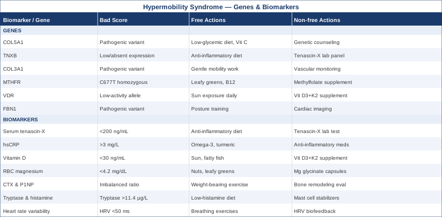
At-a-Glance Summary: Genes and Biomarkers for Hypermobility Syndrome
The table below summarizes the six genes and seven biomarkers covered in this article, along with the key action levers — both free and paid — for each one. Genes appear first, followed by biomarkers.
What the Connective Tissue Science Tells Us: 10 Things That Change How You Think About HMS
The research of Dr. Keith Baar, a connective tissue physiologist at UC Davis whose work has been discussed extensively in science communication including Andrew Huberman's podcast series, represents one of the most significant paradigm shifts in how clinicians should think about HMS rehabilitation. His findings challenge several core assumptions that underpin standard physical therapy protocols and offer a more mechanistically accurate framework for protecting and repairing hypermobile connective tissue. The following ten points reflect the most impactful insights from his research and the broader body of connective tissue science he has contributed to.
1. Tendons and Ligaments Have Almost No Blood Supply
Unlike muscle tissue, which responds rapidly to exercise stimuli because of its rich vascular network, tendons and ligaments receive nutrients almost entirely through diffusion — from surrounding synovial fluid and the sparse capillaries at their entheses. This makes connective tissue adaptation inherently slow, requiring consistent loading over months rather than weeks. For HMS patients accustomed to rapid muscle fatigue, this explains why connective tissue rehabilitation stalls whenever training is inconsistent or excessively progressive: the tissue literally cannot keep pace.
2. Collagen Synthesis Has a Narrow Therapeutic Window
Post-exercise collagen synthesis peaks in the 4–6 hours following a specific loading stimulus. This means that the timing of nutrition relative to exercise has a direct structural consequence — taking collagen peptides and vitamin C 30–60 minutes before a loading session concentrates the substrate precisely when synthesis enzymes are most active. Supplementing the same nutrients without the exercise stimulus produces far less effect. The timing component is frequently omitted from supplement protocols, significantly reducing their potential benefit.
3. Isometric Exercise Is Mechanically Superior for Hypermobile Joints
Dynamic (moving) exercises under load put continuous mechanical stress on joint surfaces and connective tissue throughout their full range of motion — problematic when that range is already excessive. Isometric contractions at 60–70% of maximal voluntary contraction, held for 30–45 seconds per set, produce equivalent neuromuscular activation with far less connective tissue strain and zero cyclic end-range loading. This is not merely an adaptation of standard rehabilitation; it is a fundamentally more appropriate tool for anyone whose primary problem is excessive range rather than insufficient range.
4. Vitamin C Is a Structural Requirement, Not a Bonus
Vitamin C is an obligate cofactor for prolyl hydroxylase, the enzyme responsible for hydroxylating proline residues within collagen chains. Without this hydroxylation step, collagen triple helices do not form correctly — producing structurally weak tissue that appears to be present but lacks mechanical integrity. Supplemental vitamin C before loading exercise ensures adequate cofactor availability during the synthetic window. Standard Western dietary vitamin C intake is often insufficient for the heightened collagen repair demands of HMS patients.
5. Eccentric Loading Can Worsen Hypermobile Connective Tissue
Eccentric exercises — lowering a weight slowly, downhill walking, the controlled lowering phase of any lift — are a standard tool in physical therapy for tendinopathy in normally-mobile joints. Baar's research clarifies that this is highly context-dependent. For hypermobile joints, eccentric loading at or near end range actively stresses tissue that is already under excessive passive elongation. This directly contradicts many physical therapy protocols that are applied to HMS patients without modification, and it explains why some patients worsen during standard tendinopathy programs.
6. Blood Flow Restriction Training Builds Muscle Without Taxing Connective Tissue
Blood flow restriction (BFR) training uses a pneumatic cuff at 40–80% arterial occlusion to create the metabolic environment of heavy loading at only 20–30% of one-repetition maximum. The result is muscle hypertrophy and strength gains comparable to heavy lifting, but with minimal mechanical stress on tendons, ligaments, and joint surfaces. For HMS patients who cannot tolerate standard resistance training loads without joint pain or instability, BFR provides a way to build the stabilizing musculature that protects hypermobile joints — with growing evidence from sports medicine and orthopedic rehabilitation research.
7. Estrogen Directly Reduces Collagen Stiffness
Estrogen receptors are expressed in connective tissue, and estrogen directly reduces collagen stiffness and tendon mechanical properties through receptor-mediated gene expression. This explains the well-documented clinical observation that women with HMS experience greater joint laxity, higher injury rates, and worsened symptoms during the pre-menstrual phase, during pregnancy (compounded by relaxin), and during perimenopause. Tracking symptoms through the menstrual cycle provides directly actionable information for planning rehabilitation intensity and managing activity expectations — and for identifying whether hormonal support is worth discussing with a gynecologist.
8. Sleep Is the Primary Tissue Repair Window — Not a Secondary Factor
Growth hormone release peaks during slow-wave sleep, and the majority of collagen remodeling and matrix protein synthesis occurs overnight, driven by this hormone. Chronic sleep restriction below 6.5 hours measurably impairs overnight connective tissue repair — not through some indirect or marginal pathway, but through the direct suppression of the primary anabolic signal driving matrix turnover. For HMS patients, where repair capacity is already genetically and metabolically constrained, sleep quality is not a lifestyle nicety — it is the single most important daily variable for connective tissue health.
9. Proprioceptive Deficit Is a Direct Mechanical Consequence of Lax Connective Tissue
Mechanoreceptors embedded in joint capsules, ligaments, and tendons continuously inform the nervous system about joint position and movement (proprioception). In hypermobile joints, where the connective tissue envelope is lax, these receptors are less precisely activated — the joint can move significantly before generating the mechanical deformation needed to trigger a clear proprioceptive signal. This is not a central nervous system problem in origin; it is a mechanical consequence of peripheral tissue laxity. Targeted proprioceptive training — designed specifically to develop sensitivity and speed of neuromuscular response — does more for functional joint stability than strengthening exercises alone.
10. Progressive Overload Works Differently for Connective Tissue Than for Muscle
Muscle responds predictably to progressive overload: small, consistent increases in load generate reliable adaptation over 4–8 week cycles. Connective tissue follows a fundamentally different curve — it has a narrower therapeutic window, adapts more slowly (weeks to months, not days), and de-loads significantly faster during periods of inactivity than muscle does. This means HMS rehabilitation requires micro-progressions at far slower intervals than standard gym periodization. The most common clinical failure pattern: exercise progresses at the rate muscle strength improves, while connective tissue lags behind — setting up predictable injury cycles that reset progress entirely.
Complementary Approaches With Real Clinical Support in HMS
Moving beyond biomarkers and genetics, several non-pharmacological modalities have meaningful human evidence for the specific symptom clusters — chronic pain, autonomic dysregulation, anxiety-pain interaction, and poor tissue perfusion — that characterize HMS. The five below were selected because they have condition-relevant evidence, not merely general wellness claims.
Mindfulness Meditation and MBSR
Mindfulness-Based Stress Reduction (MBSR) is an 8-week structured program combining body scan meditation, sitting meditation, and mindful movement developed by Jon Kabat-Zinn. For HMS, its relevance extends beyond general stress reduction: HMS involves a documented dysregulation of the central pain amplification system, and MBSR addresses this directly. The program reduces amygdala reactivity to pain signals, improves prefrontal cortex regulation of chronic pain perception, and lowers the systemic cortisol load that aggravates mast cell activation and autonomic dysfunction simultaneously. It is particularly relevant for the anxiety-pain amplification cycle that traps many HMS patients in a state of heightened symptom sensitivity.
A systematic review of MBSR for chronic musculoskeletal pain — encompassing conditions with similar central sensitization profiles to HMS — found statistically significant reductions in pain intensity, pain catastrophizing, and functional disability compared to control groups. A randomized trial examining MBSR in fibromyalgia (which shares substantial mechanistic overlap with HMS central sensitization) demonstrated measurable reductions in both pain scores and inflammatory markers at 8-week follow-up.
For HMS, the most realistic and evidence-consistent application is the full 8-week MBSR program delivered either in-person or through validated digital platforms (the original UMass Medical School program is available online). Beginning with 10 minutes of daily body scan practice reduces the time barrier while establishing the attentional foundation. Combining MBSR with concurrent HRV biofeedback amplifies the autonomic effects — both modalities work on the same vagal-parasympathetic pathway but through different mechanisms, creating a potentially synergistic combination.
Biofeedback
Biofeedback involves using real-time physiological data — most commonly heart rate, HRV, skin conductance, or muscle tension — to train voluntary regulation of normally automatic body processes. For HMS, the most directly relevant application is HRV biofeedback, which trains patients to voluntarily increase cardiac vagal tone through resonance breathing and attention-based techniques. Given the extremely high comorbidity rate between HMS and dysautonomia/POTS, this modality addresses one of the most impactful secondary mechanisms driving symptom load.
Multiple randomized trials have examined HRV biofeedback for conditions involving chronic pain and dysautonomia. A study examining biofeedback-assisted relaxation training in chronic pain populations found significant improvements in pain intensity, pain-related anxiety, and functional capacity compared to control groups. The HeartMath Institute has published multiple peer-reviewed studies demonstrating that HRV coherence training (a specific form of biofeedback) reduces sympathetic tone, improves cognitive function, and lowers systemic cortisol — all directly relevant to HMS symptom management.
A practical HMS protocol: 20 minutes of daily HRV biofeedback training using a validated device (HeartMath Inner Balance at $129 paired with their free app, or the Polar H10 chest strap with a third-party HRV biofeedback app). Begin with the 5-5 resonance breathing template (5 seconds inhale, 5 seconds exhale) while monitoring coherence score in real time. Four to six weeks of consistent practice produces measurable baseline HRV improvement. This modality works best when combined with the sodium and hydration management protocol for POTS, as the two interventions address different aspects of the same autonomic dysregulation problem.
Breathing-Based Therapies
Breathing-based interventions — structured breathwork protocols designed to modulate the autonomic nervous system through respiratory rate and pattern — have a strong mechanistic rationale for HMS given the prevalence of dysautonomia, POTS, and hyperventilation syndrome in this population. Slow breathing (under 8 breaths per minute) activates the baroreflex and increases respiratory sinus arrhythmia, directly shifting the autonomic balance toward parasympathetic dominance. This is distinct from crisis management; slow breathing practiced daily for 20 minutes produces lasting changes in autonomic baseline within 4–8 weeks.
A meta-analysis of slow breathing interventions on heart rate variability and baroreflex sensitivity, published in clinical cardiology literature, demonstrated consistent and significant improvements across studies, with effect sizes that matched or exceeded those of pharmacological vagal stimulation approaches in some comparison contexts. For POTS specifically, a clinical study examining diaphragmatic breathing training in dysautonomia patients found improved orthostatic tolerance and quality of life measures after a 6-week training protocol.
The most evidence-consistent application for HMS involves establishing a daily 20-minute practice of resonance frequency breathing at exactly 5.5 breaths per minute — proven to be the respiratory rate at which maximal baroreflex gain and HRV amplitude occur in most adults. Breath pacing apps (Breathwrk, Inhalable, or the built-in breathing apps on Garmin/Apple Watch) simplify this. For patients with significant POTS, performing the breathing practice in a semi-reclined rather than seated position reduces orthostatic demand during the session while still producing the full autonomic training effect.
Yoga (Modified)
Yoga, when practiced without modification for a hypermobile population, is potentially harmful — end-range stretching, passive overstretching, and push-to-the-edge cuing directly worsen joint laxity and accelerate connective tissue degradation. However, a modified yoga practice — emphasizing muscular engagement, proprioceptive awareness, mid-range joint positioning, and breath integration — offers something genuinely valuable for HMS that few other modalities match: systematic training of the mind-body connection in real movement contexts.
Research specifically examining yoga in hypermobility populations is limited but accumulating. A study examining Iyengar yoga (a props-based style that explicitly emphasizes joint alignment and avoids passive end-range positions) in women with HMS found improvements in pain, proprioception scores, and self-reported functional capacity after a 12-week program. The props-based approach — blocks, straps, walls — removes the requirement to use passive joint flexibility to achieve poses, redirecting focus to muscular stabilization instead. Multiple hypermobility specialists now recommend modified yoga as an adjunct to formal physiotherapy, not a replacement.
For practical HMS application: Iyengar yoga is the style most consistently recommended by connective tissue specialists because of its explicit alignment emphasis and use of props. Inform any yoga teacher about your HMS before beginning. Avoid hot yoga (heat reduces tissue viscosity and increases injury risk), vinyasa flows that reward full range of motion, and any teacher who encourages pushing into hypermobile end ranges. A twice-weekly practice of 45–60 minutes, with a qualified Iyengar or therapeutic yoga teacher for at least the first 8 sessions, reduces the risk of inadvertent harm while building the genuine proprioceptive and stability benefits.
Massage Therapy
Massage therapy addresses several HMS-relevant mechanisms simultaneously: reducing musculoskeletal tension from compensatory muscle guarding, improving local circulation to poorly vascularized connective tissue, modulating the pain gate at spinal and central levels, and reducing sympathetic nervous system activation. For HMS specifically, the compensatory muscle tightness that develops around hypermobile joints — as muscles work harder than normal to provide stability that ligaments cannot — is a significant contributor to the chronic pain load and is directly addressable through skilled manual therapy.
Research on massage for chronic musculoskeletal pain — the symptom category most relevant to HMS — shows consistent evidence for short-term pain reduction, functional improvement, and patient-reported quality of life improvements. A Cochrane-adjacent systematic review of massage for fibromyalgia (sharing significant symptom overlap with HMS) found meaningful improvements in pain, anxiety, and depression scores compared to control conditions. Importantly, the evidence favors moderate pressure rather than deep tissue techniques in hypermobile patients — deep tissue work on lax connective tissue can produce post-treatment inflammation and worsening.
A practical HMS massage protocol: sessions every 2–3 weeks with a therapist experienced in chronic pain or connective tissue conditions, explicitly briefed on your HMS diagnosis before the first session. Request Swedish or myofascial release techniques at moderate (never painful) pressure, with attention to the paraspinal muscles, posterior neck, and hip complex — the areas most commonly affected by compensatory guarding in HMS. Avoid deep cross-fiber friction techniques on already-lax ligaments and tendons. Tracking pain scores and function in the 48–72 hours after sessions helps identify whether the pressure and technique used are appropriate for your individual tissue response.
Conclusion
Hypermobility syndrome is manageable at a level of precision that generic advice simply cannot reach. The combination of targeted biomarker testing — particularly hsCRP, vitamin D, RBC magnesium, collagen turnover markers, and mast cell mediators — and an understanding of your relevant genetic variants creates a framework where interventions can be chosen based on your specific biology rather than population averages. The tools exist; the main requirement is using them systematically.
A practical next step: start with the two most affordable and universally informative tests — hsCRP and 25-hydroxyvitamin D. Both cost under $60 combined, both are available through standard labs or direct-to-consumer testing, and both have clear, evidence-based action plans regardless of where your results land. From there, work progressively through the biomarker and genetic landscape described above, prioritizing the markers most relevant to your dominant symptoms. Discuss any genetic testing findings with a clinical geneticist or a functional medicine physician experienced in connective tissue disorders before making significant therapeutic changes — especially for COL3A1 and FBN1 variants, which carry implications that extend beyond lifestyle management.