This article contains AI generated content.
· UpdatedHyperparathyroidism: 7 Genes and 7 Biomarkers to Track
Introduction
If you have been told your calcium is "slightly elevated" and your parathyroid hormone is "a bit high," and the plan is simply to monitor it, you are not alone. Many people with hyperparathyroidism — or with biochemical patterns that suggest it — feel trapped between a diagnosis that is real enough to cause symptoms and not dramatic enough to prompt immediate action. The fatigue, the kidney stones, the bone aches, the cognitive fog: these are not imagined, and they are not inevitable.
The challenge is that hyperparathyroidism is not one disease. It can arise from a single overactive gland producing a benign adenoma, from a genetic syndrome that affects multiple endocrine tissues, from years of unaddressed vitamin D deficiency, or from kidney disease creating an upstream signal that forces PTH upward. Each scenario has a different root cause, a different trajectory, and a different set of interventions that actually make sense. Generic advice — eat less calcium, take more vitamin D, come back in six months — often misses this complexity entirely.
This article takes a more precise approach. The first and deeper section identifies seven specific biomarkers that give you a real-time picture of what your parathyroid system is doing to your bones, kidneys, and mineral balance — along with what to do when each one is out of range. A second section then maps the genetic factors that shape individual susceptibility and explain why the same calcium level may mean very different things in different people. A book-and-podcast-derived framework follows, extracting the ten most impactful insights from the science of mineral metabolism. Finally, evidence-based complementary practices close out with approaches that have real mechanistic rationale for this condition.
More information, precisely targeted, leads to better questions and better decisions. That is the premise here — not that you can self-treat your way out of a parathyroid adenoma, but that you can stop being a passive observer of your own lab results.
7 Biomarkers That Reveal What Your Parathyroid System Is Actually Doing
Biomarkers are where the analysis starts. They provide a real-time picture of calcium and phosphorus balance, bone remodeling, and kidney stress — the three main systems where hyperparathyroidism exerts its damage. The following seven markers offer the clearest signal, organized from the most foundational to the more advanced.
1. Intact PTH (Parathyroid Hormone)
Why it matters
Intact PTH is the master signal of the parathyroid system. When parathyroid glands are overactive, PTH rises and initiates a cascade: it pulls calcium from bone by stimulating osteoclast activity, increases kidney reabsorption of calcium, and drives renal conversion of vitamin D to its active form to enhance gut calcium absorption. Sustained PTH elevation — even at the upper end of the normal range over many years — accelerates cortical bone loss, increases kidney stone risk, and impairs cognitive function. It is the central number in any hyperparathyroidism workup.
How to measure it
Intact PTH is measured from a standard fasting blood draw. Most labs report a reference range of 15–65 pg/mL, though clinicians focused on metabolic optimization often prefer 20–55 pg/mL as the functional target. Cost: $50–$150 USD through standard labs, often included in endocrine panels. Some direct-to-consumer labs offer it for $40–$80. Always measure PTH alongside serum calcium — interpreting either in isolation is insufficient.
If PTH is elevated — the plan without supplements
Before concluding that elevated PTH reflects primary hyperparathyroidism, rule out the most common cause first: vitamin D insufficiency. Secondary hyperparathyroidism driven by low vitamin D is far more prevalent than primary HPT and can normalize completely once vitamin D is repleted. Reduce dietary phosphate load from processed foods and phosphoric acid-containing sodas, which chronically stimulate PTH. Hydrate adequately — at least 2–2.5 liters of water daily — to offset kidney stone risk. Consistent weight-bearing exercise (30-minute walks daily, resistance training 2–3 times per week) directly counteracts the cortical bone loss driven by chronically elevated PTH.
If PTH is elevated — the plan with supplements or equipment
Vitamin D3 with K2: If 25-OH vitamin D is below 40 ng/mL, supplementing D3 to reach 40–60 ng/mL often reduces PTH significantly in secondary HPT. Starting doses of 2000–4000 IU/day are common; always pair with vitamin K2 (MK-7, 100–200 mcg/day) to direct calcium away from soft tissues. Retest at 8–12 weeks. Magnesium glycinate or citrate: Magnesium deficiency impairs PTH regulation and is extremely common; 200–400 mg/day is a reasonable starting dose, generally well tolerated continuously. Important note: If PTH elevation is confirmed as primary hyperparathyroidism due to an adenoma, cinacalcet (a calcimimetic medication requiring a prescription) can lower PTH and calcium medically for patients who are not surgical candidates or awaiting surgery.
2. Serum Calcium (Total and Ionized)
Why it matters
Serum calcium is the downstream consequence of PTH activity. Most labs report total calcium, which includes protein-bound and free fractions. Ionized calcium — the biologically active fraction — is more informative but less commonly ordered. Total calcium above 10.5 mg/dL (2.6 mmol/L) warrants investigation; sustained levels above 11 mg/dL carry increasing risk to heart rhythm, kidney function, and neurological status. A critical clinical nuance: always interpret total calcium alongside albumin. Low albumin artificially suppresses reported calcium, so corrected calcium (or ionized calcium) is essential in anyone with chronic illness or malnutrition.
How to measure it
Total serum calcium is among the least expensive lab tests available — $15–$30 USD, included in basic and comprehensive metabolic panels. Ionized calcium costs $30–$80 and requires specific sample handling, but provides greater precision. Request both when possible, particularly if serum albumin is below normal.
If calcium is high — the plan without supplements
Aggressive hydration is the most immediate free intervention — 2–3 liters of water daily reduces hypercalcemic symptoms and protects kidney function. Eliminate calcium supplements entirely (carbonate supplements in particular raise serum calcium faster than food-sourced calcium). Moderate dietary calcium at 1000–1200 mg/day from food sources — paradoxically, restriction below this level stimulates compensatory PTH secretion and worsens bone loss. If you are taking thiazide diuretics for blood pressure, discuss with your physician: thiazides reduce urinary calcium excretion and can worsen hypercalcemia meaningfully.
If calcium is high — the plan with supplements or equipment
Vitamin K2 (MK-7, 100–200 mcg/day) activates matrix Gla protein, which inhibits calcium deposition in arterial walls and soft tissue — a real concern in chronic hypercalcemia. This does not lower serum calcium but reduces one of its most significant secondary risks. Clarifying note: If serum calcium elevation is confirmed to originate from a parathyroid adenoma, no supplement protocol substitutes for surgical evaluation. Parathyroidectomy resolves hypercalcemia in over 95% of properly selected cases and should be a central part of any conversation with a specialist.
3. 25-OH Vitamin D
Why it matters
The vitamin D-PTH relationship is bidirectional and consistently misunderstood. Low 25-OH vitamin D is the leading driver of secondary hyperparathyroidism — PTH rises to compensate for reduced intestinal calcium absorption when vitamin D is insufficient. In primary hyperparathyroidism, however, the picture is more complex: the overactive gland accelerates conversion of 25-OH D to calcitriol (active vitamin D), so 1,25-dihydroxyvitamin D is often high even when total 25-OH D is low. Tracking this marker helps distinguish secondary from primary HPT and determines whether vitamin D supplementation is safe and appropriate in a given individual.
How to measure it
Standard blood test, $30–$80 USD, widely available. Functional medicine practitioners and physicians such as Peter Attia recommend targeting 40–60 ng/mL as optimal; conventional labs flag deficiency below 20 ng/mL and insufficiency below 30 ng/mL. Testing twice yearly (fall and spring) captures seasonal variation, particularly for those at northern latitudes.
If 25-OH vitamin D is low — the plan without supplements
Consistent midday sun exposure to arms and legs without sunscreen for 15–30 minutes several days per week can raise vitamin D levels by 10–20 ng/mL over several weeks, depending on skin tone, latitude, and season. Dietary sources — fatty fish, egg yolks, UV-irradiated mushrooms — contribute modestly. Prioritize time outdoors year-round as a non-negotiable baseline.
If 25-OH vitamin D is low — the plan with supplements or equipment
Vitamin D3 + K2: 2000–5000 IU of D3 daily paired with 100–200 mcg MK-7. Retest at 8–12 weeks and adjust to hit the 40–60 ng/mL target. In confirmed secondary HPT, repleting vitamin D often normalizes PTH within 3–6 months. UVB lamps: Narrowband UVB home devices (originally designed for skin conditions) also stimulate cutaneous vitamin D synthesis and are particularly useful in northern winters. Entry-level devices cost $150–$400. Caution: Do not begin aggressive vitamin D supplementation without first measuring serum calcium. If primary HPT is present, supplementing vitamin D without medical supervision can further elevate calcium.
4. Serum Phosphorus
Why it matters
PTH actively lowers phosphorus by increasing urinary phosphate excretion through the kidney — specifically via downregulation of the sodium-phosphate cotransporter NaPi-IIa. When PTH is chronically elevated, phosphorus tends to fall below range, contributing to fatigue, proximal muscle weakness, and impaired bone mineralization. Phosphorus is also a useful differential diagnosis tool: a simultaneously elevated PTH with normal phosphorus suggests pseudohypoparathyroidism (PTH resistance at the tissue level), an entirely different condition requiring different management.
How to measure it
Included in comprehensive metabolic panels. $15–$30 USD standalone. Normal range: 2.5–4.5 mg/dL. Phosphorus below 2.5 mg/dL in the context of elevated PTH is clinically significant and warrants investigation. Best measured fasting to avoid postprandial fluctuations.
If phosphorus is low — the plan without supplements
Increase dietary phosphate-rich foods: lean meats, poultry, eggs, dairy, fish, and legumes provide well-absorbed phosphorus. Avoid excess use of aluminum- or magnesium-hydroxide antacids, which bind dietary phosphate in the gut and worsen hypophosphatemia. Limit alcohol consumption, which increases urinary phosphate wasting. The root-cause approach is treating the elevated PTH — once PTH normalizes, phosphorus tends to follow.
If phosphorus is low — the plan with supplements or equipment
Oral phosphate supplementation (sodium or potassium phosphate salts) is available but should be used only under medical supervision, as incorrect dosing can dramatically shift calcium-phosphorus balance with serious consequences. This is not a self-supplementation situation. Addressing the PTH elevation — through vitamin D repletion in secondary HPT or surgical treatment in primary HPT — is the only durable fix for chronically low phosphorus in this context.
5. 24-Hour Urinary Calcium
Why it matters
The kidney bears much of the silent damage from hyperparathyroidism. Elevated PTH increases intestinal calcium absorption, and despite reducing urinary calcium excretion relative to filtered load, the absolute calcium overflow still accumulates in urine — contributing over years to kidney stones, nephrocalcinosis, and progressive chronic kidney disease. The 24-hour urinary calcium test quantifies exactly how much calcium the kidneys are clearing each day. Critically, it is the test that distinguishes primary hyperparathyroidism (elevated urinary calcium) from familial hypocalciuric hypercalcemia — a benign inherited condition where urinary calcium is paradoxically low despite elevated serum calcium and PTH. Getting this distinction right completely changes clinical management, since FHH requires no surgery and no treatment.
How to measure it
At-home urine collection in a provided container over exactly 24 hours, then lab analysis. $50–$100 USD. Normal calcium excretion: 100–300 mg/day in adults. Above 400 mg/day carries meaningfully elevated kidney stone risk. The calcium-creatinine clearance ratio (CCCR), calculated from both serum and urine calcium and creatinine simultaneously, is even more specific for distinguishing primary HPT from FHH — request it alongside the 24-hour collection.
If urinary calcium is high — the plan without supplements
High fluid intake targeting urine output above 2 liters/day is the single most evidence-supported free intervention for reducing kidney stone formation. Reduce sodium intake significantly — dietary sodium drives urinary calcium excretion through shared tubular transport, and cutting sodium from 3500 mg/day to under 2000 mg/day can reduce urinary calcium by 100+ mg/day. Limit animal protein to moderate levels (0.8–1.0 g/kg/day) to reduce urinary calcium and oxalate. Avoid supplemental vitamin C above 500–1000 mg/day, which converts to oxalate in susceptible individuals.
If urinary calcium is high — the plan with supplements or equipment
Magnesium citrate: 200–400 mg/day binds oxalate in the gut and reduces urinary oxalate — relevant for calcium oxalate stone formers. Vitamin B6 (pyridoxine): 25–100 mg/day reduces endogenous oxalate synthesis in susceptible individuals. Potassium citrate (prescription): alkalinizes urine and inhibits calcium oxalate crystallization — discuss with a nephrologist or urologist if stones are recurrent. Thiazide diuretics (prescription): directly reduce urinary calcium excretion and are sometimes used to protect kidneys in patients awaiting parathyroid surgery. These require prescriptions and monitoring.
6. Bone-Specific Alkaline Phosphatase (BSAP)
Why it matters
Total alkaline phosphatase (ALP) exists in multiple tissue isoforms — liver, bone, intestinal, and placental. The bone-specific fraction (BSAP) reflects directly the activity of osteoblasts, the cells responsible for bone formation. In hyperparathyroidism, PTH stimulates both osteoclast resorption and compensatory osteoblast formation — BSAP rises as the skeleton attempts to keep up with increased turnover. An elevated BSAP signals that active bone remodeling and potential net loss is occurring, often well before DEXA scans show measurable density reduction. Metabolic medicine specialists including Thomas Dayspring have long advocated for bone turnover markers as underused clinical tools that detect bone damage earlier than structural imaging alone.
How to measure it
A specific blood test measuring only the bone-derived ALP isoform (not total ALP, which would include liver fraction). $50–$150 USD; not included in standard panels — request it explicitly, specifying bone-specific ALP or the Ostase assay. Reference range: under 14 ng/mL for women; under 20 ng/mL for men. Measure fasting in the morning for best consistency.
If BSAP is elevated — the plan without supplements
Progressive resistance training is the most potent free tool for shifting bone turnover toward net formation. Two to four strength training sessions per week over 6–12 months measurably improve bone density even in patients with mild primary HPT. High-impact loading (jumping, tennis, jogging on firm surfaces) adds piezoelectric mechanical signals that stimulate osteoblast activity. Adequate dietary protein — 1.2–1.6 g/kg/day — supports collagen synthesis and osteoblast function. Eliminating smoking has a direct and quantifiable benefit on bone turnover markers within weeks.
If BSAP is elevated — the plan with supplements or equipment
Vitamin K2 (MK-4 or MK-7): activates osteocalcin, the protein that anchors calcium into bone matrix. MK-7 at 90–200 mcg/day has the best evidence for convenience and bioavailability; MK-4 at therapeutic doses (1.5–45 mg/day) has been studied in Japanese trials for osteoporosis. Hydrolyzed collagen peptides: 5–10 g/day supports bone matrix synthesis; best absorbed when taken with vitamin C. Whole-body vibration platforms: 15–30 minutes of low-magnitude mechanical vibration 3–5 days/week has shown modest benefit for bone density in controlled trials. Entry-level home devices cost $200–$500. Trace minerals — silica (from orthosilicic acid, 5–10 mg/day) and boron (2–6 mg/day) — have emerging evidence for supporting bone turnover regulation.
7. Bone Mineral Density (DEXA Scan)
Why it matters
DEXA (dual-energy X-ray absorptiometry) provides the structural endpoint — what has actually happened to the skeleton over time as a result of chronic PTH exposure. A key clinical detail: hyperparathyroidism preferentially damages cortical bone (hip, forearm) rather than trabecular bone (lumbar spine) — the opposite pattern from postmenopausal osteoporosis. This means standard spine T-scores can appear reassuring while cortical sites are silently deteriorating. The distal third radius (forearm) measurement is therefore essential in any HPT patient and is frequently omitted in standard osteoporosis scans. Peter Attia consistently emphasizes DEXA as one of the highest-yield longitudinal health measurements available, covering not just bone density but body composition.
How to measure it
DEXA scan at a radiology center or bone health clinic. $100–$300 USD; insurance coverage is common with an appropriate clinical indication from a physician. Request measurements at three sites: lumbar spine, total hip, and distal third radius. The forearm site is non-negotiable for HPT monitoring. Current international guidelines for asymptomatic primary HPT recommend DEXA at diagnosis and every 1–2 years thereafter.
If BMD is low — the plan without supplements
Weight-bearing and resistance exercise remain the highest-leverage free interventions for bone density. Progressive resistance training, high-impact activities, and walking all stimulate bone formation through mechanical loading. Smoking cessation is among the most impactful single changes for bone trajectory. Adequate total caloric intake and dietary protein prevent the catabolic state that worsens bone loss. Sleep optimization — consistent 7–9 hours, dark environment, consistent sleep-wake timing — supports nocturnal growth hormone secretion, one of the primary anabolic bone signals in adults.
If BMD is low — the plan with supplements or equipment
Calcium from food first: Target 1000–1200 mg/day total from dairy, sardines with bones, or leafy greens. Supplement only the gap, as excess supplemental calcium (particularly carbonate) carries cardiovascular risk signals when used at high doses. Vitamin D3 + K2 as outlined above. Collagen peptides + vitamin C for matrix support. Vibration platforms as above. In patients with post-surgical "hungry bone syndrome" — when bones rapidly absorb large amounts of calcium after successful parathyroidectomy — temporary high-dose calcium and vitamin D supplementation under medical supervision is required. Anti-resorptive medications (bisphosphonates, denosumab) may be considered for moderate to severe pre-surgical bone loss in consultation with an endocrinologist.
The biomarker picture tells you what is happening and how urgently. Understanding the genetic layer underneath helps explain why — and who in your family may need screening.
The Genetic Architecture of Hyperparathyroidism: 7 Genes That Shape Your Risk
Most hyperparathyroidism is sporadic — a somatic mutation in a single parathyroid cell, arising from causes that are still incompletely understood. But approximately 10–15% of all primary HPT cases involve germline mutations that can be inherited, and several variants in calcium-sensing and vitamin D metabolism pathways modulate disease severity even in those without a formal hereditary syndrome. Understanding these genes informs both your own management and the screening decisions for your family.
MEN1 (Multiple Endocrine Neoplasia Type 1)
What it does: MEN1 encodes menin, a tumor suppressor that regulates gene transcription and cell cycle control. Loss-of-function mutations in MEN1 cause MEN1 syndrome, the most common inherited cause of primary hyperparathyroidism. Over 90% of MEN1 carriers develop HPT — typically by age 25–30 — often with multiple gland involvement rather than a single adenoma. The syndrome also includes pituitary and pancreatic tumors, making early recognition critical. MEN1 gene entry on NCBI
If the gene has a known pathogenic variant — plan without supplements: Annual biochemical screening from age 8–10 (calcium, intact PTH, fasting glucose, prolactin, fasting gastrin) per published MEN1 guidelines. Genetic counseling and cascade testing for all first-degree relatives. Lifestyle: eliminate tobacco, limit alcohol, avoid high-dose calcium supplementation, maintain vitamin D sufficiency. Regular parathyroid ultrasound and cross-sectional imaging per endocrinologist guidance. Surgical strategy for MEN1-HPT differs from sporadic HPT — subtotal parathyroidectomy (3.5 glands removed) or total parathyroidectomy with autotransplantation is preferred over targeted single-gland removal, given inevitable multigland disease.
If the gene has a known pathogenic variant — plan with supplements or equipment: Cinacalcet (calcimimetic, prescription) can lower PTH and calcium in patients with recurrent HPT after initial surgery or those not yet meeting surgical thresholds. Maintain 25-OH vitamin D at 40–60 ng/mL to reduce compensatory PTH. Vitamin K2 MK-7 (100–200 mcg/day) alongside D3 for bone protection. DEXA every 1–2 years; consider bisphosphonates if T-score falls below -2.5 at any site.
CDC73 / HRPT2 (Cell Division Cycle 73)
What it does: CDC73 encodes parafibromin, a tumor suppressor involved in chromatin remodeling and cell cycle regulation. Loss-of-function mutations define hyperparathyroidism-jaw tumor syndrome (HPT-JT) and are identified in approximately 15–20% of parathyroid carcinomas — making CDC73 the most critical genetic marker for malignant parathyroid disease. Clinical red flags warranting CDC73 testing include very high calcium (above 14 mg/dL), markedly elevated PTH (often 5–10× normal), a palpable neck mass, fibro-osseous jaw lesions, and uterine tumors. CDC73 gene entry on NCBI
Plan without supplements: Any clinical scenario suggesting parathyroid carcinoma requires referral to a high-volume parathyroid surgery center — not a general surgeon. CDC73 variant status changes surgical planning entirely: en bloc resection with ipsilateral thyroid lobe and soft tissue is required, as opposed to simple gland removal. Germline CDC73 testing and family screening follow from a confirmed pathogenic variant.
Plan with supplements or equipment: For inoperable or recurrent carcinoma, cinacalcet (calcimimetic) provides palliative calcium control. Denosumab (RANK-L inhibitor, prescription) reduces bone resorption markers when conventional treatments are insufficient. Biochemical surveillance (calcium, PTH, 24-hour urinary calcium) every 6 months is required lifelong given high recurrence rates in carcinoma.
CASR (Calcium-Sensing Receptor)
What it does: CASR encodes the receptor on parathyroid cells and kidney tubules that detects ambient calcium concentration and suppresses PTH secretion when calcium is adequate. Loss-of-function mutations reduce receptor sensitivity, causing the parathyroid gland to perceive normal calcium as insufficient — resulting in inappropriately elevated PTH and hypercalcemia. This produces familial hypocalciuric hypercalcemia (FHH), a benign condition that biochemically mimics primary HPT but does not benefit from — and is not cured by — parathyroid surgery. Gain-of-function CASR mutations produce autosomal dominant hypocalcemia. Getting this genetic distinction right is one of the most consequential clinical decisions in the HPT workup. CASR gene entry on NCBI
Plan without supplements: The calcium-creatinine clearance ratio (CCCR) is the non-genetic screening test: a CCCR below 0.01 in a hypercalcemic individual strongly suggests FHH rather than primary HPT. If FHH is confirmed clinically or genetically, surgery is contraindicated. Annual biochemical monitoring (calcium, PTH) is all that is required. Patient education is critical: FHH carriers should carry documentation to prevent inappropriate surgical referral.
Plan with supplements or equipment: Cinacalcet is mechanistically the most rational treatment for CASR loss-of-function variants — it acts directly on the calcium-sensing receptor to increase sensitivity, lowering PTH and calcium. It is generally reserved for symptomatic cases. Avoid thiazide diuretics, which worsen hypercalcemia in FHH. Hydration and dietary sodium reduction (which reduces urinary calcium loss) apply.
CCND1 (Cyclin D1)
What it does: CCND1 encodes cyclin D1, a core driver of the G1-to-S cell cycle transition. Rearrangement of chromosomal region 11q13 places CCND1 under transcriptional control of the PTH gene promoter, causing its overexpression in parathyroid cells and driving uncontrolled proliferation. This somatic rearrangement is found in approximately 20–40% of sporadic parathyroid adenomas and represents one of the most common acquired genetic events in HPT. Unlike MEN1 or CASR, this is typically a tumor-specific change rather than an inherited germline variant — but germline CCND1 polymorphisms can contribute to parathyroid tumor susceptibility. CCND1 gene entry on NCBI
Plan without supplements: Somatic CCND1 overexpression in a confirmed adenoma has direct surgical implications: it confirms monoclonal adenoma origin rather than diffuse hyperplasia, supporting a focused minimally invasive parathyroidectomy approach over bilateral four-gland exploration in appropriate surgical candidates.
Plan with supplements or equipment: Curcumin (standardized to 95% curcuminoids, 500–1000 mg/day with piperine for bioavailability) and EGCG from green tea extract (400–800 mg/day) both demonstrate cyclin D1 downregulation activity in cell and animal studies. Human parathyroid-specific data is absent, evidence is very preliminary, and these should not substitute for surgical assessment. Side effects at these doses are generally mild; cycle off every 8–12 weeks. These are considered adjunctive lifestyle support with a reasonable safety profile, not primary treatment.
GCM2 (Glial Cells Missing 2)
What it does: GCM2 encodes a transcription factor that is essential for parathyroid gland formation and specification during embryonic development. It continues to regulate parathyroid cell identity in adult tissue. Loss-of-function variants have been associated with hypoparathyroidism, while gain-of-function variants and certain activating GCM2 mutations have recently been identified as germline predisposing factors for sporadic primary HPT, particularly in patients with multigland disease. GCM2 is an emerging gene in HPT genetics and is not yet included in most standard clinical panels. GCM2 gene entry on NCBI
Plan without supplements: Activating GCM2 variants suggest that multigland parathyroid disease is more likely than single-adenoma disease — which is surgically relevant. When GCM2 variants are identified, bilateral four-gland exploration rather than targeted unilateral surgery may be more appropriate. Genetic counseling is appropriate as clinical understanding of GCM2 evolves.
Plan with supplements or equipment: No specific supplement protocol targets GCM2 variants. General parathyroid metabolic support applies: vitamin D maintained at 40–60 ng/mL, magnesium glycinate 200–400 mg/day, calcium from food sources, avoidance of thiazide diuretics and prolonged proton pump inhibitor use (both impair calcium absorption).
VDR (Vitamin D Receptor)
What it does: VDR encodes the nuclear receptor through which active vitamin D (calcitriol) exerts its direct suppressive effect on PTH gene transcription in parathyroid cells. Polymorphisms in VDR — notably BsmI, FokI, ApaI, and TaqI variants — affect receptor function and the strength of vitamin D signaling. Individuals carrying certain VDR polymorphisms may require higher serum 25-OH vitamin D levels to achieve equivalent PTH suppression compared to those with higher-function VDR variants, making them functionally more vulnerable to secondary hyperparathyroidism even when their vitamin D level appears "sufficient" on standard criteria. VDR gene entry on NCBI
Plan without supplements: Maximize sun exposure and dietary vitamin D. Prioritize anti-inflammatory dietary patterns (Mediterranean-style, with omega-3 rich foods) that support nuclear receptor signaling pathways broadly. Optimize circadian rhythm — consistent sleep-wake cycles influence gene expression including nuclear receptor activity. Regular aerobic and resistance exercise also improves VDR expression in target tissues.
Plan with supplements or equipment: VDR polymorphism carriers may need to target 25-OH vitamin D levels of 55–70 ng/mL — higher than standard recommendations — to achieve adequate PTH suppression. Magnesium (300–400 mg/day from diet and supplements) is an essential cofactor for VDR activation and the enzymatic conversion of vitamin D to its active form. Vitamin K2 MK-7 (100–200 mcg/day) works through overlapping nuclear receptor pathways and complements VDR-mediated calcium regulation. These supplements require no cycling at these doses and are generally safe long-term.
RET (Rearranged During Transfection Proto-Oncogene)
What it does: Gain-of-function mutations in the RET proto-oncogene define multiple endocrine neoplasia type 2A (MEN2A), in which hyperparathyroidism occurs in approximately 20–30% of affected individuals — typically milder than MEN1-HPT and usually involving fewer glands. RET testing is mandatory in anyone presenting with concurrent medullary thyroid carcinoma or pheochromocytoma, as these three tumors together characterize MEN2A. Identifying a RET mutation before parathyroid surgery can be lifesaving — unrecognized pheochromocytoma in the perioperative setting can trigger a hypertensive crisis. RET gene entry on NCBI
Plan without supplements: Screening for pheochromocytoma (plasma or urinary metanephrines) is mandatory before any surgical procedure in suspected RET variant carriers. Annual surveillance includes calcitonin (medullary thyroid cancer marker), urinary metanephrines, calcium, and PTH. Thyroid ultrasound per endocrinology guidance. The HPT component of MEN2A is typically mild and single-gland — surgical approach is correspondingly more conservative than for MEN1.
Plan with supplements or equipment: RET-driven HPT is usually biochemically mild and may not require immediate treatment. Standard vitamin D, K2, and magnesium optimization applies as baseline metabolic support. Kinase inhibitor therapies prescribed for RET-positive medullary thyroid cancer do not directly treat parathyroid disease. Biochemical monitoring frequency is dictated by the overall MEN2A surveillance protocol.
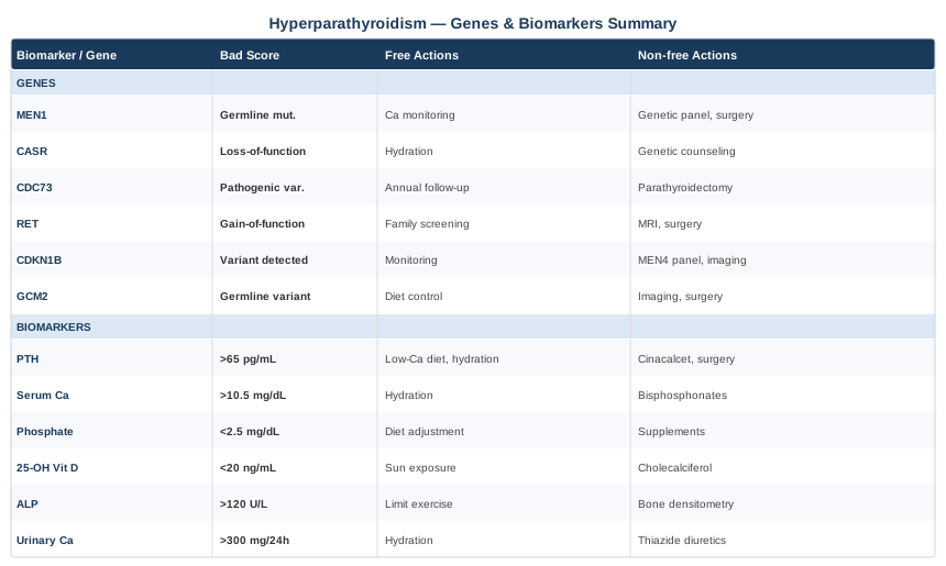
Both the biomarker and genetic layers point toward the same underlying reality: the parathyroid-calcium-vitamin D axis is a finely tuned system that rewards precision over generic advice. The scientific framework for understanding this system has been articulated compellingly in a number of educational sources — including one podcast series that has introduced mineral metabolism and hormonal health to millions of people.
Ten Things That Change How You Think About Calcium, PTH, and Bone Health
Key insights from the Huberman Lab and related research, applied to hyperparathyroidism
The Huberman Lab podcast and associated research from the Stanford neurobiology group synthesize peer-reviewed evidence on hormonal and metabolic health in a rigorous yet accessible way. What follows draws on that framework — particularly episodes on vitamin D, bone health, sleep, and hormonal regulation — translated directly into the context of hyperparathyroidism.
1. The Skin-Sun-Vitamin D Conversion Is Far More Variable Than Standard Advice Suggests
Skin exposed to UVB radiation converts 7-dehydrocholesterol to previtamin D3, which isomerizes spontaneously to vitamin D3. This process is dramatically affected by skin pigmentation, latitude, season, time of day, clothing coverage, and aging. Adults over 65 produce roughly 50% less cutaneous vitamin D from identical sun exposure compared to young adults — a factor that directly explains why secondary HPT is so prevalent in older populations even when they are nominally "getting sun." Standard supplementation doses were calibrated for average populations and routinely undershoot individual needs.
2. Blood 25-OH Vitamin D Is Not the Same as Vitamin D Activity
Active vitamin D (1,25-dihydroxyvitamin D, calcitriol) is what actually suppresses PTH transcription and drives calcium absorption. The conversion from 25-OH D to calcitriol occurs primarily in the kidneys and is controlled by PTH itself. In primary HPT, PTH has maximally upregulated this conversion — calcitriol can be elevated even when 25-OH D is low. This is why some primary HPT patients develop hypercalcemia even with seemingly normal vitamin D levels. Testing both 25-OH D and 1,25-OH2D gives the complete picture; relying solely on total 25-OH D misses this distinction.
3. Magnesium Is the Most Overlooked Factor in the Entire Vitamin D-PTH System
Magnesium serves as a cofactor for the enzymes that convert vitamin D to its active form, for PTH secretion itself, and for end-organ PTH response. Magnesium deficiency — reported in over 50% of Western adults based on dietary intake surveys — can cause PTH to become dysregulated in multiple directions. Correcting magnesium status before aggressive vitamin D supplementation is not just reasonable but physiologically sound. Adequate magnesium is also required for the VDR to function optimally.
4. There Is a Specific Vitamin D Threshold Below Which PTH Rises Sharply
Research consistently shows a non-linear relationship between 25-OH vitamin D and PTH: below approximately 30 ng/mL, PTH rises steeply; above 50 ng/mL, further increments produce diminishing additional PTH suppression. The most effective anti-HPT range for secondary forms is 40–60 ng/mL. Maintaining this range with consistent supplementation or sun exposure is achievable year-round for most people with intentional effort, and may normalize PTH entirely in secondary HPT cases.
5. Calcitonin — PTH's Opposing Hormone — Is Rarely Discussed but Practically Important
Calcitonin, secreted by thyroid C-cells, directly inhibits osteoclast activity and counteracts PTH-driven bone resorption. Physical exercise, particularly high-intensity and resistance training, acutely stimulates calcitonin release. This is one of the mechanistic reasons — beyond simple mechanical loading — why regular exercise consistently protects bone in hyperparathyroid patients. Understanding this pathway reframes exercise from a generic lifestyle recommendation into a specific, hormonally mediated intervention.
6. Sleep Is the Primary Bone-Building Window — and PTH Disrupts It
Growth hormone secretion occurs predominantly during slow-wave sleep and is one of the most potent anabolic signals for osteoblast activity in adults. PTH also follows a circadian rhythm with a nocturnal secretion peak. Chronic sleep disruption elevates overnight cortisol, blunts growth hormone pulses, and keeps nocturnal PTH elevated longer than it should be — all of which accelerate bone remodeling imbalance. Optimizing sleep duration (7–9 hours), minimizing artificial light after 10 pm, and maintaining consistent sleep-wake timing are not peripheral wellness habits in HPT — they are mechanistically connected to your bone outcomes.
7. Cortisol Directly Antagonizes Every Bone-Protective Pathway
Cortisol reduces intestinal calcium absorption by opposing vitamin D receptor signaling, increases urinary calcium excretion, suppresses osteoblast activity, and impairs collagen synthesis. Chronic psychological stress therefore amplifies every dimension of hyperparathyroid skeletal damage. This is why the mind-body interventions covered in the next section are not soft additions to HPT management — they operate through a well-characterized endocrine mechanism that directly affects the same calcium-bone pathways that PTH is disrupting.
8. Vitamin K2 Protects Against One of Hyperparathyroidism's Most Underrecognized Risks
Chronically elevated serum calcium poses a risk beyond kidney and bone: arterial wall calcification. When calcium circulates at high concentrations over years, it can precipitate in vascular smooth muscle — a process that dramatically elevates cardiovascular risk. Matrix Gla protein (MGP), activated by vitamin K2, is the primary inhibitor of vascular calcification in the arterial wall. Low vitamin K2 status — which is common in populations eating few fermented foods — leaves MGP undercarboxylated and nonfunctional. Supplementing K2 MK-7 in hyperparathyroid patients addresses a risk that is almost never discussed in standard clinical management.
9. Exercise Changes the PTH Signal Over Both Short and Long Time Horizons
A single bout of resistance exercise produces a brief PTH spike followed by a compensatory calcitonin surge — the net short-term effect is bone stimulation. Over months of consistent training, resting PTH tends to normalize, bone turnover shifts toward net formation, and bone density improves measurably even in the presence of mild primary HPT awaiting surgical assessment. There is no supplement or medication that replicates the multi-pathway bone stimulus that structured resistance training provides. This is not a secondary recommendation — it belongs at the center of any HPT management plan.
10. Consumer Genetic Testing Can Now Reveal Your Functional Vitamin D-PTH Axis
VDR polymorphisms, CYP27B1 variants (the enzyme converting 25-OH D to calcitriol), and CASR variants can all be identified through consumer genetic platforms such as 23andMe, with the raw data processed through tools like Genetic Genie or Rhonda Patrick's foundational information resources. Knowing whether you carry high-risk combinations — for example, a weak-function VDR allele alongside a CASR variant — allows you to personalize vitamin D supplementation targets and monitoring intervals rather than following population averages that may not apply to you.
Complementary and Integrative Approaches for Hyperparathyroidism
The following modalities have meaningful physiological rationale and, in several cases, clinical evidence applicable to the mechanisms driving hyperparathyroid complications. None are alternatives to appropriate medical or surgical evaluation; all are useful additions to a comprehensive management approach.
Yoga
Yoga is relevant to hyperparathyroidism through two converging mechanisms: weight-bearing postures provide the mechanical loading stimulus that PTH-driven cortical bone loss specifically requires, and breath-synchronized movement produces measurable parasympathetic activation that reduces cortisol — a direct antagonist to bone formation. Standing, balancing, and load-bearing poses (warrior series, single-leg balance poses, lunges) deliver ground reaction forces through the hip and femur, sites where cortical loss is most clinically significant in HPT.
A 2016 randomized controlled pilot trial by Motorwala and colleagues, published in the International Journal of Yoga, found that 6 months of yoga practice improved bone turnover markers and balance scores in postmenopausal women with osteopenia — a population whose bone loss mechanism overlaps meaningfully with HPT-driven cortical deterioration. A broader meta-analysis of yoga's effects on bone mineral density in postmenopausal women found consistent improvements at the lumbar spine and hip with regular practice (typically 3+ sessions per week over 6–12 months).
Practically: three to four sessions per week of a weight-bearing style (Iyengar, Vinyasa, or Power yoga) is an appropriate protocol. Avoid hot yoga environments when serum calcium is elevated — dehydration acutely worsens hypercalcemia symptoms. If DEXA has confirmed significant bone loss, begin with a modified or gentle class and avoid high-risk spinal loading (deep forward folds, unsupported spinal flexion) until bone strength is assessed. A qualified instructor with knowledge of osteoporosis modifications is valuable.
Mindfulness Meditation and MBSR
Mindfulness-Based Stress Reduction (MBSR) has a direct and well-characterized physiological rationale in hyperparathyroidism: it reduces cortisol. Cortisol reduces intestinal calcium absorption, increases urinary calcium wasting, suppresses osteoblast differentiation, and impairs collagen synthesis — all effects that amplify the bone damage driven by PTH. Chronic psychological stress is therefore not a peripheral factor in HPT but a direct co-contributor to its most damaging consequences.
Multiple controlled trials have demonstrated that 8-week MBSR programs (standardized as 2.5-hour weekly sessions plus 45 minutes of daily home practice) produce significant reductions in morning cortisol, C-reactive protein, and self-reported pain and fatigue — all outcomes directly relevant to the HPT symptom burden. While no MBSR trials have enrolled HPT-specific populations, the cortisol-calcium-PTH pathway provides a mechanistically sound bridge. A meta-analysis published in JAMA Internal Medicine examining mindfulness-based interventions and biological stress markers across diverse populations found consistent HPA axis attenuation across most study designs.
For realistic daily application, even 10–15 minutes of breath-focused attention practice (body scan, focused breathing, or open monitoring) produces measurable HRV improvements within 4 weeks. Apps such as Insight Timer, Waking Up, or Ten Percent Happier provide structured guided sessions. The full 8-week MBSR protocol, offered online and through hospital integrative medicine programs at roughly $300–$600, represents an investment with evidence support for chronic condition management. This is a low-risk, scalable intervention that requires no equipment.
Microbiome-Directed Therapies
The gut microbiome directly influences calcium absorption — an underappreciated connection in hyperparathyroidism management. Fermentative gut bacteria produce short-chain fatty acids (SCFAs) including butyrate that acidify the colon, improving the solubility and paracellular transport of calcium. Gut dysbiosis, by contrast, reduces SCFA production, impairs calcium absorption, and can drive compensatory PTH elevation in a cycle that resembles secondary HPT. There is also emerging evidence that gut bacteria modulate systemic inflammation and vitamin D metabolism, both of which influence parathyroid activity.
A randomized controlled trial published in Clinical Nutrition examining prebiotic inulin-type fructan supplementation in postmenopausal women found measurable improvement in calcium absorption and reduction in PTH levels over 8 weeks — interpreted as reflecting improved gut-mediated calcium bioavailability. The evidence base for the gut-bone axis is growing, with specific study in HPT populations still limited, and findings should be applied cautiously.
Practically: prioritize dietary fiber diversity (targeting 30+ grams daily from vegetables, legumes, whole grains, and fruit) to support SCFA-producing Bacteroidetes and Firmicutes populations. Fermented foods — kefir, full-fat yogurt with live cultures, sauerkraut, kimchi — introduce beneficial bacterial strains with each serving. A targeted synbiotic supplement combining Lactobacillus and Bifidobacterium strains with a prebiotic substrate (inulin or FOS, 5–10 g/day) is a reasonable structured addition. Avoid unnecessary antibiotic courses and minimize proton pump inhibitor use, both of which deplete beneficial gut microbiota and impair calcium absorption.
Breathing-Based Therapies
Controlled slow breathing — including resonance frequency breathing (approximately 6 breaths per minute), box breathing, and the 4-7-8 technique — activates the vagus nerve and generates large, coherent oscillations in heart rate variability (HRV). This produces downstream reductions in sympathetic tone, cortisol, and norepinephrine. In the context of hyperparathyroidism, sustained autonomic downregulation through daily breathing practice represents a practical, scalable tool for blunting the cortisol-driven acceleration of bone resorption and hypercalciuria described throughout this article.
Clinical trials examining HRV biofeedback and slow-paced breathing have consistently demonstrated reductions in cortisol, blood pressure, and inflammatory markers over 4–8 week programs. Research published in Frontiers in Psychology and Applied Psychophysiology and Biofeedback supports the physiological validity of breathing-based interventions for HPA axis regulation. Again, HPT-specific trials are lacking, but the mechanism is well characterized and the safety profile is essentially zero-risk.
Practically: 10–20 minutes of slow diaphragmatic breathing (5–6 second inhale, 5–6 second exhale) once or twice daily produces measurable HRV improvement within 2–4 weeks. This requires no equipment. Adding HRV monitoring via a wearable (Oura Ring $300–$500, Garmin or Polar devices $150–$400, or the free HRV4Training smartphone app using your phone camera) provides objective feedback on autonomic recovery status and helps calibrate whether the practice is working. Consistent practice is more important than any specific technique — choose the pattern that you will actually do daily.
Conclusion
Hyperparathyroidism rewards those who pay close attention. The seven biomarkers outlined here — intact PTH, serum calcium, 25-OH vitamin D, phosphorus, 24-hour urinary calcium, bone-specific alkaline phosphatase, and DEXA bone density — collectively give you a three-dimensional picture of where your system stands right now: how active PTH is, what it is doing to your kidneys, and how much structural impact has accumulated in your skeleton. The seven genetic factors add a layer of explanation, clarifying whether you are dealing with an inherited predisposition, a calcium-sensing variation, or a sporadic event — and what that means for how you are monitored and treated.
The most important next step is not to optimize everything at once. Start with what you can measure. If you have not recently had intact PTH, corrected serum calcium, and 25-OH vitamin D measured simultaneously, that is the right place to begin. Bring those results to an endocrinologist or internist who is comfortable managing mineral metabolism. Use this article as a framework for asking more informed questions — not for self-diagnosing or self-treating a condition that, in its primary form, often requires surgical evaluation. The clearest path forward is better data, tracked consistently, interpreted by someone with clinical expertise and full knowledge of your individual history.
Cardiovascular Endocrine & Metabolic
Musculoskeletal: Bone Conditions
Urological: Kidney Conditions
Women's Health: Hormonal Conditions