This article contains AI generated content.
· UpdatedHypersensitivity Vasculitis Genes & Biomarkers — 5 Genes And 6 Biomarkers To Track
Introduction
Hypersensitivity vasculitis — also called leukocytoclastic vasculitis or cutaneous small-vessel vasculitis — sits in an uncomfortable grey zone of medicine. Most people who develop it are told to identify the trigger, wait for resolution, and possibly take a short course of steroids. That guidance is not wrong, but for anyone dealing with recurrent flares, unclear triggers, or a condition that simply refuses to resolve, it leaves an enormous gap between what medicine offers and what the body is actually doing.
What makes this condition genuinely complex is that two people can have identical-looking purpuric lesions and completely different underlying mechanisms. For one person, the driving force is an immune complex deposited after a drug reaction that will clear in weeks. For another, a subtle deficit in complement regulation or an overactive inflammasome keeps the cycle alive indefinitely. Standard inflammatory panels rarely distinguish these scenarios, and that is precisely why many patients cycle through treatments without finding a lasting answer.
This article takes a different approach. Rather than reviewing generic advice, it focuses on the specific biological signals — both measurable laboratory markers and genetic predispositions — that can explain why hypersensitivity vasculitis persists, recurs, or responds poorly to treatment. The goal is not a diagnosis but a clearer map of what may actually be happening inside the body.
Better information leads to better decisions. Understanding which biomarkers reflect current disease activity, which ones point to underlying immune dysregulation, and which genetic variants may raise your baseline vulnerability gives you something concrete to bring to a clinician — or to act on independently between appointments. What follows covers six key biomarkers to track, five genetic factors worth investigating, a protocol from the most research-backed autoimmune framework available, and selected complementary approaches with meaningful human evidence behind them.
Summary
This article maps the biology of hypersensitivity vasculitis at two levels: what you can measure now and what may have been shaping your immune responses for years. The biomarker section covers six specific lab values — including one (complement C3/C4) that is frequently skipped in standard workups yet is arguably the most diagnostically informative. For each marker, you will find target ranges, cost estimates, and detailed plans for improving a bad score, both with and without supplements. The genetics section explores five genes — including HLA-DRB1, TNFA, and MEFV — explaining what a problematic variant means in practical terms and what compensatory strategies exist. Beyond lab work, you will find a detailed summary of Sarah Ballantyne's Autoimmune Protocol with ten of its most clinically impactful insights, plus four complementary approaches — from mindfulness to photobiomodulation — selected for their relevance to small-vessel inflammation specifically. If you have ever felt that managing your vasculitis was more trial and error than targeted strategy, the frameworks in this article may help you identify the missing piece.
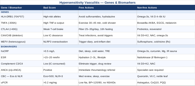
6 Biomarkers to Track in Hypersensitivity Vasculitis
Biomarkers serve two distinct purposes in hypersensitivity vasculitis: some tell you how active the inflammation is right now, while others reveal the underlying immune mechanisms that may be driving it. Tracking the right panel — not just the obvious inflammatory markers — can transform a vague clinical picture into something concrete you can monitor and influence over time.
1. High-Sensitivity C-Reactive Protein (hsCRP)
Why it matters: CRP is produced by the liver in response to interleukin-6 (IL-6), a cytokine that rises sharply during active vasculitic inflammation. In hypersensitivity vasculitis, elevated CRP is one of the most reliable real-time signals of immune complex–driven vascular inflammation. Standard CRP is largely meaningless below 5 mg/L with conventional assays, but high-sensitivity CRP (hsCRP) resolves values down to 0.1 mg/L, making it valuable for detecting smoldering, sub-clinical disease that may not produce visible lesions between flares. Peter Attia and clinicians working in precision medicine recommend hsCRP as a near-mandatory marker because chronic low-grade inflammation at any level signals ongoing tissue damage.
How to measure it: Standard blood draw, ordered specifically as hsCRP (not standard CRP). Cost: $15–45 USD at most labs. Optimal target: hsCRP below 0.5 mg/L for low inflammatory risk. During active disease, CRP can exceed 50–100 mg/L; tracking the downward trend week over week matters as much as any single reading.
If the score is bad — plan without supplements: Eliminate ultra-processed foods, refined seed oils (corn, soybean, sunflower), and trans fats. Time-restricted eating (10–12 hour eating window, supported by multiple trials on IL-6 reduction) reduces baseline inflammatory signaling. Cold thermogenesis — ending showers with 1–2 minutes of cold water — activates norepinephrine pathways that suppress inflammatory cytokines. Improving sleep quality is one of the most potent non-pharmacological hsCRP reducers available; even a one-night sleep disruption measurably elevates CRP the following morning. Aim for 7.5–9 hours with a consistent wake time.
If the score is bad — plan with supplements or equipment: Omega-3 fatty acids (EPA+DHA): 2–4g daily with food. Reduces hsCRP via specialized pro-resolving mediator pathways (resolvins, protectins). Safe for continuous use; monitor LDL particle count if on doses above 3g/day. Side effects: mild fishy aftertaste, increased bleeding time at doses above 4g/day. Curcumin with piperine: 500–1000mg curcuminoid daily. Multiple meta-analyses confirm significant reduction in CRP, with effect sizes comparable to low-dose NSAIDs in mild-to-moderate inflammation. Take with black pepper extract for bioavailability. Cycle: 12 weeks on, 4 weeks off. Side effects: GI upset at high doses, potential interaction with anticoagulants. Magnesium glycinate: 300–400mg nightly. Magnesium deficiency correlates with elevated CRP; repletion is inexpensive and carries minimal risk. Infrared sauna: 3–4 sessions per week, 20 minutes at 55–65°C. Observational data from Finnish population studies, corroborated by smaller clinical trials, support consistent reduction in inflammatory markers with regular sauna use.
2. Erythrocyte Sedimentation Rate (ESR)
Why it matters: ESR reflects the degree to which red blood cells aggregate in the presence of acute-phase proteins, particularly fibrinogen. It lags behind CRP by 24–48 hours and remains elevated longer after the acute phase, making it a useful complement: if CRP has normalized but ESR remains high, subclinical inflammation is likely still active. In serial vasculitis monitoring, a steadily falling ESR over consecutive weeks is one of the clearest objective indicators that immune complex burden is decreasing and resolution is occurring.
ESR is non-specific — it rises in infections, anemia, and aging — so it should never be interpreted in isolation. Its diagnostic value is in the trend across multiple time points, not any single reading.
How to measure it: Blood draw, Westergren method is standard. Cost: $10–25 USD. Normal reference ranges: men under 50, below 15 mm/hr; women under 50, below 20 mm/hr. Values consistently above 40 mm/hr suggest significant ongoing inflammation requiring investigation. Retest every 4–6 weeks during active disease management.
If the score is bad — plan without supplements: Address the same inflammatory lifestyle factors as hsCRP. Separately, note that ESR is disproportionately elevated by iron deficiency anemia and dysproteinemias — rule these out with a CBC and serum protein electrophoresis before attributing a high ESR exclusively to vasculitis activity. Hydration status also affects ESR; chronic mild dehydration elevates readings. Consistent intake of 2–3L of water daily is an often overlooked basic correction.
If the score is bad — plan with supplements or equipment: The interventions that lower hsCRP also lower ESR over time — omega-3s, curcumin, and lifestyle correction work through shared pathways. No supplement specifically targets ESR as a mechanism. If fibrinogen is the primary driver of elevated ESR (confirmed by a fibrinogen assay), nattokinase at 100–200mg daily (between meals, away from food) has early evidence for reducing fibrinogen levels in humans. Cycle: 8 weeks on, 4 weeks off. Caution: do not combine with anticoagulants or antiplatelet agents without physician oversight.
3. Complement C3 and C4
Why it matters: This is the biomarker most likely to be omitted from standard workups, and it is arguably the most informative in hypersensitivity vasculitis. The condition is largely driven by immune complexes — antigen-antibody aggregates that deposit in small vessel walls. When immune complexes circulate at high levels, they activate the complement system, and C3 and C4 are consumed in the process. Low C3 and C4 during an active flare is a strong direct signal of immune complex–mediated disease, distinguishing it from direct vessel wall attack or a pure cytokine-driven mechanism. Without this test, clinicians are essentially flying blind about which pathway is active.
C4 is particularly informative because it participates in the classical complement pathway — the primary route activated by immune complexes. A persistently low C4 even between flares may indicate either a genetic C4 deficiency (see the Genetics section) or ongoing low-level immune complex disease that is quietly damaging vessels in the absence of visible skin changes. Thomas Dayspring has highlighted complement testing as systematically underutilized in inflammatory workups, and in vasculitis-focused rheumatology it has long been considered part of the core diagnostic evaluation.
How to measure it: Blood draw, ordered as "complement C3 and C4." Often included in rheumatology autoimmune panels or ordered separately. Cost: $30–80 USD. Normal ranges: C3, 90–180 mg/dL; C4, 16–47 mg/dL. During active immune complex disease, both may fall significantly; C4 often drops first and more steeply than C3.
If the score is bad — plan without supplements: Low complement during a flare requires above all the identification and elimination of the trigger driving immune complex formation — the offending drug, the active infection, or the persistent antigen source. Without trigger removal, complement consumption will continue and levels will not normalize. Work systematically with a clinician through a medication elimination review, infection workup (including hepatitis B and C, HIV, streptococcal serology), and environmental antigen screening. This is the single highest-leverage action for complement-low vasculitis.
If the score is bad — plan with supplements or equipment: Vitamin D3 + K2: Vitamin D regulates complement gene expression and modulates the classical pathway. Target serum 25-OH vitamin D between 50–80 ng/mL. Dose: 4,000–6,000 IU D3 daily with 100–200mcg K2 (MK-7 form). Monitor serum levels at 3 months. Side effects: hypercalcemia is possible at very high doses above 10,000 IU/day without monitoring. N-Acetyl Cysteine (NAC): 600mg, 1–2 times daily. Reduces oxidative stress that amplifies complement activation and tissue injury from complex deposition. Cycle: 12 weeks on, 4 weeks off. Well tolerated; rare GI upset. Note: Complement component levels themselves cannot be supplemented directly — the goal is to reduce consumption (eliminate the trigger) and reduce the oxidative amplification of complement-mediated injury.
4. ANCA (Anti-Neutrophil Cytoplasmic Antibodies)
Why it matters: ANCA testing serves a critical diagnostic function: ruling out ANCA-associated vasculitis (granulomatosis with polyangiitis, microscopic polyangiitis, and eosinophilic granulomatosis with polyangiitis), all of which can present with small-vessel cutaneous lesions similar to hypersensitivity vasculitis but carry a profoundly different prognosis, organ involvement risk, and treatment requirement. A positive c-ANCA/PR3 or p-ANCA/MPO shifts the diagnosis completely and urgently requires specialist management.
Beyond the initial diagnostic workup, tracking ANCA over time in confirmed hypersensitivity vasculitis is relevant because a subset of patients initially classified as having drug-induced or idiopathic hypersensitivity vasculitis subsequently develop positive ANCA — signaling evolution toward a systemic vasculitis that demands a different treatment approach. Missing this conversion is a clinically significant oversight.
How to measure it: Blood draw; immunofluorescence screening is performed first, followed by ELISA for PR3 and MPO specificity if the screen is positive. Cost: $80–200 USD. A negative ANCA in a patient with small-vessel cutaneous purpura, no systemic symptoms, and identifiable trigger supports a hypersensitivity vasculitis classification. Retest if the clinical picture changes, symptoms extend beyond the skin, or the condition becomes refractory to standard management.
If the score is bad (ANCA positive): A positive result requires immediate rheumatology referral — this changes management substantially and is outside the scope of any self-directed protocol. There are no supplements or lifestyle interventions appropriate as primary management for ANCA-associated vasculitis; immunosuppressive therapy is standard of care and delays in treatment carry real organ-damage risk.
If ANCA is negative but inflammation persists: The diagnostic and management focus shifts to complement levels, immune complex load, and systematic trigger identification. A negative ANCA with persistent vasculitis, low complement, and no identifiable trigger should prompt investigation for cryoglobulins, hepatitis serology, and less common causes of immune complex–mediated small-vessel disease. This clinical picture warrants rheumatology involvement even in the absence of ANCA positivity.
5. Complete Blood Count with Differential — Eosinophils and Neutrophil-to-Lymphocyte Ratio
Why it matters: The CBC differential provides two distinct and underused signals in hypersensitivity vasculitis. First, peripheral eosinophilia (absolute eosinophil count above 500 cells/µL) can indicate a drug-induced or allergic hypersensitivity mechanism, as well as eosinophilic granulomatosis with polyangiitis masquerading as simpler cutaneous disease. A high eosinophil count alongside vasculitis lesions should immediately prompt a thorough medication review, since NSAIDs, antibiotics, diuretics, and antiepileptics are among the most common triggers of eosinophilic hypersensitivity vasculitis.
Second, the neutrophil-to-lymphocyte ratio (NLR) reflects the balance between innate neutrophilic and adaptive lymphocytic immunity. An elevated NLR above 3.0 correlates with more aggressive inflammatory phenotypes in immune-mediated conditions and predicts higher disease activity. In leukocytoclastic vasculitis specifically, neutrophil degranulation in vessel walls is the proximate mechanism of tissue injury — tracking NLR captures something that isolated CRP monitoring may miss.
How to measure it: Standard CBC with differential is usually already included in routine workups. Cost: $15–35 USD. Calculate NLR as absolute neutrophil count divided by absolute lymphocyte count. Normal eosinophils: 100–500 cells/µL. Optimal NLR: 1.0–2.5. Both values should be tracked at each clinical assessment during active and quiescent phases.
If the score is bad — plan without supplements: Elevated eosinophils require systematic medication elimination as the first action — maintain a detailed drug log and work with your physician to remove non-essential medications one at a time, allowing 4–6 weeks of observation after each removal. Also investigate food allergens, environmental triggers (mold, latex, pollens), and GI parasites in appropriate epidemiological contexts. For elevated NLR: consistent sleep (7–9 hours), stress reduction, removal of ultra-processed foods, and regular moderate aerobic exercise all shift the balance toward a lower NLR within 4–8 weeks of consistent practice.
If the score is bad — plan with supplements or equipment: Quercetin: 500mg, twice daily. Mast cell stabilizer and eosinophil modulator with evidence from multiple human clinical trials in allergic conditions. Reduce to 500mg/day maintenance after 8 weeks. Side effects: minimal at these doses; rare headache. Vitamin C (buffered form): 1,000–2,000mg daily in divided doses. Eosinophil degranulation generates reactive oxygen species that amplify vessel wall injury; ascorbate provides meaningful antioxidant buffering. Well tolerated; reduce dose if loose stools occur. Stinging nettle leaf extract: 300–600mg freeze-dried, twice daily. Human pilot data support eosinophil and histamine modulation. Cycle: 8 weeks on, 4 weeks off.
6. Urinary Protein-to-Creatinine Ratio (uPCR)
Why it matters: Hypersensitivity vasculitis is primarily a cutaneous condition, but immune complex deposition can also affect renal glomeruli — particularly in cases with high circulating immune complex burdens. Subclinical renal involvement may produce no symptoms whatsoever while causing measurable structural damage. Proteinuria is one of the earliest detectable signs of glomerular injury, and a spot morning urine protein-to-creatinine ratio is more practical and reproducible than a 24-hour urine collection for initial screening and serial monitoring.
Allan Sniderman and other cardiometabolic clinicians have consistently emphasized that organ involvement beyond the presenting site must be actively screened for in inflammatory conditions, because silent end-organ damage changes both prognosis and urgency of treatment fundamentally. Missing evolving glomerulonephritis in a patient managed conservatively for "simple" cutaneous vasculitis is a preventable and consequential oversight.
How to measure it: Spot urine sample (preferably first morning void), uPCR calculation. Cost: $20–40 USD. Normal uPCR: below 0.2 mg/mg. Values between 0.3 and 0.5 mg/mg suggest clinically meaningful early proteinuria; values above 1.0 mg/mg require prompt nephrology evaluation. Repeat at every major clinical reassessment, not just at initial diagnosis.
If the score is bad — plan without supplements: Reduce dietary sodium to below 2g/day, which directly reduces glomerular filtration pressure. Maintain protein intake at 0.8g/kg body weight — higher intake increases hyperfiltration in already-compromised glomeruli. Eliminate NSAIDs, which reduce renal prostaglandin synthesis and impair afferent arteriolar dilation. Blood pressure control is critical: target below 120/80 mmHg, as even modestly elevated pressure dramatically accelerates glomerular damage. Above all, aggressive trigger elimination to reduce immune complex load on the kidney is the foundational intervention.
If the score is bad — plan with supplements or equipment: Astragalus membranaceus (standardized extract): A traditional Chinese medicinal herb with small human clinical trials demonstrating reduction in proteinuria in immune-mediated nephritis. Dose: 15–30g dried root equivalent in standardized extract form daily. Duration: consult with a practitioner experienced in botanical medicine; not for concurrent use with immunosuppressants. CoQ10 (ubiquinol form): 200mg daily. Mitochondrial protectant for renal tubular cells under oxidative stress; reduces tubular injury biomarkers in preliminary human data. Side effects: minimal; may modestly lower blood pressure. PQQ (Pyrroloquinoline quinone): 20mg daily. Stimulates mitochondrial biogenesis in renal cells; early human data in oxidative stress conditions. Combine with CoQ10 for synergistic effect.
The six biomarkers above provide a practical monitoring panel that can be run through standard labs, tracked at home between clinical visits, and used to make targeted decisions rather than waiting passively. The next layer of understanding comes from what is encoded in your DNA — the upstream factors that set your baseline immune reactivity and your threshold for vasculitic disease.
5 Genes That May Shape Your Hypersensitivity Vasculitis Risk
Genetic predispositions do not cause hypersensitivity vasculitis by themselves. They influence the threshold at which immune responses become excessive, the efficiency with which inflammation resolves, and the vulnerability of specific immune pathways to environmental triggers. Genetic testing via direct-to-consumer platforms (23andMe, AncestryDNA) combined with interpretation tools such as Genetic Genie, Rhonda Patrick's SNP resources, or consultation with a functional medicine practitioner can surface variants relevant to what follows.
Gene 1: HLA-DRB1 — The Antigen Presentation Gate
What it does: HLA-DRB1 encodes a major histocompatibility complex class II protein on the surface of antigen-presenting cells. When this receptor engages certain drug-derived or infection-derived peptides and presents them to CD4+ T cells, the adaptive immune system decides whether to mount a response or tolerate the antigen. Certain HLA-DRB1 alleles — particularly the *04 and *07 supertype clusters — are associated with increased risk of drug-induced hypersensitivity reactions, including vasculitis, because they bind and present certain immunogenic peptide structures with higher affinity, triggering stronger T-cell activation.
If the gene is bad — plan without supplements: The most impactful practical implication is pharmacogenomic: if you carry high-risk HLA-DRB1 alleles, every prescribing clinician should be informed. Drug choices for other conditions should systematically avoid the highest-risk category agents when alternatives exist — particularly sulfonamide antibiotics, hydralazine, allopurinol, minocycline, and propylthiouracil, which carry the highest documented rates of drug-induced vasculitis. Maintain an annotated drug reaction history and share it at every new provider encounter.
If the score is bad — plan with supplements or equipment: HLA alleles are fixed and cannot be modified. The compensatory strategy is to reduce the downstream probability that an HLA-DRB1 risk variant translates into clinical disease by maintaining a consistently low inflammatory tone. A strong foundation — omega-3s 2–3g/day, vitamin D 4,000–6,000 IU/day, consistent sleep, and low ultra-processed food intake — reduces the background immune activation that makes T-cell priming more likely when an HLA-DRB1 risk allele presents an immunogenic peptide. No supplement specifically blocks HLA-DRB1 function, nor should it: the goal is reducing context, not eliminating immune surveillance.
Gene 2: TNFA (TNF-Alpha Promoter Polymorphisms) — The Inflammatory Amplifier
What it does: TNF-alpha is a master cytokine of acute and chronic inflammation. Promoter polymorphisms in the TNFA gene — most notably the -308 G>A variant (rs1800629) — increase the transcription rate of TNF-alpha, resulting in higher circulating and tissue levels of this cytokine both at baseline and in response to triggers. In vasculitis, elevated TNF-alpha drives neutrophil recruitment to vessel walls, increases vascular permeability, activates endothelial adhesion molecules, and sustains the inflammatory cascade that produces the histological picture of leukocytoclastic vasculitis. The -308A allele is consistently associated with more severe and persistent inflammatory responses across multiple autoimmune and hypersensitivity conditions in human genetic association studies.
If the gene is bad — plan without supplements: High TNF-alpha producers benefit disproportionately from consistent aerobic exercise. Meta-analyses confirm that 30–45 minutes of moderate-intensity aerobic activity performed 5 days per week produces meaningful reductions in circulating TNF-alpha within 8–12 weeks. Cold thermogenesis (1–2 minutes cold water at the end of showers) activates norepinephrine release, which directly inhibits TNF-alpha gene transcription via beta-adrenergic receptor signaling. Strict elimination of refined sugar and trans fats removes two of the most potent dietary activators of NF-κB, the transcription factor that drives TNFA expression.
If the score is bad — plan with supplements or equipment: Boswellic acids (AKBA form): 100–150mg of 30% AKBA extract, twice daily. Direct inhibition of 5-LOX and TNF-alpha transcription shown in multiple human studies. Cycle: 12 weeks on, 4 weeks off. Side effects: generally minimal; occasional GI upset. Green tea extract (EGCG): 400–600mg daily standardized extract. Multiple human trials demonstrate reduction in TNF-alpha and IL-6 in inflammatory conditions. Take with food to avoid nausea. Avoid doses above 800mg/day due to hepatotoxicity risk at very high doses. Melatonin (low-dose): 0.5–1mg, 30 minutes before bed. Low-dose melatonin modulates NF-κB, the transcription factor that drives TNFA expression, and has a body of human data in inflammatory conditions. Side effects: drowsiness if dose is too high; start at 0.5mg.
Gene 3: CTLA4 — The T-Cell Brake
What it does: CTLA4 encodes an inhibitory receptor on T cells that functions as a checkpoint — it competes with CD28 for B7 ligands on antigen-presenting cells and, when properly engaged, dampens T-cell activation. This prevents adaptive immune responses from becoming self-sustaining. Polymorphisms in CTLA4 — particularly the +49 A>G variant (rs231775) — reduce the efficiency of this checkpoint, leaving T cells more prone to sustained activation in response to triggers. This variant is one of the most replicated genetic associations across autoimmune and hypersensitivity conditions, including type 1 diabetes, Graves disease, and multiple vasculitic syndromes, in genome-wide association studies.
If the gene is bad — plan without supplements: CTLA4 hypofunction means the brake on T-cell activation is structurally weaker. The highest-leverage non-supplement intervention is gut microbiome health — the gut epithelium and its associated regulatory T cell (Treg) populations represent the largest peripheral immune regulatory organ in the body. Treg induction in the gut depends on microbiome diversity, prebiotic fiber intake (25–35g daily from diverse plant sources), and the presence of short-chain fatty acid–producing bacteria. Fasting periods of 16–18 hours (daily or several times weekly) promote autophagy and reduce T-cell autoreactivity. Avoidance of unnecessary antibiotic exposure is also critical, as antibiotics disproportionately deplete the Clostridiales species most responsible for gut Treg induction.
If the score is bad — plan with supplements or equipment: Multi-strain probiotics: Formulations including Lactobacillus rhamnosus GG and Bifidobacterium longum at 25–50 billion CFU daily. Human trials in autoimmune conditions demonstrate measurable increases in Treg populations with sustained probiotic supplementation. Cycle: 3 months on, 1 month off. Resveratrol: 250–500mg daily with food. Activates SIRT1, which supports Treg function and contributes to CTLA4 pathway efficiency. Cycle: 12 weeks on, 4 weeks off. Side effects: mild; estrogenic activity at high doses — use with caution in hormone-sensitive conditions.
Gene 4: C4A / C4B Copy Number Variants — The Clearance Deficit
What it does: Genetic copy number variants in the C4A and C4B genes are among the most clinically underrecognized contributors to immune complex–mediated disease, and they sit at the direct mechanistic center of hypersensitivity vasculitis. C4A is significantly more efficient than C4B at binding antigen-antibody immune complexes through its isotype-specific thioester chemistry, tagging them for clearance by complement receptor 1 on erythrocytes and phagocytes. A deletion of one or both C4A gene copies — present in an estimated 15–20% of people of Northern European descent — reduces this clearance efficiency, allowing immune complexes to persist in circulation longer and deposit in vessel walls at lower antigen concentrations. This represents a structural, genetically determined predisposition to conditions like hypersensitivity vasculitis, lupus nephritis, and IgA nephropathy.
This variant is not detectable on standard consumer genetic panels. It requires specialized complement gene copy number testing available through academic rheumatology centers or specialized reference laboratories.
If the gene is bad — plan without supplements: The functional consequence is reduced immune complex clearance capacity. Practical management priorities: avoid situations that generate high immune complex loads — treat infections promptly and completely rather than allowing them to linger; notify prescribers of your genetic status so high-risk drugs can be systematically avoided; maintain excellent oral and mucosal health (chronic subclinical infections contribute antigen load continuously); and monitor complement C3 and C4 levels regularly to track real-time clearance capacity.
If the score is bad — plan with supplements or equipment: Complement proteins cannot be directly supplemented. The focus is entirely on reducing immune complex formation (trigger elimination) and reducing the oxidative amplification of complement-mediated vessel wall injury. Vitamin D, omega-3s, NAC, and the anti-inflammatory lifestyle foundation described in the biomarker section are the appropriate tools. In severe or refractory cases, plasmapheresis — a medical procedure that mechanically reduces circulating immune complex levels — is used in clinical settings; this is not a self-directed intervention.
Gene 5: MEFV — The Autoinflammatory Threshold
What it does: MEFV encodes pyrin, a protein that regulates the NLRP3 inflammasome — the molecular platform that triggers IL-1β and IL-18 release in response to danger signals. MEFV mutations are most associated with Familial Mediterranean Fever in homozygous carriers, but heterozygous carriers — those with one mutated copy — represent a far larger, clinically underrecognized population who present with recurrent unexplained inflammatory episodes rather than classic FMF. In this carrier state, the inflammasome fires more readily in response to triggers that would not activate it in non-carriers. The clinical result can manifest as recurrent vasculitis, serositis, arthralgia, or unexplained fever episodes — a picture frequently misattributed to other conditions for years before MEFV carrier status is identified.
If the gene is bad — plan without supplements: Heterozygous MEFV carriers benefit from identifying and systematically avoiding their personal inflammatory triggers with particular diligence, since their inflammasome activation threshold is genuinely lower than average. Keep a detailed symptom and trigger diary tracking diet, stress levels, infections, sleep quality, and environmental exposures in the 48 hours before each flare. Consistent anti-inflammatory dietary and lifestyle practices (detailed above) are especially high-leverage in this population because their background inflammasome tone is elevated. Temperature extremes, emotional stress, and physical overexertion are common triggers worth tracking specifically.
If the score is bad — plan with supplements or equipment: Sulforaphane (broccoli sprout extract): Activates the Nrf2 pathway and has demonstrated NLRP3 inflammasome inhibition in human cell and early clinical studies. 30–60mg sulforaphane equivalent daily from standardized extract. Cycle: 12 weeks on, 4 weeks off. Well tolerated. Low-dose colchicine (prescription): 0.5–1mg daily is the standard of care for symptomatic MEFV-related inflammatory conditions and is increasingly used by rheumatologists for heterozygous carriers with recurrent vasculitis. This is a prescription drug — discuss with a rheumatologist familiar with autoinflammatory conditions. Side effects: GI upset is common; rarely muscle toxicity at higher doses or with statin co-administration. Low-dose naltrexone (LDN): 1.5–4.5mg nightly. Emerging evidence in autoinflammatory conditions through microglial and opioid growth factor receptor modulation of the inflammasome. Requires prescription; a functional medicine or integrative rheumatology practitioner is the appropriate entry point.
Understanding what your genes may be predisposing you to provides the deepest layer of the picture. But even with strong genetic and biomarker data in hand, long-term management of an immune-mediated condition requires a systems-level protocol — one that addresses gut health, immune regulation, nutrient status, and lifestyle in an integrated way. The following framework does exactly that.
The Autoimmune Protocol: What the Research Behind It Actually Shows
Sarah Ballantyne, PhD (known publicly as The Paleo Mom), developed the Autoimmune Protocol (AIP) as a systematized elimination and reintroduction dietary framework combined with lifestyle interventions targeting the biological mechanisms shared across autoimmune and immune-mediated conditions. Her book The Paleo Approach synthesizes over 1,200 peer-reviewed studies. While not written specifically for vasculitis, the mechanisms it addresses — intestinal permeability, immune complex formation, Th1/Th17 imbalance, complement dysregulation, and microbiome collapse — map directly onto the pathophysiology described throughout this article. AIP is always worth considering in immune-mediated inflammatory conditions, and for hypersensitivity vasculitis in particular, several of its core insights are directly applicable.
1. Intestinal Permeability Is a Gateway to Immune Complex Formation
When the gut epithelium is compromised through stress, NSAIDs, alcohol, or inflammatory foods, intact protein fragments cross into circulation and generate immune complexes. Sealing the gut is therefore not just digestive health — it directly reduces the circulating immune complex burden that drives small-vessel vasculitis. Ballantyne reviews multiple human studies showing measurable increases in serum LPS (a marker of gut barrier compromise) in autoimmune conditions, with normalization following dietary intervention.
2. Grains, Legumes, and Nightshades Are the First Elimination Targets
These foods contain lectins, saponins, and protease inhibitors that disrupt tight junction proteins in the gut epithelium. The AIP removes all of these categories during an initial elimination phase of minimum 30–60 days to identify reactivity and allow epithelial repair. This is frequently the most surprising finding for patients: a vasculitis flare pattern that correlates with dietary cycles rather than only drug exposures or infections.
3. Nutrient Density Is as Important as Elimination
The protocol explicitly emphasizes organ meats (the most nutrient-dense food category per calorie), bone broth (collagen precursors, glycine, gut-supportive amino acids), wild-caught fatty fish (EPA/DHA), and fermented vegetables (microbiome support). Deficiencies in zinc, vitamin A, vitamin K2, and B vitamins — all corrected by these foods — are consistently associated with immune dysregulation in the research literature Ballantyne reviews.
4. Sleep Is the Most Powerful Immune Modulator in the Entire Protocol
A single night of sleep restriction (4–5 hours) measurably increases intestinal permeability, elevates CRP, activates NF-κB in peripheral blood cells, and reduces Treg function — all mechanisms directly relevant to vasculitis pathophysiology. Ballantyne treats 7–9 hours of consistent, well-timed sleep as a non-negotiable therapeutic intervention, not a lifestyle preference.
5. Chronic Stress Activates the Same Pathways as Food and Drug Triggers
Cortisol dysregulation — both chronically elevated and chronically suppressed (HPA axis burnout) — disrupts Treg function and CTLA4 signaling, the exact mechanisms implicated in the CTLA4 section above. Stress management is an explicit component of the AIP protocol, with structured evidence for its impact on intestinal permeability and NF-κB activity.
6. Reintroduction Is Where Real Personalization Happens
After the elimination phase, foods are systematically reintroduced one at a time with a 5–7 day observation window per food. Ballantyne's protocol is highly specific about reintroduction order — starting with egg yolks, then fermented dairy, then seeds and nuts — and about observation criteria. This is what transforms a generic elimination diet into a personalized trigger map that can be used for years.
7. The Th1/Th17 Imbalance Is the Central Immune Target
AIP foods and nutrients are specifically selected to shift immune activity away from the Th17 pro-inflammatory phenotype toward Th1/Treg balance. Fermentable fiber, omega-3s, vitamin D, and sleep all converge on this shift through distinct but complementary mechanisms. This is directly relevant to immune complex–driven vasculitis, where excess Th17 activity amplifies neutrophil recruitment and IL-17–mediated vascular injury.
8. Exercise Dose Matters — Too Little and Too Much Both Worsen Inflammation
The optimal zone for immune benefit is moderate-intensity aerobic activity (30–45 minutes, 4–5 days per week) plus light resistance training twice weekly. High-intensity exercise performed during an active vasculitis flare is counter-productive — it acutely elevates CRP, IL-6, and neutrophil counts in ways that can prolong the inflammatory episode. Timing matters as much as frequency.
9. Alcohol Is a Category 1 Trigger
Ethanol increases intestinal permeability within hours of consumption through direct disruption of tight junction proteins — an effect documented in human intestinal biopsy studies. In someone already predisposed to immune complex formation, even moderate regular alcohol use creates a persistent low-grade permeability that continuously adds to immune complex burden. The AIP protocol eliminates alcohol entirely during the elimination phase, with careful reintroduction testing afterward.
10. Microbiome Diversity Predicts Immune Tolerance
Loss of gut microbiome diversity — from antibiotics, restrictive eating, or chronic stress — reduces the induction of Tregs in the gut-associated lymphoid tissue. Specifically, Clostridiales species are key Treg inducers, fed selectively by diverse prebiotic fiber. A microbiome collapse removes the very regulatory cell populations responsible for dampening hypersensitivity responses. This connects directly to the CTLA4 section: genetic T-cell brake deficiency and microbiome-driven Treg deficiency compound each other toward the same clinical endpoint.
The lifestyle and dietary framework above addresses the terrain in which vasculitis arises. The following approaches add targeted modality-specific tools that address particular aspects of the inflammatory and stress biology involved.
Complementary Approaches With Human Evidence
The following modalities were selected for having meaningful clinical evidence specific to the mechanisms involved in hypersensitivity vasculitis — vascular inflammation, immune dysregulation, stress-mediated cytokine activation, or gut-immune axis modulation. They are not replacements for biomarker tracking or medical care; they are additive tools.
Mindfulness Meditation / MBSR
Mindfulness-Based Stress Reduction (MBSR) is a structured 8-week program combining body scan, sitting meditation, and mindful movement originally developed by Jon Kabat-Zinn at the University of Massachusetts Medical School. Its relevance to hypersensitivity vasculitis lies in its well-documented effects on HPA axis regulation, NF-κB inflammatory gene expression, and vagal tone — all of which influence the inflammatory threshold at which vasculitis flares are triggered. Chronic psychological stress is both a recognized precipitant of hypersensitivity reactions and a perpetuating factor that maintains vascular permeability and immune complex deposition even after the primary trigger has been removed.
Multiple randomized controlled trials have examined MBSR's effects on inflammatory biomarkers. A well-replicated finding is reduction in NF-κB–linked gene expression in peripheral blood mononuclear cells, with downstream reductions in IL-6 and CRP in participants with elevated baseline inflammatory markers. The mechanism involves both reduced sympathetic nervous system activation and increased vagal tone — the parasympathetic anti-inflammatory pathway — following consistent practice. Effect sizes in inflammatory marker reduction are clinically meaningful (20–30% reductions in responders over 8 weeks) and durable at 6-month follow-up.
Practical protocol: an 8-week formal MBSR course (available in-person through major academic medical centers or via evidence-based digital platforms). The program requires approximately 45 minutes of daily practice during the 8 weeks. After completion, 20 minutes of daily maintenance practice sustains the gains. For vasculitis specifically, a body scan practice applied at the onset of early warning signs (skin sensitivity, itching, fatigue preceding lesions) may modulate the early inflammatory cascade before it becomes a full flare.
Microbiome-Directed Therapies
The gut microbiome regulates systemic immune function through multiple converging mechanisms: maintenance of gut epithelial barrier integrity, production of short-chain fatty acids (particularly butyrate, an NF-κB inhibitor), induction of regulatory T cells in the gut-associated lymphoid tissue, and modulation of systemic IL-6 and TNF-alpha levels. In immune complex–mediated conditions like hypersensitivity vasculitis, gut dysbiosis simultaneously increases the antigenic load entering the bloodstream (more immune complexes) and reduces the Treg populations needed to keep immune complex–driven inflammation in check (less resolution). The mechanistic rationale is exceptionally strong; direct intervention trials in hypersensitivity vasculitis specifically are limited, but the evidence base in related immune-complex conditions (IgA nephropathy, lupus) is accumulating.
Research in Clinical Immunology has documented consistent associations between microbiome dysbiosis and vasculitis disease activity scores, with depletion of Lactobacillus and Bifidobacterium species correlating with higher inflammatory burden. Separately, interventional studies in systemic lupus erythematosus — mechanistically the closest well-studied analog — have shown reductions in anti-dsDNA antibodies and complement consumption with targeted probiotic supplementation, consistent with reduced immune complex formation.
Practical application: stool microbiome testing (commercially available from Viome, Genova Diagnostics, or Doctor's Data) provides a personalized baseline. From there, targeted prebiotic fiber — resistant starch, inulin, and arabinoxylan, titrated from 5g to 25g/day over 4–6 weeks to minimize GI distress — feeds the keystone Treg-inducing species. Multi-strain probiotics (Lactobacillus rhamnosus, Bifidobacterium longum, Lactobacillus reuteri) and daily fermented foods (kefir, kimchi, sauerkraut, plain yogurt) support colonization. Broad-spectrum antibiotics should be avoided unless strictly necessary; discuss targeted alternatives with your prescriber whenever an infection arises.
Breathing-Based Therapies
Slow diaphragmatic breathing at approximately 5–6 breaths per minute (resonance frequency breathing) activates the parasympathetic nervous system through the vagus nerve and directly engages the cholinergic anti-inflammatory pathway, a mechanism described extensively in the work of Kevin Tracey at the Feinstein Institutes. Vagal nerve stimulation suppresses peripheral TNF-alpha production through alpha-7 nicotinic acetylcholine receptors on macrophages — one of the most direct physiological connections between conscious breathing practice and measurable inflammatory cytokine reduction. This pathway is not hypothetical; vagal nerve stimulation devices have been studied in rheumatoid arthritis with clinically significant reductions in inflammatory markers.
Human studies using slow resonance breathing (0.1Hz, approximately 5-second inhale / 5-second exhale cycles) demonstrate increased heart rate variability (HRV) and reduced salivary and serum inflammatory markers including IL-6 and TNF-alpha. Heart rate variability itself is now recognized as a valid surrogate marker for systemic inflammatory tone — low HRV correlates with higher baseline CRP and IL-6. HRV biofeedback devices (Polar H10 heart rate monitor combined with apps like Elite HRV or HeartMath Inner Balance) provide real-time feedback for acquiring the technique efficiently.
Practical protocol: 10–15 minutes of resonance frequency breathing (5 seconds inhale through the nose, 5 seconds exhale through pursed lips) twice daily — once in the morning and once during any period of elevated stress or early flare warning signs. A biofeedback device accelerates skill acquisition but is not required; apps such as Breathwrk or a simple timer are sufficient. Measurable improvements in HRV and inflammatory markers typically appear after 4–6 weeks of consistent practice at this frequency.
Low-Level Laser Therapy / Photobiomodulation
Photobiomodulation (PBM) uses specific wavelengths of red (630–670nm) and near-infrared (810–850nm) light to stimulate cytochrome c oxidase in the mitochondrial electron transport chain, increasing local ATP production, reducing reactive oxygen species, and activating anti-inflammatory gene expression pathways. Its relevance to hypersensitivity vasculitis lies in its documented NF-κB suppression, reduction of TNF-alpha and IL-1β production in irradiated tissues, and acceleration of inflammatory resolution in the skin and vasculature. These effects are particularly relevant at the dermal level, where leukocytoclastic vasculitis produces its primary pathology — neutrophil infiltration, fibrinoid necrosis, and immune complex deposition in small vessel walls.
Evidence from meta-analyses across multiple inflammatory skin conditions confirms anti-inflammatory effects of PBM — including reduction in erythema, inflammatory cell infiltration, and local cytokine production — histological features that overlap substantially with the leukocytoclastic picture of hypersensitivity vasculitis. Direct randomized trials in hypersensitivity vasculitis specifically are not yet available; the evidence is extrapolated from related dermal inflammatory conditions and the well-documented mechanism of action. This should be held with appropriate caution and used as an adjunct, not a primary treatment.
Practical application: red and near-infrared combination devices (FDA-cleared home panels such as those from Joovv, Mito Red Light, or similar brands) allow treatment over affected skin areas. Suggested protocol during active disease: 10–15 minutes per session at a distance of 15–20cm, 4–5 days per week. Near-infrared at 850nm penetrates more deeply into the vascular dermis where immune complex deposition occurs; combination devices delivering both wavelengths are preferable. Cost: $300–2,000 for a home panel, depending on size. Contraindications: avoid irradiation directly over cancerous or suspicious lesions, over an active thyroid, or on recently photosensitized skin from medication.
Conclusion
Hypersensitivity vasculitis is rarely a simple story. For most people dealing with it — whether in a single prolonged episode or a pattern of recurrent flares — the path forward requires more precision than waiting and hoping the trigger becomes obvious. The six biomarkers in this article (hsCRP, ESR, complement C3/C4, ANCA, CBC differential, and urine protein-to-creatinine ratio) form a practical, accessible monitoring panel that translates directly into actionable clinical decisions. The five genetic factors (HLA-DRB1, TNFA, CTLA4, C4 copy number variants, and MEFV) provide a framework for understanding why your immune system responds the way it does to the same triggers that others tolerate without incident.
None of this replaces qualified clinical oversight. Vasculitis can evolve, can involve organs beyond the skin, and can mask systemic conditions that require specialist management. But precision monitoring — tracking the right signals, understanding the underlying biology, and choosing targeted interventions over generic ones — turns passive waiting into an active strategy. The next concrete step is not doing everything in this article at once. It is requesting one biomarker panel from your clinician this week, making one dietary change this month, and bringing the genetic and complement data to a rheumatologist willing to test and track rather than simply observe. Better information is already within reach.
Cardiovascular: Vascular Conditions
Skin: Inflammatory Skin Conditions
Autoimmune: Inflammatory Conditions
Urological: Kidney Conditions