This article contains AI generated content.
· UpdatedHypertrophic Osteoarthropathy Genes Biomarkers - 3 Genes and 6 Biomarkers to Track
Introduction
Hypertrophic osteoarthropathy (HOA) is one of those conditions that sits at the edge of routine clinical practice. The symptoms — deep periosteal aching, painful joint swelling, and the unmistakable thickening of the fingertips — are real and often debilitating. Yet many people with this diagnosis leave their appointments with little more than a label and a painkiller, without any clear picture of what is actually happening at the molecular level or what they can do about it.
Most conversations around HOA stop at the structural level: imaging confirms periosteal thickening, an underlying condition is identified (or not), and management focuses on symptom suppression. What is rarely discussed is the specific biological machinery driving the disease — the prostaglandin pathways, the angiogenic signaling, and the bone turnover imbalances that produce HOA's characteristic features. Without understanding these mechanisms, even well-intentioned interventions remain blunt instruments.
This article takes a different approach. Rather than repeating generic anti-inflammatory advice, it focuses on the specific biomarkers that reflect disease activity in HOA — measurable, trackable signals that change with treatment and lifestyle — and on the three key genes that explain why some people are biologically predisposed to this condition in the first place. Both layers of information point toward more targeted, personalized action.
Better information leads to better decisions. The biomarker section covers the six most clinically useful markers to monitor, with specific guidance on how to measure them and what to do when they are out of range. The genetics section explains the molecular roots of primary HOA and what interventions make the most sense for each gene variant. Together they form a more complete and more useful picture of this condition than you will find in most clinical summaries — and a foundation for more productive conversations with your care team.
6 Biomarkers That Reveal What Is Actually Happening in HOA
The power of the biomarker approach is real-time feedback. Genes are fixed; biomarkers change. They respond to treatment, lifestyle, and disease activity in ways that can be tracked over weeks and months, giving you something actionable rather than just explanatory. In HOA, the central biological mechanism involves excess prostaglandin E2 (PGE2), but the downstream consequences — elevated angiogenic signaling, disordered bone turnover, and systemic inflammation — leave measurable traces that go beyond any single marker. Tracking the right panel gives you a multi-dimensional picture of your current disease state.
1. Urinary PGE2 Metabolites (PGEM)
PGE2 is the central molecular driver of primary HOA. Under normal conditions, this prostaglandin is rapidly degraded by the enzyme 15-hydroxyprostaglandin dehydrogenase. When that degradation system fails — whether due to genetic mutation or overwhelming production — PGE2 accumulates and drives the periosteal, digital, and skin changes that define the condition. The most reliable way to quantify systemic PGE2 activity without the instability of direct serum measurements is through its urinary metabolites, collectively called PGEM (primarily 13,14-dihydro-15-keto-PGE2).
Why it matters: Elevated PGEM is not only a feature of primary HOA — it correlates with disease activity over time. In published case series, PGEM levels tracked closely with joint symptoms and periosteal progression, and dropped meaningfully in patients who responded to COX-2 inhibitor therapy. It is the closest thing HOA has to a disease-specific activity marker.
How to measure it
Urinary PGE2 metabolite assays are available through specialty reference laboratories such as ARUP or Mayo Clinic Labs. The test typically involves either a 24-hour urine collection or a spot urine corrected for creatinine. Cost ranges from approximately $80 to $200 depending on the laboratory and country. Request "urinary prostaglandin E metabolites" or "PGE2 urinary screen" specifically — standard prostaglandin assays may not isolate the PGEM fraction.
If the score is high, the plan without supplements
The most impactful non-supplement action is reducing dietary arachidonic acid — the omega-6 fatty acid that is the direct precursor to PGE2. Fatty cuts of red meat, grain-fed poultry, and egg yolks are the primary sources. Replacing these with cold-water fatty fish (sardines, mackerel, wild salmon — three to four servings per week) shifts the eicosanoid balance toward less inflammatory products. A whole-food Mediterranean pattern consistently reduces systemic prostaglandin output without pharmacological intervention. Cold water immersion (10–15 minutes at 10–15°C, three to four times per week) has been shown in small human studies to reduce systemic prostaglandin levels. Eliminating vegetable seed oils (sunflower, corn, soybean) and replacing them with olive oil removes a significant source of omega-6 substrate from the diet.
If the score is high, the plan with supplements or equipment
Omega-3 EPA+DHA: 3–4 g per day of combined EPA and DHA (fish oil or algal oil). This is the single most important supplement for PGEM reduction — omega-3s directly compete with arachidonic acid for COX enzyme access, reducing PGE2 output at the source. Take with the largest meal. Monitor platelet function if on anticoagulants. Cycle: 12 weeks on, retest PGEM; adjust dose based on response. Side effects: fishy aftertaste, loose stools at high doses.
Curcumin (phytosome or with piperine): 500–1000 mg twice daily. Inhibits COX-2 at the transcriptional level. Take with fat-containing meals for absorption. Cycle: 8–12 weeks on, reassess. Side effects: occasional GI upset; avoid with gallbladder disease.
COX-2 selective inhibitors (celecoxib, etoricoxib): These require a prescription and represent the most directly effective pharmacological strategy for reducing PGE2 in HOA. Celecoxib 200 mg daily has been reported to reduce both PGEM and clinical symptoms in multiple HOA case reports. Long-term cardiovascular and renal monitoring is mandatory — discuss with your rheumatologist before initiating.
2. VEGF (Vascular Endothelial Growth Factor)
VEGF is one of the most consistently elevated markers in HOA research, found in both primary and secondary forms of the disease. It is believed to contribute to periosteal vascularity, abnormal bone deposition, and digital clubbing through excessive angiogenesis in the periosteum and fingertip soft tissue. Elevated PGE2 directly stimulates VEGFA gene expression, making VEGF a key downstream effector of the prostaglandin excess at the core of HOA.
Why it matters: In secondary HOA, VEGF elevation is often driven by tissue hypoxia from the underlying condition (lung cancer, cyanotic heart disease, chronic pulmonary infection). In primary HOA, elevated PGE2 is the trigger. Tracking VEGF helps distinguish active disease from structural damage that has already occurred, and serves as a useful response marker when treatment is initiated.
How to measure it
Serum VEGF is available from most major diagnostic laboratories. Cost: approximately $50–$120. Platelet-poor plasma gives more consistent results than serum in some assay formats, since platelets store VEGF and release it during clotting, which can artificially elevate serum values. Most labs report values above 500 pg/mL as elevated; confirm the assay format and reference range with your laboratory.
If the score is high, the plan without supplements
Identifying and treating underlying hypoxia is the most powerful lever here. Obstructive sleep apnea is a particularly common and underdiagnosed driver of chronically elevated VEGF — CPAP therapy consistently reduces VEGF in sleep apnea patients. Smoking cessation has a dramatic effect on VEGF. Moderate-intensity aerobic exercise (150 minutes per week) normalizes chronically elevated VEGF over time, while extreme endurance training can transiently raise it further. Achieving and maintaining a healthy body weight reduces adipose-tissue VEGF contributions. Avoid prolonged exposure to high-altitude environments without proper acclimatization.
If the score is high, the plan with supplements or equipment
EGCG (green tea extract): 400–800 mg per day of standardized EGCG. One of the most studied natural anti-angiogenic compounds, with consistent anti-VEGF activity across cellular and human studies. Take away from meals and separate from iron supplements (EGCG chelates iron). Cycle: 8 weeks on, 4 weeks off. Side effects: potential hepatotoxicity above 800 mg/day long-term — monitor liver enzymes with extended use.
Quercetin: 500–1000 mg per day. Inhibits VEGF signaling and complements omega-3 supplementation. Well tolerated at standard doses. Cycle alongside omega-3 supplementation.
Melatonin: 3–10 mg at bedtime. Beyond sleep regulation, melatonin has demonstrated anti-VEGF signaling properties in multiple tissue studies. Particularly relevant when sleep quality is poor, since poor sleep independently elevates VEGF. Generally safe for three-month cycles of continuous use.
3. Bone-Specific Alkaline Phosphatase (BSALP)
Total alkaline phosphatase is a familiar lab marker, but in HOA the bone-specific fraction is far more informative. BSALP reflects osteoblast activity — the rate at which new bone is being laid down at the periosteum. In active HOA, this process is excessive and often painful, producing the characteristic lamellar periosteal thickening visible on long bone radiographs.
Why it matters: BSALP is a direct read on current bone formation intensity. High levels signal active periosteal disease. Normalization over time, following treatment or lifestyle changes, suggests the process is slowing. It is far more specific for skeletal activity than total ALP, which rises with liver disease and other non-skeletal conditions — making it essential to order the bone-specific isoform separately.
How to measure it
BSALP can be ordered as a specific assay (immunoassay format preferred over heat denaturation methods) from major reference labs. Cost: approximately $40–$100. Normal range is typically 7–20 U/L for adults; values above 20 U/L in the absence of Paget's disease or primary bone tumors warrant attention in the HOA context. Morning samples are preferred for consistency.
If the score is high, the plan without supplements
The relationship between mechanical loading and periosteal bone formation requires careful calibration in HOA. Excessive compressive loading can worsen pain and drive further periosteal activity, while complete immobility accelerates bone quality decline. Aquatic exercise — particularly pool-based resistance training or hydrotherapy — provides musculoskeletal stimulus without compressive loading on the periosteum. Daily sunlight exposure (15–20 minutes, arms and face) supports natural vitamin D synthesis, which calibrates osteoblast activity. Ensuring adequate dietary protein (1.2–1.6 g/kg body weight daily) provides bone matrix building blocks without overstimulating formation.
If the score is high, the plan with supplements or equipment
Vitamin K2 (MK-7 form): 90–180 mcg per day. MK-7 activates osteocalcin, directing calcium into bone matrix rather than soft tissue, and has been shown to modulate osteoblast activity in human bone turnover studies. Take with a fat-containing meal daily. Reassess BSALP every three months. Side effects: minimal; caution with warfarin therapy.
Vitamin D3: Correct any deficiency (target serum 25-OH vitamin D of 40–60 ng/mL). Dose depends on baseline — 2,000–5,000 IU per day is typical for those starting below 30 ng/mL. Test-based dosing is important; excessive vitamin D can paradoxically increase bone resorption markers.
Bisphosphonates (pamidronate, zoledronic acid): Intravenous bisphosphonates have been reported to reduce both bone formation markers and periosteal pain in HOA case reports and small series. This is a specialist-managed option; discuss with a rheumatologist or endocrinologist when BSALP is persistently and significantly elevated.
4. P1NP (Procollagen Type 1 N-terminal Propeptide)
P1NP is one of the most sensitive and specific markers of bone formation available in clinical practice. When osteoblasts produce new bone, they cleave the N-terminal propeptide from type 1 procollagen and release it into blood, where it circulates as a direct indicator of collagen synthesis rate. In HOA, where periosteal bone formation is the defining pathological process, P1NP can be dramatically elevated — often more sensitively than BSALP alone.
Why it matters: P1NP is recommended as a first-line bone formation marker by the International Osteoporosis Foundation and is highlighted by Peter Attia as one of the two most informative bone turnover markers for longitudinal tracking (alongside CTX-I). It is sensitive to treatment changes within three to six months, making it particularly useful for monitoring response to any intervention.
How to measure it
P1NP requires a morning, fasting blood draw — bone formation markers have modest diurnal variation, and fasting standardizes the result for serial comparisons. Available from most major labs; cost $50–$120. Normal adult values are roughly 15–80 ng/mL, but reference ranges vary by laboratory and sex. Values consistently above 80 ng/mL warrant further assessment; in active HOA, levels several times this threshold have been reported.
If the score is high, the plan without supplements
Sleep is a critical and often overlooked regulator of bone turnover. Most physiological bone formation occurs during sleep, tied to growth hormone pulsatility during slow-wave sleep stages. Chronic sleep disruption — whether from pain, apnea, or poor sleep hygiene — dysregulates this nocturnal bone metabolism. Prioritizing seven to nine hours of quality sleep and treating sleep disorders creates a more regulated hormonal environment for bone metabolism. Moderate resistance exercise (two to three sessions per week) supports bone quality without pushing excessive periosteal formation. Chronic cortisol elevation from psychological stress directly dysregulates bone turnover — the stress management tools discussed in the complementary section below have genuine relevance here.
If the score is high, the plan with supplements or equipment
Magnesium glycinate: 300–400 mg per day. Required as a cofactor in bone matrix mineralization and has regulatory effects on osteoblast function. Magnesium deficiency — common in the general population — amplifies disordered bone turnover. Take in the evening for combined sleep and bone benefits. Daily use long-term is safe; glycinate is better tolerated than oxide forms.
Collagen peptides: 10–15 g per day of hydrolyzed type I collagen peptides taken with vitamin C. A randomized controlled trial by Zdzieblik and colleagues demonstrated reductions in bone resorption markers and improvements in bone formation marker quality with this combination. Cycle: 12 weeks on, then reassess markers.
Bisphosphonates or anti-resorptive agents: When P1NP is dramatically elevated and symptomatic periosteal disease is active, rheumatologist-directed pharmacotherapy consistently produces both marker and clinical improvements in published HOA reports. Not a first-line self-directed approach but worth discussing when P1NP remains persistently high despite lifestyle optimization.
5. High-Sensitivity CRP (hsCRP)
C-reactive protein, measured with a high-sensitivity assay, is the most accessible and widely recognized systemic inflammation marker. In HOA, inflammation is not always the primary driver in the way it is in rheumatoid arthritis, but it is frequently elevated — particularly in secondary HOA where an underlying condition is actively driving the clinical picture.
Why it matters: hsCRP provides essential context. Markedly elevated hsCRP in secondary HOA suggests systemic inflammatory activity from the underlying condition that may be amplifying periosteal symptoms. Normal hsCRP in primary HOA suggests that inflammation is more localized and prostaglandin-driven rather than globally inflammatory — a distinction that shapes the treatment approach. Additionally, hsCRP is a surrogate for chronic inflammatory signaling that independently elevates PGE2 production through upregulation of COX-2, creating a feed-forward loop in HOA.
How to measure it
hsCRP is inexpensive ($10–$30) and universally available. It should be measured during clinical stability, not during an acute infection, which causes transient CRP spikes unrelated to chronic inflammatory status. Optimal value: below 1 mg/L. Values of 1–3 mg/L indicate moderate inflammatory and cardiovascular risk; values above 3 mg/L indicate significant systemic inflammation that warrants investigation and intervention.
If the score is high, the plan without supplements
A Mediterranean dietary pattern (extra-virgin olive oil, fatty fish three times per week, abundant vegetables, legumes, minimal ultra-processed foods) reduces hsCRP by 20–30% in multiple randomized controlled trials — among the most consistently documented dietary effects in inflammation research. Sleep quality directly regulates CRP: even one night of insufficient sleep acutely raises CRP. Aerobic exercise at 150 minutes per week reduces chronic CRP through multiple pathways. Eliminating smoking and reducing alcohol to below two standard drinks per day produce some of the most potent CRP reductions available without medication.
If the score is high, the plan with supplements or equipment
Omega-3 EPA+DHA: 2–4 g per day reduces hsCRP by a clinically meaningful margin in patients with elevated baseline values. Cycle every three months with testing.
Curcumin (bioavailable form): 500–1000 mg twice daily of phytosome or piperine-enhanced curcumin reduces hsCRP in multiple RCTs. Effect size is modest to moderate but consistent.
Magnesium: Magnesium deficiency is independently associated with elevated CRP in population studies. Correcting deficiency (glycinate or malate forms, 300–400 mg/day) reduces hsCRP across multiple studies. Low cost and broad safety profile make it one of the most accessible interventions for chronically elevated hsCRP.
6. CTX-I (Cross-Linked C-Telopeptide of Type I Collagen)
Where P1NP measures bone formation, CTX-I measures bone resorption — the rate at which osteoclasts are actively degrading existing bone matrix. In HOA, formation typically dominates, but in some patients — particularly those with secondary HOA from malignancy or those with long-standing primary disease — resorption can also be abnormal. The formation-resorption balance, not either marker in isolation, is what informs the full clinical picture.
Why it matters: A high P1NP with a suppressed CTX-I suggests predominantly formative excess — the more typical HOA pattern. High P1NP with elevated CTX-I suggests rapid, disorganized bone turnover, which carries a different set of implications and responds differently to treatment. Measuring both together, as Peter Attia consistently recommends in his bone health framework, gives you information that neither marker provides alone.
How to measure it
CTX-I requires a morning fasting blood draw — this is not optional, as CTX-I falls by up to 25% after eating. A single non-fasting result can look falsely reassuring. Collect at the same time of day for all serial comparisons to ensure valid longitudinal tracking. Cost: $40–$100. Normal values in adults: approximately 0.10–0.57 ng/mL, with significant age and sex variation (higher post-menopause in women).
If the score is high, the plan without supplements
Brief bouts of impact loading — ten minutes of weight-bearing activity (brisk walking on firm surfaces, gentle jumping) twice daily — stimulate bone formation and reduce net resorption through a well-characterized mechanosensory pathway. This applies even in a painful condition like HOA, where the goal is not intensity but regularity. Adequate dietary protein (1.2–1.6 g/kg body weight) provides amino acids for bone matrix repair. Reducing alcohol intake, which is directly osteolytic at more than two drinks per day, is one of the most high-leverage free actions available.
If the score is high, the plan with supplements or equipment
Vitamin D3 + K2 combination: Together, these normalize calcium handling and bone matrix quality. D3 ensures calcium is absorbed and available; K2 (MK-7, 90–180 mcg daily) directs it into bone rather than vasculature. This combination consistently improves bone resorption markers in supplementation studies.
Collagen peptides + vitamin C: As noted above, 10–15 g hydrolyzed collagen with 50 mg vitamin C shifts the formation-resorption balance toward quality bone matrix. Take with or around exercise for enhanced uptake.
Denosumab or bisphosphonates: When CTX-I is dramatically elevated — particularly in secondary HOA driven by malignancy or metabolic bone disease — specialist-directed anti-resorptive therapy has well-established evidence in the relevant populations. Strictly a physician-managed decision.
The Genetic Foundation: 3 Key Genes Behind HOA
The biomarker framework gives you real-time feedback on what your body is doing. But understanding the genetic architecture of HOA explains why the prostaglandin and VEGF systems are dysregulated in a given individual — and why standard inflammation management may need to be more targeted. Primary HOA is a genetically defined condition. Two genes account for the great majority of confirmed primary HOA cases; a third key molecular actor shapes severity and expression through its interaction with prostaglandin signaling.
HPGD: The Prostaglandin Degradation Gene
What it does: HPGD encodes 15-hydroxyprostaglandin dehydrogenase (15-PGDH), the primary enzyme responsible for degrading prostaglandin E2. This enzyme is the body's main mechanism for terminating PGE2 signaling. When both copies of HPGD carry loss-of-function mutations — the condition is autosomal recessive — this degradation fails entirely. PGE2 accumulates to chronically elevated levels, driving the periosteal, digital, and skin changes that define primary HOA (also known as pachydermoperiostosis).
The connection is not subtle. Individuals with confirmed biallelic HPGD mutations have urinary PGEM levels that can be five to ten times the normal upper limit. The condition was formally linked to HPGD mutations in a landmark genetic study by Uppal and colleagues published in Nature Genetics in 2008. Since then, dozens of additional family studies have confirmed the relationship across multiple populations worldwide, making HPGD the most clearly established genetic cause of primary HOA.
If the gene is bad, the plan without supplements
A biallelic HPGD loss-of-function variant cannot be corrected — but its downstream consequence, excess PGE2, can be substantially managed. The most impactful dietary intervention is reducing arachidonic acid intake, since this omega-6 fatty acid is the direct precursor to PGE2 through the COX pathway. Eliminating fatty red meat, grain-fed poultry, and processed meat products removes the largest dietary substrate source. Replacing these with three to four servings per week of oily fish shifts the eicosanoid balance toward EPA-derived products, which are significantly less potent PGE2 sources.
Stress management matters here more than it does for most musculoskeletal conditions. Cortisol and catecholamines directly upregulate COX-2, increasing PGE2 production at a step upstream of the failed HPGD degradation. A person with HPGD mutations and chronic psychological stress will produce substantially more PGE2 than one with a well-regulated stress response. Daily low-impact aerobic exercise — 30–45 minutes of walking, swimming, or cycling — maintains physical function without aggravating periosteal pain and supports anti-inflammatory hormonal signaling. Eliminating vegetable seed oils from cooking entirely, and replacing them with extra-virgin olive oil, removes a major hidden source of arachidonic acid precursors.
If the gene is bad, the plan with supplements or equipment
Omega-3 EPA+DHA: 3–4 g per day. This is the most important single supplement for HPGD mutation carriers. By competing with arachidonic acid for COX enzyme binding, omega-3s directly reduce PGE2 production at its source — compensating partially for the failed degradation downstream. Monitor if on anticoagulants. Cycle: 12 weeks on, retest PGEM, adjust dose accordingly.
Quercetin: 500–1000 mg per day. Inhibits COX-2 at the transcriptional level, reducing PGE2 generation before it even reaches the degradation step that is blocked by HPGD mutation. Take with bromelain to improve absorption. Cycle: 8 weeks on, 4 weeks off.
Boswellic acids (AKBA-standardized extract): 200–400 mg per day of AKBA-standardized boswellia. Inhibits 5-lipoxygenase, a parallel inflammatory enzyme that shares substrate with COX and whose products amplify PGE2 signaling. Daily use; GI side effects are rare. Take with food.
COX-2 inhibitors (celecoxib, prescription): The pharmacological strategy most directly compensating for HPGD loss. Celecoxib 200 mg daily reduces PGE2 production at its source, partially bypassing the degradation failure. Reported in multiple HOA case reports with consistent positive clinical responses. Long-term use requires mandatory cardiovascular and renal monitoring under physician supervision — this is not a supplement to self-prescribe.
SLCO2A1: The Prostaglandin Transport Gene
What it does: SLCO2A1 encodes the prostaglandin transporter (PGT), a membrane protein that allows cells to take up circulating PGE2 — a necessary step before 15-PGDH can degrade it intracellularly. Without functional PGT, PGE2 cannot enter cells to be metabolized, leaving it stranded extracellularly and biologically active, producing an effect that is functionally similar to HPGD mutation despite occurring at a different step.
SLCO2A1 mutations were identified as a second genetic cause of primary HOA around 2012, in work from research groups in China and Japan. Interestingly, some SLCO2A1 variants cause a syndrome that also includes chronic intestinal inflammation — referred to as CEAS (chronic enteropathy associated with SLCO2A1 gene) — suggesting that when the prostaglandin transporter fails, the gut epithelium may be independently affected by excess PGE2 signaling. Inheritance can be autosomal recessive or, in some families, shows a dominant pattern with variable expressivity.
If the gene is bad, the plan without supplements
The core dietary and lifestyle approach overlaps substantially with the HPGD protocol — both result in PGE2 excess, and reducing production is the shared strategy. The additional dimension in SLCO2A1 carriers is gut health. Since the intestinal epithelium may be independently affected by dysfunctional PGE2 transport, a gut-supportive approach is warranted: avoiding ultra-processed foods, minimizing NSAIDs (which have direct GI effects compounding the PGE2-driven mucosal changes), and incorporating fermented foods (yogurt, kefir, sauerkraut, kimchi) as a daily practice. If GI symptoms accompany the musculoskeletal picture — unexplained abdominal pain, chronic diarrhea, GI bleeding — investigation for CEAS is warranted before initiating anti-inflammatory medications, which could mask or worsen intestinal involvement.
If the gene is bad, the plan with supplements or equipment
The core supplement stack from the HPGD section applies: omega-3s at 3–4 g per day, quercetin at 500–1000 mg per day, and COX-2 inhibitors under physician guidance. Given the gut dimension of SLCO2A1 disease, two additions are particularly relevant:
Multi-strain probiotic: Dysbiosis amplifies gut PGE2 effects through bacterial lipopolysaccharide stimulation of COX-2. A probiotic containing Lactobacillus and Bifidobacterium species (10–50 billion CFU daily) may support intestinal barrier integrity in those with CEAS features. Evidence is indirect — no CEAS-specific probiotic trial exists — but the gut-PGE2 mechanism is biologically well-grounded. Use for 8–12 week cycles.
L-glutamine: 5–10 g per day on an empty stomach. This amino acid is the primary fuel source for enterocytes and is one of the most consistently used agents for supporting intestinal barrier function in inflammatory gut conditions. Cycle: 8 weeks on, reassess GI symptoms.
VEGFA: The Angiogenesis Amplifier
What it does: VEGFA encodes vascular endothelial growth factor A, the principal driver of angiogenesis in the body. In HOA, VEGF is a downstream mediator of PGE2's effects on periosteal tissue and fingertip vascularity — elevated PGE2 directly stimulates VEGFA gene expression through PGE2-EP receptor signaling. However, individuals carrying genetic variants that independently increase VEGF production — such as the rs2010963 C-allele, associated with higher VEGFA transcriptional activity — may experience more pronounced angiogenesis, more aggressive clubbing, and more active periosteal disease even with the same PGE2 load as individuals with lower-expression variants.
This is an area where human evidence in HOA specifically is early-stage. VEGFA polymorphisms have been examined in small case series and limited genetic association studies in HOA populations. The evidence is biologically plausible and consistent with what is known about VEGF's role in the condition, but should be treated as an informative signal for heightened vigilance rather than a definitive diagnostic marker.
If the gene is bad, the plan without supplements
Avoiding chronic hypoxia is the most powerful non-supplement lever for VEGF control. Hypoxia is the single strongest physiological stimulus for VEGFA transcription. This means treating obstructive sleep apnea — CPAP therapy is one of the most consistently effective VEGF-lowering interventions documented in human studies. Smoking cessation dramatically reduces both hypoxia-driven and direct inflammation-driven VEGF. Regular moderate-intensity aerobic exercise (150 minutes per week) — but not extreme endurance training, which chronically elevates VEGF through hypoxic muscle signaling — improves vascular efficiency and reduces the baseline hypoxic drive for VEGF. Maintaining a healthy body composition is relevant because adipose tissue is an active VEGF source.
If the gene is bad, the plan with supplements or equipment
EGCG (green tea extract): 400–600 mg of standardized EGCG daily. One of the most studied natural anti-angiogenic compounds. Cycle: 8 weeks on, 4 weeks off. Liver enzyme monitoring with extended high-dose use. Separate from iron supplementation.
Resveratrol: 250–500 mg of trans-resveratrol daily. Anti-VEGF activity demonstrated in cellular and animal studies; human evidence is limited but the compound is widely used in preventive medicine contexts with a favorable safety profile. Take with a fat-containing meal. Cycle: 12 weeks on, reassess.
Cold exposure protocol: Brief cold water immersion (3–10 minutes at 10–15°C) or daily cold showers (ending with 60–90 seconds of cold) stimulates norepinephrine release, which has transient anti-VEGF effects. More practically, regular cold exposure consistently reduces systemic inflammatory markers that amplify VEGF signaling. This is one of the most accessible zero-cost tools for individuals with high VEGFA expression variants — feasible as a daily morning practice.
Summary at a Glance
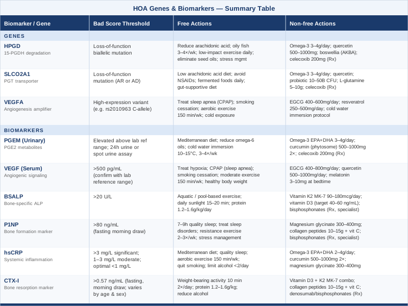
The table below consolidates both the genetic and biomarker frameworks discussed above, offering a quick reference for action across each dimension.
What Peter Attia's Bone Health Framework Reveals About HOA
Beyond individual markers, some of the most actionable insights come from frameworks that connect prostaglandin biology, bone metabolism, and inflammation into a coherent monitoring strategy. Peter Attia's work on longevity medicine — particularly as outlined in his book Outlive: The Science and Art of Longevity (2023) and in hundreds of hours of content on The Drive podcast — has done more than almost any other popular source to elevate the conversation around bone biomarkers. While Attia's primary focus on bone health centers on fracture prevention in aging, his framework for paired longitudinal bone marker tracking applies with surprising precision to HOA monitoring.
1. Always Measure Both Sides of Bone Turnover
Attia argues consistently that measuring only one bone marker gives you half the picture. P1NP reflects formation; CTX-I reflects resorption. In HOA, looking at both together is essential — you need to know whether periosteal bone formation is occurring in a coupled or uncoupled state relative to resorption, because the distinction changes the treatment picture entirely.
2. The Formation-Resorption Ratio Is Clinically Meaningful
High P1NP with suppressed CTX-I is a very different clinical picture from high P1NP with elevated CTX-I. The former suggests controlled formative excess; the latter suggests rapid and disorganized turnover. Attia uses the concept of "coupling" to describe healthy synchrony between formation and resorption — in HOA, this coupling is broken in predictable but variable ways across patients.
3. Fasting Morning Blood Draws Are Non-Negotiable for CTX
CTX-I falls by up to 25% after eating. Attia emphasizes that serial bone marker comparisons are meaningless unless collection conditions are identical every time. For HOA patients tracking bone markers over months and years, this standardization is non-negotiable — a single post-meal CTX result can appear falsely reassuring and misdirect treatment decisions.
4. Vitamin D Is a Prerequisite, Not an Optional Add-On
Attia targets serum 25-OH vitamin D at 40–60 ng/mL as the foundation beneath all other bone interventions. Deficient vitamin D dysregulates parathyroid hormone, accelerates resorption, and undermines the effectiveness of other bone-directed therapies. In HOA patients with dysregulated bone formation, an adequate vitamin D baseline is particularly foundational — deficiency creates a second competing layer of metabolic dysregulation on top of the prostaglandin-driven one.
5. K2 Amplifies D3 — Don't Use One Without the Other
Attia is one of the more prominent voices emphasizing that vitamin D supplementation without K2 can paradoxically increase soft tissue calcification risk. MK-7 (90–180 mcg/day) ensures that calcium mobilized by vitamin D is directed into bone matrix rather than vasculature or other soft tissue — critical in a condition where abnormal periosteal deposition is already the central problem.
6. Protein Intake Is Underappreciated in Bone Metabolism
Attia prescribes higher protein than standard guidelines (targeting 1.6–2.2 g/kg/day in many patients) as part of musculoskeletal longevity. Dietary protein provides amino acid substrate for type I collagen — the dominant bone matrix protein. In HOA, where type I collagen is being overproduced at the periosteum, ensuring the quality of what is being formed, rather than adding further substrate deficit, requires adequate protein supply.
7. Resistance Exercise Sends the Bone Signal — Do Not Omit It
Bone formation responds to mechanical loading through well-characterized mechanosensory pathways. Attia argues that resistance training (compound movements loading the axial and appendicular skeleton) is the most potent stimulus for healthy bone modeling. In HOA, this needs to be calibrated — significant periosteal pain limits loading tolerance — but aquatic resistance training (pool-based exercises with resistance bands) provides stimulus without compressive pain.
8. Sleep Is a Non-Negotiable Bone Health Lever
Growth hormone — the primary anabolic signal modulating the IGF-1 axis in bone — is released predominantly during slow-wave sleep. Chronic sleep disruption reduces GH pulsatility, undermines healthy bone formation signaling, and amplifies inflammatory cytokine production. For HOA patients managing nocturnal pain, sleep quality optimization serves simultaneously as a symptom strategy and a bone biology intervention.
9. Biomarkers Should Be Tracked Quarterly, Not Annually
Standard practice is to recheck bone markers annually at best. Attia's approach is to track quarterly when actively intervening, then extend to every six months once stability is achieved. For HOA patients assessing treatment response, quarterly P1NP, CTX-I, VEGF, and PGEM tracking provides genuinely meaningful feedback — enough time to see real change, not so long that you persist with an ineffective protocol for a year before recognizing it.
10. Effective Pharmacology Is Underused in Rare Bone Conditions
Attia speaks candidly about the underuse of effective bone pharmacology — bisphosphonates, anti-resorptive agents, anabolic therapies — in patients who clearly need them. In HOA, where periosteal disease can be severely disabling, clinician reluctance to prescribe agent-directed therapy is sometimes a barrier. Patients who understand their biomarker picture and can articulate why their situation warrants escalation are better positioned to have productive conversations with their care team about treatment options that exist but are rarely offered.
Complementary Approaches Worth Considering
The molecular and biomarker frameworks above address the underlying biology of HOA. Complementary modalities offer something distinct: relief from the chronic pain experience, reduction in localized swelling, and quality-of-life improvements that are often independent of disease mechanism. Evidence for complementary therapies specifically in HOA is limited by the rarity of the condition — dedicated clinical trials do not exist for most modalities. The three below are selected because they have meaningful human evidence in conditions that share key features with HOA (periosteal pain, joint synovitis, chronic inflammatory musculoskeletal disease) and because the mechanisms are biologically plausible for HOA.
Low-Level Laser Therapy (Photobiomodulation)
Low-level laser therapy (LLLT), also called photobiomodulation, uses specific wavelengths of red and near-infrared light (typically 630–1000 nm) to modulate cellular energy metabolism, reduce local inflammation, and support tissue repair. In HOA, where periosteal inflammation and joint synovitis produce persistent and often deep-seated pain, LLLT offers a non-pharmacological option for localized symptom management. The mechanism involves mitochondrial photostimulation via cytochrome c oxidase, increasing ATP production and reducing oxidative stress at the tissue level — effects that are particularly relevant to inflamed periosteal tissue.
A 2022 systematic review published in Photobiomodulation, Photomedicine, and Laser Surgery evaluated LLLT for inflammatory arthritis across multiple joint conditions and found consistent evidence of pain reduction and functional improvement. Typical clinical protocols use 20–100 mW devices at 660–830 nm wavelengths, three to five sessions per week for four to six weeks. Home devices at 100–200 mW (commercially available at $100–$400 from reputable manufacturers) allow maintenance between clinical sessions. Evidence is strongest for knee and shoulder joint involvement, which maps directly to common HOA joint targets.
For HOA-specific application, a handheld photobiomodulation device applied five to ten minutes per site (tibial shaft, wrist, ankle) three to four times per week is a feasible home protocol for managing periosteal pain. Begin with a six-week trial, monitoring pain levels weekly using a standardized scale. No significant side effects are expected at recommended parameters. Avoid direct eye exposure; do not apply directly over areas of suspected active malignancy in secondary HOA patients.
Mindfulness Meditation and MBSR
Mindfulness-based stress reduction (MBSR) is an eight-week structured program combining sitting meditation, body scanning, and gentle movement, originally developed by Jon Kabat-Zinn at the University of Massachusetts. In HOA, chronic periosteal pain — often described as a deep, relentless aching — has a central sensitization component that pharmacological anti-inflammatory therapy does not directly address. MBSR targets the pain experience and the stress response simultaneously, which is directly relevant to HOA because cortisol and sympathetic activation upregulate COX-2 and increase PGE2 production — creating a direct feedback loop between unmanaged psychological stress and disease activity.
A landmark randomized controlled trial by Cherkin and colleagues, published in JAMA in 2016, demonstrated that MBSR was as effective as cognitive behavioral therapy for chronic musculoskeletal pain, with benefits persisting at 26-week follow-up and with improvements in pain, function, and quality of life compared to usual care. Multiple additional studies have documented that MBSR reduces serum CRP and inflammatory cytokines in chronic pain populations — directly relevant to the hsCRP biomarker discussed above and to the cortisol-PGE2 link in HOA pathophysiology.
A realistic entry protocol for HOA patients: begin with 10–15 minutes of body-scan meditation daily for four weeks using a structured app (Insight Timer, Waking Up, or the MBSR free curriculum available through the Center for Mindfulness at UMass Medical School). Progress to 20–30 minutes. A certified MBSR course — available in person or online — is the gold standard for those who want the full protocol. The relevant framing for HOA is that chronic pain has both peripheral (tissue) and central (brain) components, and MBSR meaningfully addresses the latter while complementing, rather than replacing, pharmacological or biomarker-directed treatment.
Massage Therapy
Massage therapy in HOA addresses two distinct symptom domains: joint edema and synovial effusion, and the periosteal pain and soft tissue changes (particularly skin thickening in pachydermoperiostosis) that accompany active disease. Manual lymphatic drainage (MLD) specifically targets joint and limb swelling by stimulating lymphatic return — relevant in HOA where abnormal periosteal vascularity and local inflammation impair normal fluid clearance. Swedish and clinical massage techniques address the secondary muscle guarding and myofascial tension that develops around chronically painful joints and long bones.
A Cochrane review on massage for osteoarthritis found consistent reductions in pain and improvements in function with massage for knee and hip joint involvement — conditions that share the joint inflammation and movement restriction of HOA's articular features. MLD has established evidence for reducing limb edema in lymphedema and post-surgical conditions; the mechanism is applicable to HOA-related ankle and wrist swelling. Protocols typically involve 45–60 minute sessions one to two times per week for six to eight weeks, transitioning to monthly maintenance.
For HOA specifically: seek a therapist trained in both clinical massage and MLD. In the acute phase with significant joint effusion, focus sessions on gentle MLD for edema management rather than deep tissue work, which can aggravate actively inflamed joints. Between sessions, self-massage of the lower legs — light effleurage strokes moving from the foot upward toward the knee, five minutes per leg twice daily — can help manage the periosteal discomfort and swelling that commonly affects the tibial region in HOA. Avoid deep pressure directly over areas of active periosteal swelling or cortical thickening visible on imaging. Cost: $60–$120 per session.
Conclusion
Hypertrophic osteoarthropathy is a condition where molecular detail matters — both for understanding why symptoms arise and for identifying where meaningful action is possible. The six biomarkers covered here (PGEM, VEGF, BSALP, P1NP, hsCRP, and CTX-I) give you a current, trackable picture of disease activity. The three genes (HPGD, SLCO2A1, and VEGFA) provide the underlying context that explains biological predisposition and helps prioritize which interventions are most likely to be relevant for a given person.
The most useful next step for most people reading this is a structured baseline biomarker panel, ordered in consultation with a rheumatologist or internal medicine specialist with bone metabolism experience. Track it quarterly when actively intervening; use each result to refine your approach rather than waiting for annual check-ins. Combine the biomarker and lifestyle strategies most relevant to your personal picture, and you move from passive recipient of a diagnosis to active participant in managing it.
Start with what you can measure. Act on what you find. Bring the data into your clinical conversations — and keep those conversations going.
Musculoskeletal: Bone Conditions Joint Conditions
Autoimmune: Inflammatory Conditions