This article contains AI generated content.
· UpdatedHypervitaminosis A — 7 Biomarkers And 5 Genes To Track
Introduction
Vitamin A toxicity is one of those conditions that can hide in plain sight for months or years. The symptoms—persistent headaches, unexplained bone and joint pain, dry or peeling skin, fatigue, and blurred vision—are easy to misattribute. Most people who eventually receive a diagnosis of hypervitaminosis A had the problem dismissed multiple times before anyone thought to check their vitamin A status. And even when a basic serum retinol test is run, it often comes back within range, which is misleading, because one number does not capture the full picture of how much vitamin A your body is actually carrying or how it is affecting your organs.
What makes this condition especially inconsistent is how differently it presents from person to person. Two people can take the same high-dose supplement for the same duration and have completely different outcomes—one develops liver stress and bone loss within six months, while the other shows no measurable harm for years. That variability is not random. It reflects real differences in the genes governing how your body converts, transports, stores, and clears vitamin A and its metabolites. Without accounting for those differences, any advice about safe intake levels or recovery timelines is at best an average, and averages don't describe any particular person.
Generic guidance—"stop taking your supplements and wait"—is not wrong, but it leaves an enormous amount of information on the table. Knowing which organs are currently stressed, how full your hepatic stores actually are, and how your individual genetics influence your sensitivity changes the quality of every decision you make from here forward. It can be the difference between a targeted, confident recovery plan and years of vague symptom management.
This article takes two complementary approaches. The first section covers the seven biomarkers most useful for understanding the real state of vitamin A in your body and tracking your recovery over time—many of which are rarely ordered but make a substantial difference. The second section covers the five genes with the most documented influence on how your body handles vitamin A, and what those variants mean for practical decisions. Beyond the labs and genetics, you will find a synthesis of some of the most important independent research on this topic, along with a few complementary approaches that have genuine evidence behind them for managing specific symptoms.
Summary
This article walks through seven specific biomarkers—starting with the widely ordered serum retinol and moving into fasting retinyl esters, liver enzyme patterns, bone turnover markers, lipid dynamics, retinol-binding protein 4, and the often-overlooked vitamin D interaction—explaining what each one actually reveals, how to get it measured, and what to do when results come back unfavorable, both with and without supplements. The genetics section covers five genes—including BCO1 for conversion efficiency, CYP26A1 for retinoic acid clearance, STRA6 for cellular uptake, and RARB for receptor sensitivity—that help explain why identical vitamin A exposures produce wildly different outcomes in different people. The article also covers ten of the most impactful insights from Chris Masterjohn's deep research on fat-soluble vitamin interactions, several of which directly challenge what mainstream medicine currently tells patients about vitamin A safety and recovery. If you have ever been told your vitamin A is "fine" despite symptoms that match the toxicity profile, or if you want to understand your actual risk before it becomes a clinical problem, the information here is built for that conversation.
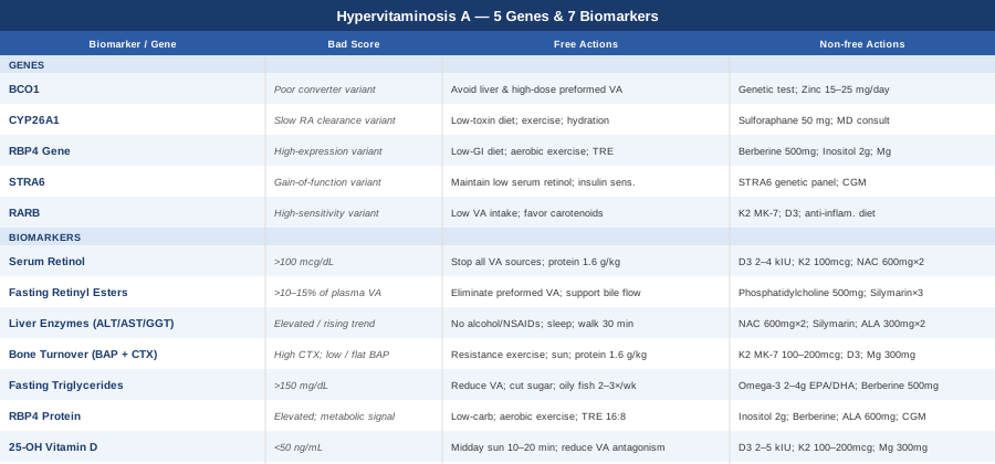
7 Biomarkers to Track If You Suspect Vitamin A Toxicity
The biggest mistake made in evaluating vitamin A status is stopping at a single number. Serum retinol on its own misses the liver-storage picture, the bone impact, the liver enzyme trend, and the important downstream effects on lipid metabolism and other fat-soluble vitamins. The seven biomarkers below give a layered understanding of what vitamin A excess is actually doing in your body—and together they create a baseline from which you can monitor change.
Biomarker 1: Serum Retinol
Why it matters and what it reveals: Serum retinol measures the circulating level of retinol, the active alcohol form of vitamin A, bound to retinol-binding protein in the bloodstream. The normal reference range for adults is approximately 30–80 mcg/dL (1.0–2.8 µmol/L). Levels above 100 mcg/dL are consistently associated with chronic toxicity risk, and acute toxicity from a single massive dose can spike values far higher. The test is useful as a screening tool and for trend monitoring, but its primary limitation is significant: the liver regulates retinol release tightly, releasing only what it needs to maintain circulating levels. This means serum retinol can appear normal even when hepatic stores are severely overloaded—a critical point that leads many clinicians to falsely reassure patients.
How to measure it: A fasting blood draw analyzed by HPLC (high-performance liquid chromatography). Widely available through any major commercial laboratory. Cost range: $50–$150, sometimes covered as part of a micronutrient panel.
If the score is elevated — without supplements
The immediate priority is eliminating all sources of preformed vitamin A: retinol and retinyl ester supplements, high-dose cod liver oil, and frequent liver consumption (beef and chicken liver can contain 15,000–70,000 IU per serving). Stop any multivitamin containing retinol or retinyl palmitate. Allow 6–12 weeks before retesting, as the liver will begin recalibrating once the excess intake stops. Prioritize adequate protein intake (1.6–2.0 g per kg of body weight), which supports the synthesis of retinol-binding protein and helps normalize retinol transport dynamics.
If the score is elevated — with supplements or equipment
Vitamin D3 at 2,000–4,000 IU daily (depending on baseline 25-OH vitamin D levels) helps offset some of the bone and immune consequences of vitamin A excess by competing at the Retinoid X Receptor. Vitamin K2 as MK-7 at 100–200 mcg daily supports calcium metabolism and bone health during the recovery period. N-acetyl cysteine (NAC) at 600 mg twice daily with food replenishes hepatic glutathione, supporting the liver's ability to process the retinoid burden. Take these daily throughout the recovery period—no cycling needed. Retest retinol every 8–12 weeks. Side effects: high-dose D3 can cause hypercalcemia; check serum calcium and 25-OH D at each retest.
Biomarker 2: Fasting Retinyl Esters
Why it matters and what it reveals: This is the most sensitive direct indicator of vitamin A overload, yet it is almost never ordered in routine clinical care. Retinyl esters are the storage form of vitamin A, esterified and held primarily in the liver's stellate (Ito) cells. In healthy, fasted individuals, retinyl esters typically account for less than 5–10% of total plasma vitamin A. When hepatic stores are saturated beyond their capacity, retinyl esters spill into circulation—and in chronic hypervitaminosis A, this fraction can rise to 30%, 50%, or higher. This ratio is far more indicative of true hepatic overload than serum retinol alone, and it is the biomarker most likely to be abnormal in someone who has been told their vitamin A is "normal." Penniston and Tanumihardjo documented this relationship in their widely cited 2006 review of vitamin A toxicity mechanisms, and it remains one of the clearest diagnostic signals available.
How to measure it: A fasting blood draw; the test must specifically request "retinyl esters" or ask for a "retinol and retinyl ester ratio." Not all standard labs offer this—specialty laboratories such as Mayo Clinic Laboratories are more reliable. Fasting for at least 8 hours before the draw is essential, as eating can temporarily elevate circulating retinyl esters even in healthy individuals. Cost: $100–$300.
If the score is elevated — without supplements
A fasting retinyl ester fraction above 10–15% is a signal that the liver's storage capacity has been exceeded. The primary intervention is complete elimination of all preformed vitamin A dietary sources. Beyond that, supporting bile flow helps: bile acids are the vehicle through which fat-soluble compounds are excreted. Maintaining adequate fat in the diet (from non-vitamin-A sources like olive oil, nuts, and avocado) stimulates bile secretion and supports hepatic clearance. Avoid caloric extremes—severe restriction impairs liver function, while caloric excess adds metabolic stress.
If the score is elevated — with supplements or equipment
Phosphatidylcholine at 500–1,000 mg daily supports hepatocyte membrane integrity and assists in liver lipid handling, which is relevant because retinyl esters are stored in lipid droplets within stellate cells. Milk thistle (standardized silymarin extract, 140 mg three times daily) has established hepatoprotective properties and is one of the few supplements with direct evidence in toxic hepatic insult models. These are supportive measures, not accelerators of clearance—realistic expectations matter here. Full recovery from severe hepatic vitamin A overload can take 12–36 months. Retest retinyl esters every 8–12 weeks.
Biomarker 3: Liver Enzymes — ALT, AST, and GGT
Why it matters and what it reveals: The liver is the primary storage site for vitamin A, and it is the organ most directly and persistently damaged by chronic excess. Alanine aminotransferase (ALT) and aspartate aminotransferase (AST) are released from hepatocytes into the bloodstream when liver cells are damaged or stressed. Gamma-glutamyl transferase (GGT) is particularly sensitive to oxidative stress in the liver and to retinoid-mediated cellular disruption. In hypervitaminosis A, the key pathological process is hyperplasia and activation of hepatic stellate cells—when these cells become overwhelmed with retinyl ester storage, they begin depositing collagen and initiating fibrosis. Early in this process, ALT and AST may be only mildly elevated or even normal, but the trend over months of continued exposure is informative. GGT elevation may appear earlier. Monitoring the full pattern rather than a single snapshot is more useful.
How to measure it: ALT and AST are included in a standard comprehensive metabolic panel (CMP), available at any laboratory. GGT requires a specific add-on. The full panel including GGT is sometimes called a liver function test (LFT) or hepatic panel. Cost: $20–$80.
If the score is elevated — without supplements
Remove the primary hepatic stressor—preformed vitamin A—immediately and completely. Eliminate alcohol and avoid unnecessary medications, particularly acetaminophen and NSAIDs taken regularly, as these add to the hepatic detoxification burden. Prioritize sleep: liver regeneration occurs predominantly during deep sleep, and sleep deprivation directly impairs hepatic recovery. Gentle aerobic activity (walking 30 minutes daily) improves hepatic blood flow and supports metabolic function without adding oxidative stress. Recheck liver enzymes every 6–8 weeks.
If the score is elevated — with supplements or equipment
NAC at 600 mg twice daily is the most direct hepatoprotective supplement, replenishing glutathione that becomes depleted under toxic retinoid load. Silymarin (milk thistle extract, 140–175 mg standardized to 70–80% silymarins, three times daily) has the most robust evidence among over-the-counter hepatoprotectives, including reduction of hepatic fibrosis markers in clinical studies. Alpha-lipoic acid at 300 mg twice daily provides complementary antioxidant support and supports mitochondrial function in stressed hepatocytes. These should be taken consistently daily, not cycled, until enzymes normalize and confirm two consecutive normal readings before discontinuing. Side effect note: NAC can cause mild nausea at higher doses; take with food.
Biomarker 4: Bone Turnover Markers — BAP and CTX
Why it matters and what it reveals: Vitamin A excess is one of a short list of nutritional conditions with a well-established, direct mechanism of skeletal harm. Retinoic acid, the oxidized active metabolite of vitamin A, stimulates osteoclast differentiation and activity (increasing bone resorption) while simultaneously suppressing osteoblast function. The result is a resorption-dominant state where bone is broken down faster than it is rebuilt—a pattern that accelerates bone loss and significantly increases fracture risk, even before DEXA imaging shows dramatic changes in bone mineral density.
Bone-specific alkaline phosphatase (BAP) is a direct enzyme marker of osteoblast activity and bone formation. CTX (cross-linked C-terminal telopeptide of type I collagen) measures the rate of bone collagen breakdown by osteoclasts. In vitamin A toxicity, the characteristic pattern is elevated CTX with flat or suppressed BAP—resorption exceeding formation. This pattern is more sensitive for detecting early skeletal impact than DEXA alone and gives real-time feedback on whether the bone microenvironment is stabilizing as vitamin A is reduced.
How to measure it: Both are serum tests. CTX must be collected in the early morning in a fasted state, as it shows significant diurnal and prandial variation. Available through hospital laboratory systems and specialty labs. Cost: $100–$300 for both markers combined.
If the score is abnormal — without supplements
Weight-bearing resistance exercise—squats, lunges, deadlifts, or equivalent bodyweight movements—is the single most effective non-pharmaceutical stimulus for osteoblast activity and bone formation. Three to four sessions per week, with progressive loading, has strong evidence for reversing adverse bone turnover patterns. High-impact activities such as jumping also stimulate bone remodeling. Adequate dietary protein (at minimum 1.6 g/kg body weight) provides the amino acid substrate for bone matrix synthesis. Optimizing sun exposure for endogenous vitamin D production directly supports osteoblast function.
If the score is abnormal — with supplements or equipment
Vitamin K2 as MK-7 at 100–200 mcg daily is the highest-priority supplement for bone health in hypervitaminosis A. K2 activates osteocalcin (which anchors calcium into bone) and activates matrix Gla-protein (which prevents arterial calcium deposition). Vitamin D3 at 2,000–4,000 IU daily is essential given the A-D antagonism. Magnesium glycinate or malate at 300–400 mg nightly is a critical cofactor for bone formation and vitamin D activation. DEXA scanning annually or semi-annually provides a complementary structural picture. For cases involving significant bone density loss, discuss bisphosphonate therapy with your physician—these prescription medications specifically target osteoclast activity and may be appropriate if CTX remains markedly elevated despite removing vitamin A and adding K2 and D3. Side effects: very high D3 requires monitoring of serum calcium; K2 is well-tolerated in clinical ranges.
Biomarker 5: Fasting Triglycerides
Why it matters and what it reveals: Retinoids exert direct and potent effects on hepatic lipid metabolism. Retinoic acid activates both retinoic acid receptors (RARs) and retinoid X receptors (RXRs), which in combination regulate hepatic triglyceride synthesis and secretion into VLDL particles. The result of this retinoid activation is that triglyceride synthesis is upregulated while peripheral clearance is reduced. This effect is most dramatic with pharmaceutical retinoids such as isotretinoin (where severe hypertriglyceridemia is a known, monitored side effect), but it also occurs with prolonged dietary or supplemental vitamin A excess. Elevated fasting triglycerides in someone with high vitamin A intake should prompt investigation rather than be attributed solely to diet. Severe hypertriglyceridemia above 500 mg/dL carries independent risk for acute pancreatitis.
How to measure it: A standard fasting lipid panel. Requires 10–12 hours of fasting. Available at any laboratory, commonly covered by insurance. Cost: $20–$50.
If the score is elevated — without supplements
Reducing preformed vitamin A is the foundational step for retinoid-mediated hypertriglyceridemia—this distinguishes it from dietary hypertriglyceridemia where refined carbohydrate reduction is the first intervention (though both interventions help). Eliminating added sugars and refined grains from the diet reduces hepatic de novo lipogenesis and complements the retinoid reduction strategy. Increasing consumption of EPA/DHA-rich whole foods—sardines, mackerel, wild salmon two to three times weekly—provides natural triglyceride-lowering effect without adding supplemental retinol risk.
If the score is elevated — with supplements or equipment
Omega-3 fatty acids as EPA and DHA at 2–4 grams per day from fish oil are among the most consistently evidence-supported interventions for elevated triglycerides, with multiple meta-analyses confirming 20–30% reductions. If triglycerides exceed 500 mg/dL, prescription-grade icosapentaenoic acid (Vascepa) should be discussed with a physician given the pancreatitis risk. Berberine at 500 mg two to three times daily with meals has evidence for lipid-lowering effects and modest triglyceride reduction. Berberine should be cycled (8 weeks on, 4 weeks off) if used long-term, as it may affect gut microbiome composition with prolonged continuous use. Side effects: GI discomfort with berberine is common initially—titrate up from 250 mg and take with meals.
Biomarker 6: Retinol-Binding Protein 4 (RBP4)
Why it matters and what it reveals: RBP4 is the primary transport protein for retinol in circulation, secreted mainly by the liver and adipose tissue. It circulates in a 1:1 molar complex with retinol, delivering it to target tissues. When the hepatic retinol burden is high, RBP4 secretion and circulating levels reflect this excess transport demand. But RBP4's significance extends beyond vitamin A transport. A substantial body of research has established RBP4 as an independent biomarker of insulin resistance, adipose inflammation, and metabolic syndrome. High circulating RBP4 impairs insulin signaling in skeletal muscle and fat cells through mechanisms involving STRA6 receptor activation and downstream JAK-STAT signaling. In hypervitaminosis A, elevated RBP4 may therefore simultaneously reflect vitamin A overload and contribute to metabolic dysfunction—a compounding effect that is not captured by liver tests alone.
How to measure it: A serum RBP4 test, not typically part of routine panels. Available through hospital laboratory systems and specialty diagnostic laboratories. Cost: $80–$250.
If the score is elevated — without supplements
Reducing vitamin A intake addresses the transport-overload component of elevated RBP4. For the insulin resistance component, the most evidence-backed approach is combining dietary carbohydrate reduction with consistent aerobic exercise. Time-restricted eating (TRE) in a 16:8 or 14:10 window has demonstrated reductions in RBP4 in studies of metabolic syndrome patients, likely through improvements in hepatic insulin sensitivity. Regular aerobic activity—30 or more minutes five times per week—reduces adipose RBP4 secretion and improves insulin signaling in skeletal muscle.
If the score is elevated — with supplements or equipment
Myo-inositol at 2 grams daily has preliminary evidence for improving cellular insulin sensitivity and may indirectly reduce RBP4 levels driven by the insulin resistance pathway. Berberine (500 mg twice daily, cycled 8 weeks on/4 weeks off) and alpha-lipoic acid (600 mg daily) both improve hepatic and peripheral insulin signaling. A continuous glucose monitor (CGM), worn for 2–4 weeks, can identify specific glycemic patterns—postprandial spikes, fasting hyperglycemia—that are driving the insulin resistance component of RBP4 elevation. This makes it a useful diagnostic tool rather than just a monitoring device. Side effects: inositol is generally well tolerated; GI softening at doses above 4 grams is the primary concern.
Biomarker 7: 25-OH Vitamin D
Why it matters and what it reveals: Vitamins A and D share a striking degree of biochemical overlap. Both use the Retinoid X Receptor (RXR) as a signaling partner, meaning they compete directly for receptor occupancy and downstream gene activation. Chronic vitamin A excess effectively blunts vitamin D signaling at the cellular level—even when serum 25-OH vitamin D looks adequate, vitamin D's actual biological activity may be significantly impaired. This mechanism, documented in both animal and human studies, means that many of the symptoms attributed purely to vitamin A toxicity—bone pain, immune dysregulation, fatigue, depression—are partly or substantially driven by functionally impaired vitamin D action. Assessing 25-OH vitamin D levels as part of a hypervitaminosis A workup is essential, not optional, for both diagnostic clarity and recovery planning.
How to measure it: A serum 25-OH vitamin D test, the standard clinical measure of vitamin D status. Widely available, often covered by insurance with appropriate diagnostic code. The Vitamin D Council, Peter Attia, and Rhonda Patrick consistently recommend maintaining levels between 50–70 ng/mL for comprehensive functional benefit. Cost: $40–$100. The NIH Office of Dietary Supplements vitamin A fact sheet addresses this interaction.
If the score is suboptimal — without supplements
Midday sun exposure—10 to 20 minutes of direct UVB on large skin surface areas (back, legs, arms) between 10 AM and 2 PM—provides physiologically regulated vitamin D synthesis without creating a supplemental imbalance. This approach avoids any risk of oversupplementation and provides additional benefits through nitric oxide release and circadian light signaling. Skin type, latitude, and season will determine the effective dose. Reducing vitamin A excess simultaneously removes the functional antagonism that was blunting vitamin D's cellular effects.
If the score is suboptimal — with supplements or equipment
Vitamin D3 at 2,000–5,000 IU daily, paired with vitamin K2 (MK-7, 100–200 mcg daily) to direct calcium appropriately, is the standard supplemental approach for restoring 25-OH vitamin D. Target the 50–70 ng/mL range and retest every 8–12 weeks while supplementing. Magnesium glycinate at 300–400 mg daily is an essential cofactor: without sufficient magnesium, the conversion of 25-OH vitamin D to its active form (1,25-dihydroxyvitamin D) is impaired. Side effects: doses above 5,000 IU D3 daily require monitoring of serum calcium and urine calcium to detect early hypercalcemia; keep serum calcium below 10.5 mg/dL.
5 Genes That Determine How Your Body Handles Vitamin A
The wide variability in how people respond to the same vitamin A intake is not arbitrary. Much of it traces back to specific genetic differences in how efficiently the body converts plant precursors to vitamin A, how quickly it clears the active metabolite retinoic acid, how avidly cells take up retinol, and how sensitively nuclear receptors respond to retinoic acid signaling. The five genes below are the most clinically meaningful in this picture. Understanding your variants, ideally through consumer or clinical whole-genome sequencing, can reframe how cautiously you approach supplementation and how aggressively you need to monitor the biomarkers above.
BCO1 — The Conversion Gatekeeper
What this gene does: BCO1 (beta-carotene oxygenase 1, also called BCMO1) encodes the enzyme responsible for the initial step in converting beta-carotene and other carotenoids into retinal, the precursor to retinol. This single enzymatic step determines how efficiently you convert plant-derived provitamin A into the active form your body uses. Two well-documented single-nucleotide polymorphisms—rs7501331 (T allele) and rs12934922 (C allele)—reduce enzyme activity by approximately 32–69% in carriers, depending on how many copies of the variant alleles are present. Studies estimate that 45–49% of women and a similar proportion of men carry at least one of these variants.
The implications are counterintuitive. Poor converters who take beta-carotene supplements or eat large amounts of orange/yellow plant foods accumulate carotenoids in fat without converting them efficiently to retinol—leading to carotenodermia (yellow-orange skin) but not to vitamin A toxicity per se. However, the same poor-converter individuals, when supplementing or eating abundant preformed retinol (animal liver, cod liver oil, high-dose supplements), face the same or greater toxicity risk as efficient converters, because BCO1 activity is irrelevant to preformed retinol metabolism. This has led some practitioners and influencers to incorrectly advise poor converters to take high-dose preformed vitamin A—which can backfire significantly.
If this variant is present — without supplements
If you are a poor BCO1 converter, your risk from carotenoid-rich plant foods is low. Your risk from preformed vitamin A (animal liver, retinyl ester supplements, cod liver oil) is the same as for any efficient converter—and you may be more vulnerable because you may have been told to take more preformed A to compensate for your poor conversion. Reduce or eliminate high-dose preformed vitamin A supplements. Moderate liver consumption (once every 2–3 weeks rather than weekly). Do not interpret carotenemia (yellow skin) from vegetables as vitamin A toxicity—these are distinct situations.
If this variant is present — with supplements or equipment
Genetic testing through consumer platforms such as 23andMe or Ancestry (followed by analysis through tools like Genetic Lifehacks or Promethease) can identify your BCO1 rs7501331 and rs12934922 status. No specific supplement corrects BCO1 function. The implication is primarily about avoiding inappropriate supplementation. Zinc is a cofactor that supports overall retinoid metabolism and may modestly support conversion pathways; 15–25 mg elemental zinc daily is a reasonable baseline if dietary zinc is low. Do not cycle zinc unnecessarily—maintain it as a consistent dietary or supplemental baseline.
CYP26A1 — The Retinoic Acid Clearance Rate
What this gene does: CYP26A1 encodes a cytochrome P450 enzyme that is the primary mechanism by which the body inactivates and clears all-trans retinoic acid (atRA), the most potent active metabolite of vitamin A. Retinoic acid has powerful effects on gene expression, immune function, skin, embryogenesis, and bone—but these effects require tight concentration control. CYP26A1 is induced by retinoic acid itself (an elegant feedback system), catalyzing its hydroxylation to 4-hydroxy-retinoic acid, 4-oxo-retinoic acid, and ultimately polar metabolites that are excreted. Variants that reduce CYP26A1 expression or activity mean that even a moderate vitamin A intake generates disproportionately high and prolonged retinoic acid exposure at the cellular level.
Research into CYP26A1 polymorphisms is still maturing compared to BCO1, but several variants have been associated with altered retinoid sensitivity, including variations in promoter regions that reduce transcriptional response. Individuals with reduced CYP26A1 function may experience toxicity-like symptoms at doses that would be harmless for the general population, and may have dramatically prolonged recovery timelines once toxicity develops.
If this variant is present — without supplements
Reduced CYP26A1 function means a lower threshold for retinoic acid bioaccumulation. The practical implication is to treat even moderate-dose vitamin A supplementation (anything above 3,000–5,000 IU of preformed retinol per day) with caution. Limit high-vitamin-A foods to once every 2–4 weeks rather than regularly. Recovery from excess will be slower, as clearance is impaired. Focus on supporting hepatic detoxification broadly through adequate hydration, regular aerobic exercise, and a low-toxin diet that does not add to the CYP450 system burden.
If this variant is present — with supplements or equipment
There is no direct supplement to upregulate CYP26A1. However, compounds that support broad cytochrome P450 function include sulforaphane (from broccoli sprouts, 50–100 mg of glucoraphanin/sulforaphane equivalent daily), which upregulates Nrf2-mediated antioxidant and detoxification pathways. Genetic testing confirming a CYP26A1 variant should prompt discussion with a functional medicine physician or geneticist about safe upper thresholds for vitamin A supplementation in your case. Avoid synthetic retinoids (isotretinoin, tretinoin) except under close medical monitoring if CYP26A1 function is reduced.
RBP4 Gene — Baseline Transport Capacity
What this gene does: The RBP4 gene encodes the liver-secreted retinol-binding protein that carries retinol safely through the bloodstream, preventing its free (unbound) circulation which would be cytotoxic. Variations in the RBP4 gene affect baseline expression levels of this transport protein, independently of current vitamin A intake. RBP4 gene variants have been associated with elevated baseline RBP4 protein expression, which is linked to insulin resistance and a higher metabolic burden from circulating retinol. Unlike the biomarker reading (which fluctuates with intake and metabolic status), the gene determines your constitutional tendency for transport capacity—meaning some people will have higher circulating retinol even at the same dietary intake, and will show RBP4-related metabolic effects more readily.
Several RBP4 promoter variants and haplotypes have been associated with significantly higher circulating RBP4 and greater likelihood of metabolic syndrome in population studies, including work from the International Journal of Obesity.
If this variant is present — without supplements
High constitutional RBP4 expression means metabolic vigilance is warranted alongside monitoring vitamin A status. A dietary pattern emphasizing low glycemic index foods, adequate dietary fiber (30+ grams per day), and consistent aerobic exercise (which reduces adipose RBP4 secretion specifically) is the most effective lifestyle framework for managing RBP4-associated insulin sensitivity. Intermittent fasting, particularly TRE in a 14:10 or 16:8 window, has clinical evidence for reducing RBP4 in metabolic syndrome contexts.
If this variant is present — with supplements or equipment
Berberine (500 mg twice daily with meals, cycled 8 weeks on/4 off), myo-inositol (2–4 grams daily in divided doses), and magnesium (300–400 mg daily) form a well-supported metabolic support stack for insulin sensitivity, which is the key downstream concern in RBP4 variants. A CGM worn for 2–4 weeks per year provides objective feedback on insulin response and helps calibrate the dietary approach. Side effects: start berberine at 250 mg once daily and titrate up over 1–2 weeks to minimize GI discomfort.
STRA6 — Cellular Retinol Uptake Sensitivity
What this gene does: STRA6 (stimulated by retinoic acid 6) encodes a membrane receptor expressed in many tissue types that specifically binds the RBP4-retinol complex and mediates cellular uptake of retinol from circulation. Beyond retinol uptake, STRA6 also activates an intracellular signaling cascade through JAK2 and STAT5, linking extracellular retinol availability to intracellular gene expression in ways that are still being characterized. Gain-of-function variants in STRA6 can lead to increased cellular retinol uptake, heightening the intracellular retinoic acid burden even when circulating retinol levels appear moderate. Loss-of-function mutations in STRA6 cause Matthew-Wood syndrome (a rare condition involving eye and lung malformations), but subtle variants in the general population influence cellular sensitivity without causing this severe presentation.
If this variant is present — without supplements
Increased STRA6-mediated uptake amplifies cellular retinoic acid exposure. This means that tissue-level vitamin A effects may be more potent than blood tests suggest. For individuals with known STRA6 gain-of-function variants, maintaining serum retinol in the lower half of the normal range (30–50 mcg/dL) rather than aiming for mid-range or higher is a reasonable precautionary approach. Avoid combining high vitamin A foods with high RBP4-raising factors (insulin resistance, obesity, liver stress) that increase the circulating retinol available for STRA6-mediated uptake.
If this variant is present — with supplements or equipment
No direct supplement modulates STRA6 function. The practical management focuses on reducing the substrate available for uptake—lower circulating retinol through dietary moderation. Lifestyle factors that improve insulin sensitivity (as above) reduce RBP4 in circulation, which indirectly reduces the retinol-RBP4 complex available for STRA6 uptake. STRA6 status is best determined through clinical whole-genome sequencing or research genetic panels; it is not currently part of consumer platforms like 23andMe.
RARB — Retinoic Acid Receptor Sensitivity
What this gene does: RARB encodes Retinoic Acid Receptor Beta, one of three nuclear receptors (RARA, RARB, RARG) that directly bind retinoic acid and regulate transcription of downstream target genes. Variations and epigenetic silencing in RARB affect how sensitively specific tissues respond to retinoic acid signaling. Promoter hypermethylation of RARB is well-studied in oncology (where it silences an anti-proliferative signal), but in the context of hypervitaminosis A, variants that increase RARB expression or function may amplify the pro-osteoclastic, pro-inflammatory, and lipogenic effects of retinoic acid at the tissue level—meaning more severe systemic consequences from the same serum concentration.
The RARB gene also influences how the immune system responds to retinoic acid, which has implications for autoimmune activity and the mucosal immune balance that vitamin A regulates.
If this variant is present — without supplements
Heightened RARB sensitivity essentially raises the effective potency of every unit of dietary or supplemental retinoic acid in the tissues that express this receptor. The conservative approach is to aim for vitamin A intake at the lower end of the adequate intake range (700–900 mcg RAE for adults) and to emphasize carotenoid sources over preformed retinol wherever possible. Monitor bone turnover markers and liver enzymes even at vitamin A intake levels that would be considered safe for the general population.
If this variant is present — with supplements or equipment
No direct supplement modulates RARB sensitivity. Supporting the downstream consequences of RARB activity is the more practical strategy: vitamin K2 and D3 (as described under the bone biomarker section) help offset the pro-resorptive bone effects of heightened RAR signaling. Anti-inflammatory dietary patterns (Mediterranean-style, rich in omega-3, polyphenols, and low in processed foods) reduce the baseline inflammatory state that heightened RAR signaling can amplify. RARB polymorphism data is increasingly available through clinical genetics platforms and research-grade genome sequencing.
What Chris Masterjohn's Research Reveals About Vitamin A and Fat-Soluble Vitamin Balance
Chris Masterjohn, PhD, is a nutritional biochemist whose extensive independent research on fat-soluble vitamins—particularly vitamins A, D, and K—has produced some of the most nuanced and evidence-grounded analysis available outside of academic journals. His work, published across his Mastering Nutrition podcast and Substack, has challenged several widely held clinical assumptions about vitamin A safety, toxicity thresholds, and recovery, drawing on mechanistic biochemistry and primary literature in ways that differ markedly from standard dietary guidance. The following ten points capture the most impactful insights from this body of work as they apply to hypervitaminosis A.
Vitamins A, D, and K2 Form a Triad — You Cannot Optimize One in Isolation
Masterjohn emphasizes repeatedly that A, D, and K2 compete and cooperate through shared nuclear receptors and shared protein activation systems. Excess vitamin A impairs vitamin D function; deficient K2 allows calcium to drift toward soft tissues; optimizing D3 without K2 creates its own risks. Managing hypervitaminosis A without simultaneously assessing and supporting D and K status leaves part of the problem unaddressed.
Moderate Liver Consumption Carries Real, Underappreciated Risk for Sensitive Individuals
A single 100-gram serving of beef liver contains roughly 26,000 IU of preformed vitamin A. Masterjohn's analysis of the toxicology literature suggests that frequent consumption—more than once weekly over months to years—can accumulate toward toxicity in individuals with genetic susceptibility, particularly those with reduced CYP26 clearance or elevated RARB sensitivity. The popular notion that food-sourced vitamin A is categorically safe does not hold under mechanistic scrutiny.
Traditional Diets Balanced Fat-Soluble Vitamins Precisely Because They Combined Food Sources
Masterjohn points to the nutritional anthropological observation that traditional diets incorporating organ meats also included fermented dairy (K2-rich), regular sun exposure (D synthesis), and seafood (D, EPA/DHA). Modern patterns of selectively supplementing only retinol or eating only liver without these balancing factors recreate an imbalance that traditional foodways never produced.
Retinoic Acid Has a Hundred Times the Potency of Retinol—The Conversion Matters
Masterjohn stresses that most toxicity discussions conflate different vitamin A forms. Retinol, retinal, and retinoic acid have dramatically different potencies and target tissues. Retinoic acid—the oxidized form that cannot be reduced back—is the primary driver of gene expression effects and the primary toxicity mechanism. Factors that accelerate retinol-to-retinoic acid conversion (alcohol, certain liver conditions, high-iron status) increase toxicity risk from a given retinol intake independent of dose alone.
Zinc and Protein Status Fundamentally Alter Vitamin A Requirements and Risk
RBP4 synthesis requires adequate zinc and protein. When these are depleted, the liver cannot export retinol efficiently, and it accumulates internally. Masterjohn's synthesis of the literature suggests that zinc deficiency combined with excess vitamin A intake creates a particularly dangerous scenario, as hepatic stores rise while transport is impaired. Assessing zinc status (through plasma zinc or alkaline phosphatase as a functional proxy) is relevant alongside vitamin A biomarkers.
Signs of Vitamin A Toxicity Can Mimic Deficiency—and Vice Versa
One of the most practically important observations in Masterjohn's work is that excess and deficiency of vitamin A share overlapping symptom profiles. Both can cause dry skin, immune dysregulation, and mood disruption. The distinction requires biomarker testing, not symptom assessment alone. Treating apparent vitamin A deficiency with supplementation in someone who is already toxic can dramatically worsen the condition.
Alcohol Dramatically Amplifies Retinoic Acid Accumulation
Ethanol shares metabolic pathways with retinol in the liver, competing for the same enzymes (ADH, ALDH) that convert retinol through retinal to retinoic acid and then catabolize it. This competition means that alcohol both impairs retinoic acid clearance and may alter the retinol-to-retinoic-acid conversion rate in ways that amplify cellular exposure. Regular alcohol consumption in the context of high vitamin A intake—even moderate amounts—substantially increases hepatic and systemic retinoic acid burden.
Thyroid Status Modulates Vitamin A Metabolism Significantly
Hypothyroidism slows the conversion of beta-carotene to retinol via BCO1 (already slow in genetic poor converters) and may alter hepatic retinoid metabolism broadly. Masterjohn's analysis suggests that thyroid optimization is a relevant factor in vitamin A management—suboptimal thyroid function can either impair conversion of carotenoids (lowering effective vitamin A status from plant sources) or alter the retinol-retinoic acid balance in the liver. TSH and free T3/T4 are worth including in a comprehensive workup.
Recovery from Chronic Toxicity Often Takes 12–36 Months, Not Weeks
Masterjohn is clear that mainstream reassurances about rapid recovery after stopping supplements are not well-supported by the hepatic retinol biology. The liver can store years of excess vitamin A in stellate cells, releasing it slowly and maintaining elevated tissue retinoic acid levels long after dietary intake is eliminated. Patience, consistent biomarker monitoring, and sustained dietary modification—not a 4-week intervention—are what the evidence actually supports.
The Interaction with Vitamin K2 for Bone Protection Is Underappreciated and Underused
Masterjohn has written specifically about the mechanism by which vitamin A excess drives bone resorption through RANK-L upregulation, and how K2-mediated activation of osteocalcin and MGP provides a direct structural counterbalance. The clinical application—always pairing vitamin A reduction with K2 supplementation during the recovery period—is not standard practice but has a sound mechanistic basis that practitioners tracking individual biomarkers should incorporate.
Complementary Approaches Worth Exploring
Beyond the direct interventions described above, several evidence-informed complementary modalities can help manage the symptoms most commonly reported by individuals with hypervitaminosis A—particularly chronic headache, musculoskeletal pain, and the cognitive disruption associated with elevated intracranial pressure and liver stress. The following three approaches have reasonable clinical evidence for at least some of these specific manifestations.
Microbiome-Directed Therapies
The gut microbiome plays a more significant role in fat-soluble vitamin metabolism than is commonly appreciated. Intestinal bacteria influence bile acid composition and recycling through the enterohepatic circulation, and bile acids are the primary vehicles for fat-soluble compound excretion. Dysbiosis—particularly overgrowth of bacteria that deconjugate bile salts—can increase reabsorption of fat-soluble compounds, including retinoids, rather than allowing their excretion. Additionally, certain bacterial species express retinoid-metabolizing enzymes and can influence the retinol-to-retinoic acid conversion in intestinal cells. Restoring a diverse, healthy microbiome may modestly support the reduction in vitamin A enterohepatic recirculation during recovery.
A specific protocol supported by clinical data is the use of Lactobacillus rhamnosus GG and Bifidobacterium longum in combination, studied in the context of bile metabolism and hepatic inflammation. A 2019 randomized trial published in Alimentary Pharmacology and Therapeutics demonstrated reductions in hepatic inflammatory markers with multispecies probiotic supplementation in non-alcoholic fatty liver disease—a condition sharing stellate cell activation pathways with vitamin A toxicity. 10–20 billion CFU per day of a multispecies probiotic containing Lactobacillus and Bifidobacterium strains is a reasonable protocol.
To apply this for hypervitaminosis A recovery, a high-quality multispecies probiotic (10–20 billion CFU/day), combined with prebiotic fiber (15–25 grams daily from diverse plant sources), and fermented foods (2–3 servings of plain yogurt, kefir, or sauerkraut per day) forms a practical 3-month protocol. This is a supportive measure—it is not a direct treatment for vitamin A toxicity, and the evidence for this specific combination is modest and indirect. Side effects: initial bloating with increased fiber is common and typically resolves within 1–2 weeks.
Mindfulness Meditation and MBSR
Chronic headache—including headaches driven by mildly elevated intracranial pressure, which can occur in hypervitaminosis A—is one of the symptom domains with the strongest evidence base for mindfulness-based intervention. Mindfulness-Based Stress Reduction (MBSR), the 8-week structured program developed by Jon Kabat-Zinn, has been evaluated in multiple randomized controlled trials for chronic pain, headache, and anxiety, with consistent findings of significant symptom reduction and improved quality of life. The mechanism is not merely placebo: MBSR demonstrably reduces activity in the brain's default mode network, lowers cortisol, reduces neuroinflammatory signaling, and improves pain tolerance thresholds.
A meta-analysis published in JAMA Internal Medicine (Goyal et al., 2014) covering 47 trials found that mindfulness meditation programs demonstrated moderate evidence for improvement in pain, anxiety, and depression. While not studied specifically in hypervitaminosis A, the headache, mood disruption, and cognitive fog symptoms of this condition are precisely the manifestations where mindfulness practice has demonstrated benefit.
The practical approach is to begin with the formal 8-week MBSR program, available through local hospital programs, university wellness centers, or the Palouse Mindfulness free online curriculum. Daily practice of 30–45 minutes for the duration of the program, transitioning to 15–20 minutes daily thereafter. Specific attention to body-scan techniques is particularly useful for managing diffuse bone and joint pain. No equipment is required. This approach is appropriate as an adjunct to medical treatment, not a substitute.
Massage Therapy
Musculoskeletal pain—including bone pain, joint aching, and myalgia—is among the most disruptive and least medically addressable symptoms of chronic hypervitaminosis A. NSAIDs, which are the usual first-line pharmacological approach to musculoskeletal pain, add hepatotoxic burden in a condition where the liver is already compromised—making them a particularly poor choice in this context. Massage therapy offers a non-hepatotoxic, evidence-supported alternative for musculoskeletal symptom management.
Clinical evidence for massage in chronic musculoskeletal pain is consistent and substantial. A Cochrane Review (Furlan et al.) of massage for low back pain demonstrated short-term benefit for pain and function, and multiple trials have shown efficacy for myofascial and soft-tissue pain generally. The mechanism involves reduced tension in surrounding soft tissue, improved local blood flow and lymphatic drainage, reduced sympathetic nervous system tone, and endogenous endorphin release. All of these are relevant to the diffuse musculoskeletal discomfort of vitamin A toxicity.
For practical application, Swedish or light deep-tissue massage once to twice weekly during the acute symptomatic phase of recovery is a reasonable protocol. Communication with the massage therapist about areas of bone pain is important—direct pressure over inflamed periosteum or joints with active resorption should be avoided; surrounding muscle and soft tissue is the more appropriate target. Myofascial release techniques for areas of referred pain (particularly around the spine and hips, common sites of vitamin A-related bone pain) are worth requesting specifically. Side effects: mild post-session soreness is normal; communicate any increase in bone pain immediately so the technique can be adjusted.
Conclusion
Hypervitaminosis A is a condition where precision matters more than general advice. Knowing that your serum retinol is elevated tells you a fraction of what you need to know. Knowing your fasting retinyl ester ratio, your liver enzyme trend, your bone resorption markers, and your vitamin D status gives you a functional map. Knowing your BCO1, CYP26A1, or STRA6 variants tells you whether you were predisposed to this outcome and what recovery will realistically require. Together, these layers of information transform what often feels like a vague, frustrating health mystery into a set of specific, trackable, addressable targets.
The most important next step is simply to start. Request fasting retinyl esters alongside serum retinol at your next blood draw. Ask for a liver enzyme panel including GGT. Add a vitamin D test and, if accessible, bone turnover markers. Bring this article to a conversation with a knowledgeable clinician—a functional medicine physician, integrative internist, or hepatologist—who can interpret these results in context and support a structured recovery plan. Genetics testing, if not already available to you, can sharpen the picture further. Better information, systematically gathered, is what makes a meaningful difference in this condition.
Musculoskeletal: Bone Conditions
Neurological: Headache & Migraine
Digestive: Liver & Gallbladder Conditions
Endocrine & Metabolic: Metabolic Syndrome