This article contains AI generated content.
Hypocalcemia Decoded: 7 Biomarkers and 6 Genes to Track
Introduction
If you have landed here, there is a good chance you have already felt something the standard advice does not quite name: the tingling around your lips and fingertips that comes and goes, the muscle cramps that arrive without warning, the twitch under your eye, or a lab report that simply says your calcium is "a little low" with no real explanation of why. You are not imagining it, and you are not being difficult for wanting a clearer answer than "eat more dairy."
Generic guidance tends to stop at the surface. It treats hypocalcemia as if it were always a shortage of dietary calcium, when in reality low blood calcium is usually a downstream signal of something else entirely — a parathyroid gland that is not signaling correctly, a magnesium level too low for your hormones to work, a vitamin D deficiency, a kidney or gut absorption problem, or occasionally an inherited variation in how your body senses calcium at all. Telling everyone with the same number to do the same thing ignores the fact that the same low reading can have very different engines behind it.
This article takes the harder but more useful road. Instead of one blanket recommendation, it maps the specific measurements that reveal why your calcium is low, and the genetic factors that can quietly set the stage — so that the plan you build actually matches the mechanism you have.
There is real reason for grounded optimism here. Hypocalcemia is one of the more reversible metabolic problems when you understand its cause, because most of the levers — vitamin D, magnesium, phosphate balance, and the calcium–parathyroid loop — respond well to correction. Below you will find, first, the seven biomarkers worth tracking and exactly what to do when each one is off; then a shorter tour of six genes that shape parathyroid function; a study-backed podcast summary; and a careful look at complementary approaches that can help with symptoms. Each layer is designed to move you from "my calcium is low" to "I know which lever to pull."
Summary
Hypocalcemia is rarely a calcium problem in isolation — it is a reporting error from a whole regulatory system. This article opens by tracking the seven biomarkers that tell you which part of that system has slipped: albumin-corrected calcium, ionized calcium, parathyroid hormone (PTH), 25-hydroxyvitamin D, magnesium, phosphate, and 24-hour urine calcium. You will learn why a "normal" calcium can still be dangerously low if your albumin is high, why chasing calcium without fixing magnesium is a common dead end, and why the PTH-to-calcium relationship is the single most revealing pair of numbers you can order.
For every biomarker, you get a concrete plan when the score is bad — first without supplements (food, sunlight, breathing, medication review), then with supplements or equipment (which forms of calcium and vitamin D actually work, how to dose magnesium, when a thiazide or PTH analog enters the picture) — always with frequency, cycling, and side effects spelled out.
After the biomarkers, the article reverses the genetics: six genes — CASR, GNA11, GCM2, GATA3, AIRE, and TBX1 — that can predispose you to low calcium, what each one changes, and how each is compensated for in practice. Then comes a ten-point summary of a study-dense podcast on the micronutrients that quietly govern calcium, and a cautious review of breathing, relaxation, and movement practices that can blunt the tingling and cramps while you fix the root cause. Keep reading — the biomarker section alone may explain a lab result no one has fully explained to you yet.
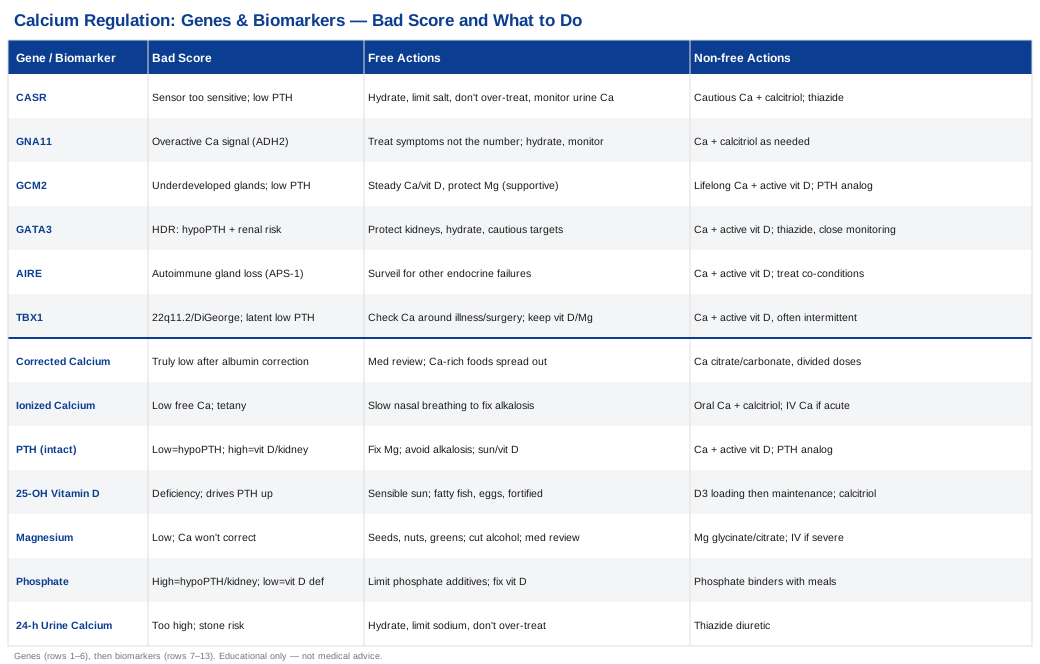
The 7 Biomarkers That Explain Why Your Calcium Is Low
Blood calcium is one of the most tightly defended numbers in your body, held within a narrow band by a feedback loop between your parathyroid glands, your kidneys, your bones, and the vitamin D system. When calcium drifts low, the useful question is never just "how low" but "which part of the loop failed." The following seven markers, read together, answer that question. As a foundation, the clinical picture and causes of low calcium are well summarized in the StatPearls review of hypocalcemia, which underlies much of the framing below.
Track these as a panel, not in isolation. A single calcium value tells you almost nothing on its own; the same 8.2 mg/dL means completely different things depending on what PTH, magnesium, and vitamin D are doing alongside it.
1. Albumin-Corrected Total Calcium
Why it matters. Roughly half of the calcium in your blood is bound to the protein albumin. A standard "total calcium" test measures both bound and free calcium, so if your albumin is low (common in illness, malnutrition, or liver disease), your total calcium reading will look falsely low even when the biologically active calcium is fine. Correcting for albumin prevents both false alarms and missed diagnoses. A widely used correction adds 0.8 mg/dL to the measured calcium for every 1.0 g/dL that albumin sits below 4.0.
How to measure it. It comes on any basic metabolic panel or comprehensive metabolic panel, which reports both calcium and albumin. Cost is roughly $10–$40 without insurance, and it is often bundled into routine bloodwork you already get.
If the score is bad — the plan without supplements. First confirm it is truly low by correcting for albumin and, ideally, checking ionized calcium (below). Review medications that lower calcium — proton pump inhibitors, loop diuretics, bisphosphonates, and some anticonvulsants — with your prescriber. Increase calcium-rich whole foods: dairy, canned sardines and salmon with bones, tofu set with calcium, leafy greens, and fortified foods. Space these across the day, since absorption is best in doses under about 500 mg at a time.
If the score is bad — the plan with supplements or equipment. Oral calcium is the workhorse. Calcium citrate (about 500 mg elemental, once or twice daily) absorbs well with or without food and is gentler if you have low stomach acid; calcium carbonate is cheaper but needs food and can cause bloating and constipation. Frequency is typically one to three divided doses daily; there is no "cycling" for maintenance, but doses are titrated to your labs. Side effects to watch: constipation, gas, kidney stones, and — if overdone — high urine calcium. Never push high-dose calcium without checking urine calcium (marker 7).
2. Ionized Calcium
Why it matters. Ionized (free) calcium is the fraction your nerves, muscles, and heart actually respond to. It is the most accurate measure of your true calcium status and does not depend on albumin. When symptoms and total calcium disagree, ionized calcium settles the argument. It also shifts with blood pH — alkalosis (for example, from hyperventilation) drives more calcium onto albumin and can trigger tetany symptoms even when total calcium is normal.
How to measure it. A dedicated ionized calcium test, usually run on fresh, carefully handled blood. Cost is roughly $30–$90. Because sample handling affects the result, it is often done in a clinic or hospital lab rather than a mail-in kit.
If the score is bad — the plan without supplements. If you get tingling and cramps during stress or panic, learn to recognize hyperventilation as a trigger and use slow nasal breathing to correct the alkalosis (see the breathing section later). Otherwise, the food and medication-review steps from marker 1 apply.
If the score is bad — the plan with supplements or equipment. Persistently low ionized calcium with symptoms is a medical situation, not a supplement experiment. Standard therapy pairs oral calcium with an active vitamin D compound (calcitriol) under supervision. Acute, symptomatic drops (carpopedal spasm, seizures, cardiac changes) require intravenous calcium in a monitored setting — this is the one biomarker whose "bad score" can be an emergency.
3. Parathyroid Hormone (Intact PTH)
Why it matters. PTH is the master regulator of calcium. When calcium falls, healthy parathyroid glands should raise PTH to pull calcium from bone, reclaim it in the kidney, and activate vitamin D. So the combination of low calcium with PTH tells you the mechanism: low calcium with low or "inappropriately normal" PTH points to hypoparathyroidism (the gland is not responding), while low calcium with high PTH points to vitamin D deficiency, kidney disease, or resistance to PTH. This single pairing is the most informative test in the whole panel.
How to measure it. An intact PTH blood test, ideally drawn at the same time as calcium so the pair can be interpreted together. Cost is roughly $40–$100.
If the score is bad — the plan without supplements. There is no food that "raises PTH" directly, but you can remove suppressors: correct low magnesium (marker 5), which is required for PTH to be secreted at all, and avoid the respiratory alkalosis that blunts calcium signaling. If PTH is high because of vitamin D deficiency, sunlight and dietary vitamin D address the upstream cause.
If the score is bad — the plan with supplements or equipment. True hypoparathyroidism (low PTH) is managed with calcium plus active vitamin D, and in selected patients with PTH analog therapy (recombinant PTH). A meta-analysis comparing PTH replacement versus oral calcium and active vitamin D found PTH therapy can reduce supplement burden but is reserved for those poorly controlled on standard therapy. These are prescription treatments with real monitoring needs — frequency and dose are set by your endocrinologist, and side effects include swings toward high calcium.
4. 25-Hydroxyvitamin D
Why it matters. Vitamin D is what lets your gut absorb calcium in the first place. Low 25-hydroxyvitamin D is one of the most common and most fixable causes of low calcium, and it typically drives PTH upward (secondary hyperparathyroidism). The relationship is well documented: PTH rises as vitamin D falls, as shown in this study of the PTH response to vitamin D deficiency in healthy individuals.
How to measure it. The 25-hydroxyvitamin D blood test is the correct one to order (not 1,25-dihydroxyvitamin D for routine screening). Cost is roughly $30–$70, and reliable at-home finger-prick kits exist in a similar range.
If the score is bad — the plan without supplements. Sensible sun exposure (short, regular, non-burning) supports natural production. Add dietary sources: fatty fish, egg yolks, and fortified foods. These help maintain a decent level but rarely correct a genuine deficiency on their own.
If the score is bad — the plan with supplements or equipment. Vitamin D3 (cholecalciferol) is the standard. A common repletion approach is a higher daily or weekly dose for 8–12 weeks, followed by a lower maintenance dose, with retesting to guide the target. Take it with a fat-containing meal for absorption. Cycling is not needed for maintenance, but the loading phase is time-limited by design. Side effects are rare at sensible doses; excessive, unmonitored high-dose vitamin D can cause calcium to climb too high. In hypoparathyroidism, plain D3 is often insufficient and calcitriol (active vitamin D) is used instead, because the failing step is the activation of vitamin D itself.
5. Magnesium (Serum and RBC)
Why it matters. This is the marker most often missed. Magnesium is required both for your parathyroid glands to release PTH and for your tissues to respond to it. When magnesium is very low, you can develop hypocalcemia that simply will not correct with calcium and vitamin D until magnesium is restored — a phenomenon described in the report on suppressed PTH in severe magnesium depletion and in a broader review of inadequate PTH secretion from hypomagnesemia. If your calcium refuses to budge, check magnesium.
How to measure it. Serum magnesium is cheap and routine (roughly $10–$40) but underestimates true status because most magnesium is inside cells. A red blood cell (RBC) magnesium test (roughly $50–$100) reflects intracellular stores more faithfully and is worth requesting if serum is "low-normal" but symptoms persist.
If the score is bad — the plan without supplements. Prioritize magnesium-rich foods: pumpkin seeds, almonds, cashews, spinach, black beans, and dark chocolate. Reduce heavy alcohol intake, and review medications that waste magnesium — proton pump inhibitors and loop/thiazide diuretics are common culprits.
If the score is bad — the plan with supplements or equipment. Use a well-absorbed form such as magnesium glycinate or citrate (commonly 200–400 mg elemental daily, in divided doses). The main side effect is loose stools, which is dose-limiting — back off if that happens. No cycling is needed, but avoid oral magnesium if you have significant kidney impairment, since excretion is reduced. Severe, symptomatic depletion is corrected intravenously in a clinical setting, after which calcium often normalizes on its own.
6. Serum Phosphate
Why it matters. Calcium and phosphate move in a see-saw. Low calcium combined with high phosphate is a fingerprint of hypoparathyroidism or kidney failure, whereas low calcium with low phosphate points toward vitamin D deficiency. Phosphate therefore helps you distinguish which mechanism is driving the calcium down, and it is a critical safety number, because giving vitamin D while phosphate is high can worsen soft-tissue calcification.
How to measure it. Included on a comprehensive metabolic panel or ordered as a standalone phosphate test, roughly $10–$40.
If the score is bad — the plan without supplements. If phosphate is high, moderate very high-phosphate processed foods and cola-type soft drinks (which use phosphate additives), and address the underlying kidney or parathyroid cause. If phosphate is low alongside low calcium, the fix is usually correcting vitamin D, not adding phosphate.
If the score is bad — the plan with supplements or equipment. High phosphate in the setting of hypoparathyroidism or kidney disease may call for phosphate binders taken with meals and a controlled-phosphate diet, under medical supervision. These are prescription-guided; side effects depend on the binder used. This is a marker to interpret with a clinician rather than self-manage.
7. 24-Hour Urine Calcium
Why it matters. Once you start treating low calcium, the danger shifts. Because people with hypoparathyroidism lack the PTH that normally reclaims calcium in the kidney, treatment can push large amounts of calcium into the urine, raising the risk of kidney stones and kidney damage — sometimes while blood calcium still looks only low-normal. Tracking urine calcium is how you treat safely rather than blindly. Standard care aims to keep blood calcium in the low-normal range precisely to avoid excess urinary calcium, as detailed in reviews of treatment options in hypoparathyroidism.
How to measure it. A 24-hour urine collection analyzed for calcium, roughly $40–$120. It is periodic monitoring rather than a one-off; typically checked at least yearly, or within weeks of a treatment change.
If the score is bad (too high) — the plan without supplements. Ensure adequate hydration, moderate sodium (high salt drives calcium into urine), and avoid over-treating your blood calcium — the target is the low end of normal, not the middle. Reassess whether your calcium and vitamin D doses are higher than needed.
If the score is bad (too high) — the plan with supplements or equipment. A thiazide diuretic is the classic tool: it reduces urinary calcium loss and is prescribed specifically for this purpose in hypoparathyroidism. It is taken daily under monitoring; side effects include low potassium, low sodium, and dehydration, so electrolytes are tracked. This is a good example of "equipment" (in the form of a targeted medication) solving a problem that supplements alone cannot.
With the measurable picture in hand, it helps to understand what can bias these numbers from birth — which is where genetics comes in.
The 6 Genes Behind Low Calcium — and How Each Is Compensated For
Most hypocalcemia is acquired, but a meaningful minority is written into the genome, especially when low calcium starts young, runs in families, or refuses to make sense. This is exactly the territory that genomics researchers like Ali Torkamani argue we should read more carefully, and that popularizers like Gary Brecka frame as "know your blueprint before you optimize." A broad, well-organized overview of these conditions is the review of genetic disorders of parathyroid development and function, which anchors the summaries below. One honest caveat up front: these are, in most cases, uncommon single-gene disorders diagnosed clinically and genetically — the "plans" here are about managing the downstream calcium problem the gene creates, because you cannot yet edit the gene itself.
1. CASR — the Calcium Thermostat Set Too Sensitive
The CASR gene encodes the calcium-sensing receptor, the sensor your parathyroid glands use to read blood calcium. Activating (gain-of-function) mutations make the thermostat too sensitive, so the glands "think" calcium is high and hold back PTH, producing autosomal dominant hypocalcemia type 1 — as described in this CASR mutation case study and literature review. It often causes mild low calcium with a tendency to spill calcium into the urine.
If the gene is bad — the plan without supplements. Because these individuals are prone to hypercalciuria and kidney stones, the emphasis is on not over-treating: keep well hydrated, moderate sodium, treat only enough to relieve symptoms rather than to fully normalize blood calcium, and monitor 24-hour urine calcium closely.
If the gene is bad — the plan with supplements or equipment. Calcium and active vitamin D (calcitriol) are used cautiously, and a thiazide diuretic is often added to limit urinary calcium loss. Dosing is titrated to keep calcium at the low end of normal; frequency is daily with periodic urine monitoring, and the main risk is triggering kidney stones by aiming too high.
2. GNA11 — the Sensor's Downstream Amplifier
GNA11 encodes a G-protein that relays the calcium-sensing receptor's signal inside the cell. Gain-of-function variants cause autosomal dominant hypocalcemia type 2, a close cousin of the CASR disorder; a large screen described in the report on GNA11 variants in hypercalcemia and hypocalcemia found affected patients tend to have normal magnesium and less hypercalciuria than the CASR group.
If the gene is bad — the plan without supplements. Management mirrors CASR: treat symptoms rather than the number, hydrate, and monitor. Because urinary calcium tends to be less of a problem here, the tightrope is a little wider, but the principle of gentle, symptom-guided treatment stands.
If the gene is bad — the plan with supplements or equipment. Calcium plus calcitriol as needed, dosed to relieve tingling and cramps while keeping blood calcium low-normal. As with CASR, over-correction is the enemy, and periodic labs guide the dose rather than a fixed prescription.
3. GCM2 — the Gland That Never Fully Formed
GCM2 is a master transcription factor for parathyroid development. Loss-of-function variants can leave the glands underdeveloped, causing familial isolated hypoparathyroidism with low PTH and low calcium. The compensation problem here is simple: there is too little PTH to do its job.
If the gene is bad — the plan without supplements. Non-supplement steps are supportive only — maintaining steady dietary calcium and vitamin D, protecting magnesium status, and avoiding hyperventilation-driven symptom flares. They cannot replace the missing hormone.
If the gene is bad — the plan with supplements or equipment. This is lifelong calcium plus active vitamin D, and in poorly controlled cases, PTH analog therapy — the same framework validated in the hypoparathyroidism treatment literature. Doses are individualized and taken daily; monitoring includes blood calcium, phosphate, and urine calcium to keep the treatment safe over decades.
4. GATA3 — the Trio of Parathyroid, Hearing, and Kidney
GATA3 mutations cause HDR (Barakat) syndrome — hypoparathyroidism, sensorineural deafness, and renal dysplasia — because the gene is needed to build all three organs. Over 90 percent of affected people have hypoparathyroidism, per the parathyroid-development review cited above. The calcium problem is a genuine PTH deficiency, complicated by the fact that the kidneys may already be vulnerable.
If the gene is bad — the plan without supplements. Protect the kidneys: careful hydration, blood pressure control, and cautious calcium targets, since kidney involvement raises the stakes of any hypercalciuria. Hearing support and regular kidney monitoring are part of the bigger picture.
If the gene is bad — the plan with supplements or equipment. Calcium and active vitamin D, dosed conservatively with close urine-calcium and kidney monitoring given the renal risk. A thiazide may help limit urinary calcium loss. Frequency is daily with more frequent lab checks than usual because the kidneys have less margin for error.
5. AIRE — When the Immune System Attacks the Gland
AIRE mutations cause autoimmune polyendocrine syndrome type 1 (APS-1), in which the immune system destroys the parathyroid glands (among others). Hypoparathyroidism is one of the most common features, as shown in the analysis of the spectrum of AIRE mutations causing APS-1. Here the low calcium is a consequence of autoimmune gland loss, often alongside adrenal and other endocrine problems.
If the gene is bad — the plan without supplements. The non-supplement priority is surveillance: because APS-1 unfolds over years, regular screening for the associated adrenal, thyroid, and other endocrine failures allows early treatment. Steady nutrition and symptom awareness support the calcium side.
If the gene is bad — the plan with supplements or equipment. Calcium and active vitamin D for the hypoparathyroidism, coordinated with treatment of the other autoimmune deficiencies (which can change calcium dynamics — for example, treating adrenal insufficiency). This demands a team approach, with dosing and frequency set against the whole endocrine picture rather than calcium alone.
6. TBX1 — the 22q11.2 (DiGeorge) Connection
TBX1 is the gene most responsible for the parathyroid features of 22q11.2 deletion syndrome (DiGeorge). Affected people may have underdeveloped parathyroid glands, causing hypocalcemia that can appear in infancy or emerge later under stress, illness, or surgery. The calcium problem ranges from latent to overt.
If the gene is bad — the plan without supplements. Anticipation is the strategy: knowing the vulnerability exists means checking calcium proactively around illness, pregnancy, and surgery, when a borderline gland can tip into symptomatic hypocalcemia. Maintaining vitamin D and magnesium sufficiency reduces the odds of a crisis.
If the gene is bad — the plan with supplements or equipment. Calcium and active vitamin D during hypocalcemic periods, sometimes only intermittently if the deficiency is latent, and more consistently if the gland cannot keep up. Because needs vary over a lifetime, this is one gene where treatment genuinely cycles with circumstance rather than running at a fixed dose.
Knowing your labs and your genetic risk is powerful, but it helps to see how the newest thinking about micronutrients ties these threads together — which the next section does.
The Micronutrient Conversation That Reframes Calcium: Huberman Lab with Dr. Rhonda Patrick
One of the most study-dense discussions of the nutrients that quietly govern calcium is the Huberman Lab episode with Dr. Rhonda Patrick on micronutrients for health and longevity. It is not a "hypocalcemia" episode, but it repeatedly circles the exact levers — vitamin D, magnesium, and the way "normal" lab ranges can hide functional shortfalls — that decide whether your calcium loop runs smoothly. Here are ten takeaways most relevant to low calcium, one idea per point.
1. "Normal" Is Not the Same as "Optimal"
A recurring theme is that population reference ranges are built around avoiding overt disease, not around ideal function. A vitamin D or magnesium level that is technically "in range" can still be too low for your parathyroid–calcium system to work its best — which is why symptoms can persist despite "normal" labs.2. Vitamin D Behaves Like a Hormone, Not Just a Vitamin
Vitamin D is converted into a steroid hormone that regulates a large slice of the genome, including genes involved in calcium transport. This reframes vitamin D repletion from "topping up a nutrient" to "restoring a signaling system" — and explains why deficiency ripples so widely.3. Magnesium Is the Silent Partner of Calcium
The discussion emphasizes how common subclinical magnesium insufficiency is, and how magnesium sits upstream of countless enzymatic reactions. For calcium specifically, magnesium is the cofactor without which PTH cannot be secreted or heard — the practical reason "calcium won't correct until magnesium does."4. The "Triage Theory" of Micronutrients
A central concept is that when a nutrient is scarce, the body prioritizes short-term survival over long-term maintenance. Mild, chronic shortfalls therefore cause slow damage that does not show up as a dramatic lab abnormality — a useful mental model for why low-grade deficiencies matter.5. Food Matrix Beats Isolated Doses
Nutrients from whole foods often come packaged with cofactors that improve their use. For calcium and its helpers, this argues for building a food foundation first, then supplementing to fill genuine gaps rather than relying on pills alone.6. Vitamin D Needs Its Cofactors
The episode underscores that vitamin D does not act alone — magnesium is required to activate it, and adequate status of related nutrients shapes where calcium ultimately goes. Repleting vitamin D while ignoring magnesium is a half-measure.7. Sun Exposure Is a Real Input
Sensible sunlight remains a meaningful driver of vitamin D status, with the caveat that skin tone, latitude, season, and age all change how much you make. For anyone with borderline calcium, this is a free, controllable lever worth using.8. Genetics Personalize Your Needs
Common gene variants can change how efficiently you convert and use nutrients like vitamin D and folate, meaning two people can eat identically and end up with different blood levels. This dovetails with the genetics section: your baseline needs are partly inherited.9. Test, Don't Guess
A consistent practical thread is measuring your levels and adjusting, rather than assuming. For calcium regulation, that means checking vitamin D and magnesium, not just calcium, before deciding what to change.10. Small, Consistent Corrections Compound
The optimistic close is that these systems are responsive: modest, sustained improvements in vitamin D, magnesium, and diet tend to pay off over months. For a reversible problem like most hypocalcemia, that is exactly the message that matters.Ideas are only as good as their execution, so it is worth closing with hands-on practices that can ease symptoms while you correct the underlying chemistry.
Complementary Approaches That Can Ease the Symptoms
Complementary practices do not raise blood calcium, and it would be dishonest to suggest otherwise. What some of them can do — with reasonable physiological rationale and, in a few cases, supportive human evidence — is reduce the symptoms of low calcium (tingling, cramps, tetany-like sensations) and help you cope with a chronic condition. The evidence here is more mechanistic and indirect than the biomarker section, and I will flag that honestly for each one.
Breathing-Based Therapies
The classic trigger that turns borderline calcium into visible symptoms is hyperventilation. Breathing too fast lowers carbon dioxide and raises blood pH, which pushes calcium onto albumin and drops the active, ionized fraction — producing the tingling and carpopedal spasm of tetany even when total calcium is only mildly low. Breathing practices target this directly.
The specific technique is slow, nasal, diaphragmatic breathing (for example, extending the exhale so that breaths slow to roughly six per minute), which reverses respiratory alkalosis and restores ionized calcium toward normal. This is the same principle behind the long-standing clinical observation that hyperventilation provokes latent tetany and that slowing the breath relieves it; the relationship between alkalosis and ionized calcium is well established in the physiology described in the hypocalcemia literature above.
Practically, if you notice tingling around the mouth or in the fingers during stress or anxiety, treat it as a cue to slow your breathing rather than breathe harder. It is safe, free, and immediately available — but it manages an acute symptom, not the cause, so it belongs alongside, not instead of, correcting your labs.
Relaxation Training and Progressive Muscle Relaxation
Because anxiety drives the over-breathing that precipitates tetany symptoms, techniques that lower physiological arousal can indirectly reduce symptom flares. Progressive muscle relaxation — systematically tensing and releasing muscle groups — and general relaxation training both dampen the sympathetic "stress" response that fuels hyperventilation.
The supporting evidence here is strongest as a general anxiety and stress intervention rather than as a hypocalcemia treatment specifically; relaxation training has broad human trial support for reducing anxiety symptoms, which is the mechanism most relevant to symptom-triggered tetany. I am flagging clearly that direct hypocalcemia trials are lacking, so this is a reasonable adjunct, not a proven calcium therapy.
To apply it, practice a short daily routine (10–15 minutes) so the skill is available when symptoms threaten, and pair it with the breathing technique above. There is essentially no downside, which makes it a sensible low-risk addition for anyone whose symptoms cluster around stressful moments.
Tai Chi
Many people with chronic low calcium also carry low vitamin D and reduced bone strength, which raises the risk of falls and fractures. Tai chi is relevant here not because it changes calcium chemistry but because it is one of the better-studied movement practices for balance and fall prevention in older and deficiency-prone adults.
The human evidence for tai chi improving balance and reducing fall risk is reasonably robust in older-adult populations, and it is frequently recommended in that context; its benefit for hypocalcemia is entirely indirect, through protecting a skeleton that low calcium and vitamin D may have weakened. That distinction matters, and I want to be explicit that no trial shows tai chi correcting a calcium level.
In practice, a twice-weekly class or guided routine is a gentle, sustainable way to build balance and lower-body strength, especially if your low calcium accompanies vitamin D deficiency or bone concerns. It is safe for most people and pairs naturally with the vitamin D and calcium work at the core of this article.
Mindfulness Meditation / MBSR
Living with a chronic, sometimes frightening symptom like tetany or recurrent cramps has a psychological cost, and heightened anxiety can worsen the hyperventilation cycle. Mindfulness-based stress reduction (MBSR) addresses the coping and arousal side of the equation.
The evidence base for MBSR is solid for reducing stress and anxiety across many chronic conditions, though again there is no hypocalcemia-specific trial — its relevance is through the well-documented anxiety pathway that intersects with symptom-triggered tetany. I am noting that limitation deliberately so expectations stay realistic.
Realistically, an 8-week MBSR course or a consistent app-based practice can lower baseline stress reactivity, making symptom flares less frequent and less alarming. Like the other approaches here, it is a supportive layer that makes the medical correction easier to live with, not a substitute for it.
Conclusion
The single most useful shift you can make with hypocalcemia is to stop treating it as a calcium shortage and start treating it as a signal. The seven biomarkers in this article — corrected and ionized calcium, PTH, 25-hydroxyvitamin D, magnesium, phosphate, and urine calcium — turn a vague "low calcium" into a specific, addressable mechanism, and the six genes explain the cases where that mechanism was partly set at birth. In the great majority of situations, once you identify why calcium is low, the correction is both possible and durable.
The practical throughline is consistent: fix magnesium and vitamin D before assuming you need more calcium, interpret PTH alongside calcium rather than alone, treat to the low end of normal to protect your kidneys, and use breathing and relaxation to manage symptoms while the chemistry is being corrected.
Your next smart step is small and concrete. Order the biomarker panel described above (or dig out recent results and re-read them as a set), note the pattern of symptoms and their triggers, and bring both to a qualified clinician who can interpret the calcium–PTH–magnesium picture with you. Better information really does lead to better decisions — and with a condition this responsive to the right lever, that information is often the difference between guessing and getting well.
This article is for general education and is not medical advice. Symptomatic or severe hypocalcemia can be a medical emergency; always discuss testing and treatment with a qualified healthcare professional before changing supplements or medications.