This article contains AI generated content.
· UpdatedHypochondroplasia: 2 Genes and 6 Biomarkers to Track
If you or your child has been diagnosed with hypochondroplasia, you have probably already noticed a gap between the paperwork you were handed at diagnosis and the questions that actually keep you up at night. The clinical summary explains what the condition is called. It rarely explains what to check next, how often, or which numbers actually change the plan.
Generic short-stature advice makes this worse, not better. Most of it is written for garden-variety constitutional short stature or growth hormone deficiency, conditions with a very different biology from a skeletal dysplasia driven by an overactive growth-plate receptor. Following that advice literally, chasing higher growth hormone levels as if the bottleneck were hormone supply, misses the actual mechanism and can create false reassurance or false alarm at the wrong moments.
A more useful approach starts from the actual biology: which gene is involved, what that gene does at the growth plate, and which specific measurements tell you whether things are on track. That is a narrower, more technical conversation than most parenting forums have, but it is also the one that lets you ask sharper questions at the genetics and endocrinology appointments you already have scheduled.
None of this replaces your medical team, and there is no version of this condition that gets "reversed" by information alone. But better information changes which questions get asked, which tests get ordered at the right time, and which treatment conversations happen before a growth window closes rather than after. This article walks through the two genes with the clearest evidence in hypochondroplasia, the six measurements worth tracking over time, a physiology detour that reframes what growth hormone can and cannot do here, and a short, honest look at complementary approaches that have real (if indirect) supporting evidence.
Summary
Roughly seven in ten cases of hypochondroplasia trace back to a specific, well-characterized variant in a single gene, FGFR3, which acts as a brake on cartilage growth at the growth plate. A smaller, less certain slice of families shows linkage to the IGF1 region, and for about three in ten people, no gene is found at all yet, which changes what a "management plan" can realistically promise. Below, each gene gets its own breakdown of what it does, how it is tested, and what a monitoring and treatment plan looks like with and without prescription-level intervention.
Beyond the genetics, six specific measurements, from growth velocity to spinal imaging, form the backbone of ongoing surveillance, and each one has its own cost range, testing cadence, and action plan when the number comes back outside the expected range. A closer look at the growth hormone axis itself, drawn from a well-known physiology deep dive, also reframes a common misconception: in hypochondroplasia, the limiting factor usually isn't how much growth hormone is available, it's how the growth plate responds to it. Finally, a short section covers which complementary approaches have any real human evidence worth knowing about, and which don't.
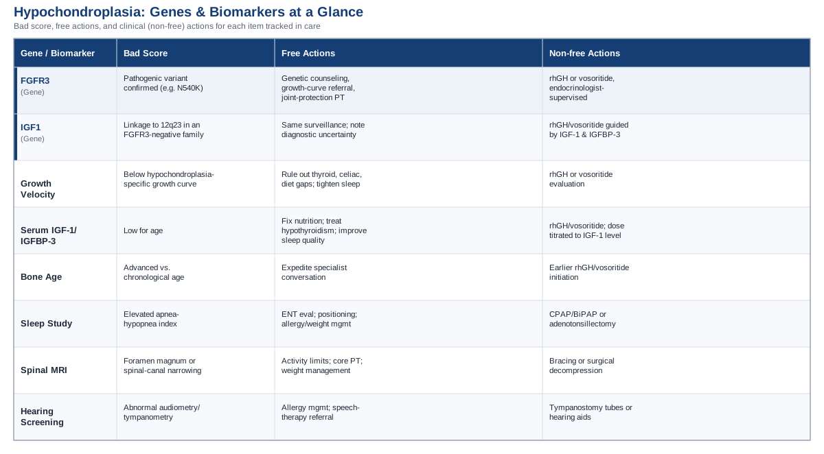
The Genes Behind Hypochondroplasia
Hypochondroplasia is a skeletal dysplasia, which is a different category of condition from the multifactorial, lifestyle-modifiable conditions that most "genes and biomarkers" content is written about. It is not caused by a mix of many small genetic risk factors interacting with diet and exercise. In most people, it comes down to a single, specific change in a single gene that alters how cartilage cells behave at the growth plate. That difference matters for how you should think about "management": there is no set of habits that offsets a growth-plate signaling defect the way exercise might offset an unfavorable cholesterol variant. What genetics testing offers here is precision about diagnosis, recurrence risk, and which medical interventions are biologically appropriate, not a menu of lifestyle levers.
How the Diagnosis Gets Confirmed
Hypochondroplasia is usually suspected clinically first, based on disproportionate short stature, a stocky build, short and broad hands and feet, mild joint laxity, and a head circumference that is large relative to height. The clinical picture overlaps with achondroplasia and several other skeletal dysplasias, which is exactly why molecular confirmation matters: it distinguishes hypochondroplasia from its look-alikes and gives a concrete answer for genetic counseling. According to GeneReviews' clinical summary of hypochondroplasia, testing typically starts with targeted analysis of the known FGFR3 hot-spot variants, and moves to a multigene skeletal dysplasia panel or exome/genome sequencing if that first test is negative and the clinical picture still fits.
This matters practically because a negative single-gene test is not the end of the story. It changes the next test ordered, not the diagnosis itself, if the clinical and radiographic picture remains convincing.
FGFR3 — The Gene Behind Most Cases
FGFR3 (fibroblast growth factor receptor 3) codes for a receptor that sits on the surface of cartilage cells and acts as a brake on their growth. In healthy growth plates, FGFR3 signaling is switched on just enough to keep chondrocyte proliferation in check as bones lengthen. In hypochondroplasia, a heterozygous gain-of-function variant, most commonly the p.Asn540Lys (N540K) change in the receptor's tyrosine kinase domain, keeps that brake partially engaged all the time. The cartilage cells that should be multiplying and organizing into new bone are held back, which produces the disproportionately short, stocky limb pattern seen in the condition. This is a milder version of the same mechanism that causes achondroplasia, where a different, more disruptive FGFR3 variant (G380R) produces a more severe phenotype, as detailed in the GeneReviews entry on achondroplasia.
FGFR3 variants account for roughly seven in ten diagnosed cases of hypochondroplasia. The condition is autosomal dominant, meaning one altered copy of the gene is enough to produce the phenotype. Most cases arise from a new (de novo) variant in a child with unaffected parents, but when a parent carries the variant, each child has a fifty percent chance of inheriting it.
If the FGFR3 Variant Is Confirmed: The Plan Without Supplements or Equipment
The first, non-pharmacological layer of management is about structure, not correction. Genetic counseling clarifies recurrence risk for future pregnancies and helps extended family decide whether testing makes sense for them. A referral to a pediatric genetics or skeletal dysplasia clinic ensures growth is plotted on hypochondroplasia-appropriate (rather than general population) growth curves, since a "flat" trajectory on a standard chart can look alarming when it is actually typical for the condition.
On the nutrition side, the goal is sufficiency, not maximization: age-appropriate protein and calcium intake, and a vitamin D level confirmed by blood test rather than assumed. There is no evidence that megadosing any nutrient improves growth-plate output in FGFR3-driven short stature, and excess vitamin D or calcium carries its own risks. Weight management deserves more attention than it usually gets, because in a body plan with shorter limbs, excess weight loads the joints and spine more than it would in an average-proportioned body, and it can also obscure whether growth velocity is genuinely slowing or just look that way on a chart. A structured physical therapy program, typically one to two sessions per week with a home routine in between, focused on core stability and joint protection, helps manage the mild joint laxity and lumbar lordosis that are common in this condition. None of this changes the underlying receptor biology, but it reduces the number of downstream problems that compound on top of it, and it keeps the surveillance schedule (annual genetics and endocrinology follow-up, with imaging added as flagged) running on time.
If Growth Remains Limited: The Plan With Supplements or Equipment
Everything in this section is prescription-only and requires ongoing supervision by a pediatric endocrinologist; none of it is something to start independently.
Recombinant human growth hormone (rhGH) is the most studied pharmacological option in hypochondroplasia, though the evidence base is smaller and older than the equivalent literature for growth hormone deficiency. A meta-analysis of height outcomes after rhGH treatment in children with hypochondroplasia found a clear first-year catch-up in growth velocity that diminishes in subsequent years, with final adult height remaining below average despite treatment. Dosing in the reviewed studies clustered around 0.25 mg/kg per week, given as daily subcutaneous injections, typically in the evening to approximate the body's natural nocturnal growth hormone pulse. There is no legitimate cycling protocol for pediatric rhGH the way there might be in other performance-oriented contexts; treatment is continuous and daily, adjusted by dose rather than by on/off periods, and continued for as long as growth potential remains and the response justifies it, based on data such as that in a long-term follow-up study of growth hormone therapy in hypochondroplasia. Side effects to expect and monitor for include injection-site reactions, transient fluid retention and joint discomfort, headache, and, rarely, benign intracranial hypertension; bloodwork tracking IGF-1 every three to six months keeps the dose from drifting into supraphysiologic territory.
Vosoritide, a C-type natriuretic peptide analog given as a daily subcutaneous injection, is approved for achondroplasia and has more recently been studied specifically in hypochondroplasia. A phase 2 trial of vosoritide in children with hypochondroplasia found an increase in annualized growth velocity of roughly 1.8 cm per year compared with each child's own pre-treatment baseline, with a good safety profile; injection-site reactions were common (over 80% of participants) and transient drops in blood pressure shortly after dosing were monitored but not treatment-limiting. It's worth being precise here: this is trial-stage evidence for hypochondroplasia specifically, so outside of a study, access currently depends on a specialist's judgment about off-label use rather than a standard, universally available prescription.
For adolescents or adults with more limited response to medical therapy, surgical limb-lengthening with external fixation equipment is a separate, more invasive option, generally considered later and only in coordination with an orthopedic surgeon experienced in skeletal dysplasia. It carries a longer recovery and a different risk profile than any of the above and is a decision made case by case, not a default next step.
IGF1 — A Weaker Signal in a Subset of Families
Not every case of hypochondroplasia traces back to FGFR3, and the search for the remaining genetic cause has an interesting, if inconclusive, thread. In the early 1990s, linkage studies in a small number of multi-generation families without an FGFR3 variant found that hypochondroplasia co-segregated with markers near the IGF1 gene locus on chromosome 12q23, as described in the original genetic linkage study of IGF1 and hypochondroplasia. This is worth knowing about, but it should be held loosely: it is old evidence, based on a handful of families, and it has not been confirmed as a direct causal mechanism by modern sequencing methods. It suggests the IGF-1 axis may play a role in a genetic subgroup, not that IGF1 is a confirmed second gene for hypochondroplasia the way FGFR3 is.
If This Family Pattern Applies: The Plan Without Supplements or Equipment
The practical management for this subgroup does not differ meaningfully from the FGFR3 pathway at the lifestyle level: the same surveillance schedule, the same nutrition-sufficiency approach, the same physical therapy for joint protection. What does change is the framing of one specific blood test, IGF-1 itself, which becomes doubly informative here, both as a general endocrine marker and as a potential clue to the underlying biology in families where FGFR3 testing has come back negative. Genetic counseling should note the uncertainty explicitly rather than presenting IGF1 as a settled diagnosis.
If Growth Monitoring Suggests a Problem: The Plan With Supplements or Equipment
The pharmacological options are the same as described for FGFR3-related cases, rhGH or, in trial settings, vosoritide, since treatment decisions in practice are driven by growth pattern and endocrine testing rather than by which specific genetic subgroup a family falls into. The one meaningful difference is that in families with this linkage pattern, baseline and follow-up IGF-1 and IGFBP-3 levels are worth discussing with the endocrinologist as part of the initial workup, since they may carry more diagnostic weight here than in a straightforward FGFR3-confirmed case.
The Genes We Haven't Found Yet
For close to a third of people with a clinical and radiographic diagnosis of hypochondroplasia, comprehensive FGFR3 testing comes back negative, and no other specific gene has been consistently identified across families. This is described as genetic heterogeneity: the same clinical picture likely arises from variants in more than one gene, most of which haven't been mapped yet. If you're in this group, it does not change your child's clinical management, which is driven by growth pattern and physical findings, not by which gene is confirmed. It does mean genetic counseling has to be a little more honest about uncertainty in recurrence risk, and it's a reasonable point to ask about enrolling in a research registry or exome/genome sequencing study, both because it may eventually yield an answer for your family and because it contributes to the pool of cases that will eventually identify the remaining genes.
The genetics, in other words, explain the mechanism and shape which treatments are biologically sensible. The next layer, ongoing measurement, is what actually tells you and your medical team whether the plan in place is working.
Biomarkers and Growth Parameters Worth Tracking
Because hypochondroplasia doesn't have a metabolic panel the way cardiovascular or metabolic conditions do, "biomarkers" here means a mix of blood tests, imaging, and functional measurements that together tell you whether growth, skeletal alignment, and related systems are on the expected track for this specific condition. Each one below has a distinct reason for being tracked, a realistic cost range, and a different action plan depending on whether it comes back as expected or not.
Growth Velocity and Height Standard Deviation Score
A single height measurement tells you almost nothing on its own; the trend over time is what matters. Growth velocity, plotted against a hypochondroplasia-specific reference curve rather than a general population chart, is the single most useful ongoing measurement because it's what actually triggers escalation to medical therapy or additional workup.
How to measure it: Standing height measured at routine pediatric visits every three to six months, ideally by the same method and, when possible, the same measurer, to reduce noise. This is typically included in a standard visit at no extra cost; a dedicated growth-clinic evaluation runs roughly $100 to $300 out of pocket where insurance doesn't cover it.
If growth velocity is below expected, the plan without supplements or equipment: Before assuming the slowdown is purely the underlying condition, rule out unrelated but common causes of poor growth, thyroid dysfunction, celiac disease, chronic illness, and inadequate caloric intake. Tightening sleep habits and confirming adequate protein and calorie intake for age is a reasonable first step, and visit frequency is often increased to three-month intervals to get a clearer trend line.
If it's confirmed as a true slowdown, the plan with supplements or equipment: This is the trigger point for the rhGH or vosoritide conversation described above, timed against bone age (below) to make sure there is still enough growth window left for treatment to matter.
Serum IGF-1 and IGFBP-3
IGF-1 is the downstream messenger through which growth hormone exerts most of its growth-promoting effect, and IGFBP-3 is its main carrier protein. Together they give a more stable read on growth hormone axis activity than a single growth hormone level, which fluctuates in sharp pulses throughout the day and is much harder to interpret from a random blood draw.
How to measure it: A standard blood draw, no fasting typically required, costing roughly $100 to $250 if not bundled into a broader endocrine panel covered by insurance. This is checked at baseline for anyone being evaluated for growth-promoting therapy, then every three to six months during treatment to titrate dose safely.
If IGF-1 is low, the plan without supplements or equipment: Confirm nutritional adequacy, since chronic undernutrition suppresses IGF-1 independent of any underlying condition, and screen for and treat any coexisting hypothyroidism, which also lowers IGF-1. Prioritizing consistent, adequate sleep matters here too, since growth hormone release is concentrated in slow-wave sleep, and poor sleep quality blunts that pulse regardless of genetics.
If it's confirmed low despite addressing the above, the plan with supplements or equipment: This supports moving forward with prescription rhGH or, where appropriate and available, vosoritide, with IGF-1 itself used as the safety ceiling, dosing is adjusted specifically to avoid pushing IGF-1 above the normal range for age, since supraphysiologic exposure raises the risk of side effects without adding proportional benefit.
Bone Age (Hand and Wrist X-ray)
Bone age estimates skeletal maturity relative to chronological age by looking at growth plate ossification patterns in the hand and wrist. It matters because it estimates how much growth window remains, which directly affects the urgency and expected payoff of any growth-promoting treatment.
How to measure it: A single hand and wrist X-ray, read against standard reference atlases, costing roughly $100 to $300. It's typically repeated every one to two years while growth-related treatment decisions are active.
If bone age is advancing faster than chronological age, the plan without supplements or equipment: This is primarily a signal to move up the timeline of specialist conversations rather than something to address through habits, since a narrowing growth window is the main practical consequence.
If bone age is confirmed advanced and growth potential is genuinely limited, the plan with supplements or equipment: This finding is used to prioritize earlier initiation of rhGH or vosoritide discussions, since these therapies work by extending the productive growth period, not by working after growth plates have already fused.
Sleep Study for Obstructive Sleep Apnea
Skeletal dysplasias that affect cranial base and midface structure carry some risk of upper airway crowding. Per surveillance guidance in the GeneReviews hypochondroplasia summary, this complication is less common in hypochondroplasia than in achondroplasia, but it's still worth actively screening for, especially if snoring, restless sleep, or witnessed pauses in breathing are reported.
How to measure it: An in-lab polysomnography study runs roughly $1,000 to $3,000; a home sleep apnea test is a more affordable alternative at roughly $150 to $500, though it captures less detail and isn't appropriate for every case. Either requires a physician referral.
If the apnea-hypopnea index is elevated, the plan without supplements or equipment: An ENT evaluation for adenotonsillar hypertrophy, a common and treatable contributor, sleep positioning changes, allergy management, and weight management where relevant.
If it's confirmed and structural, the plan with supplements or equipment: CPAP or BiPAP therapy, titrated by a sleep specialist, or adenotonsillectomy surgery where an ENT identifies enlarged tonsils or adenoids as the primary driver, followed by a repeat sleep study to confirm resolution.
Craniocervical and Spinal MRI
Surveillance imaging of the area where the skull meets the spine (the foramen magnum) in infancy, and of the lumbar spine later in life, catches narrowing that can compress the spinal cord or nerve roots before it causes lasting neurologic symptoms. This risk is lower in hypochondroplasia than in achondroplasia but is still part of standard surveillance, particularly if there are clinical signs like leg weakness, altered reflexes, or new back and leg pain.
How to measure it: MRI of the brain and cervical spine in infancy if clinical signs warrant it, or lumbar spine MRI later if back or leg symptoms develop, typically costing $500 to $3,000 depending on region and insurance coverage.
If narrowing is found, the plan without supplements or equipment: Activity modification to avoid high-impact loading of the spine, physical therapy focused on core stability and maintaining a neutral lumbar position, and weight management to reduce mechanical load.
If it's progressive or symptomatic, the plan with supplements or equipment: Bracing in select cases, or surgical decompression coordinated with a neurosurgeon if there's evidence of progressive nerve compression; this is not a common outcome in hypochondroplasia specifically, but it's the reason the imaging surveillance exists.
Audiology and Hearing Screening
Recurrent middle ear involvement and speech-language delay are part of standard surveillance in skeletal dysplasias generally. Catching a hearing problem early protects speech and language development at an age when that development is happening quickly.
How to measure it: Audiometry and tympanometry through a pediatric audiologist or ENT, roughly $100 to $300, recommended if speech is delayed or ear infections are frequent.
If results are abnormal, the plan without supplements or equipment: Management of allergies or eustachian tube dysfunction, and a speech-language therapy referral if delay is already present.
If it's confirmed and persistent, the plan with supplements or equipment: Tympanostomy tubes for recurrent middle ear fluid, or hearing aids if a sensorineural component is identified, with follow-up audiology every six to twelve months to track progress.
Taken together, these six measurements form a surveillance rhythm rather than a one-time checklist, most of them repeat on a schedule tied to age and treatment status rather than a single baseline test. Understanding why growth hormone therapy works the way it does, and where its limits are, makes the growth-velocity and IGF-1 pieces of that rhythm easier to interpret.
What a Deep Dive Into Growth Hormone Physiology Actually Reveals
A lot of the anxiety around hypochondroplasia treatment options centers on growth hormone, and a lot of that anxiety is built on a subtly wrong assumption: that if a child just had more growth hormone, they'd grow more. The Huberman Lab episode on thyroid and growth hormone physiology is a useful, non-clinical resource for understanding how this hormone actually behaves in the body, and re-reading it with hypochondroplasia specifically in mind reframes several common assumptions. None of the following is medical advice or a substitute for endocrinology guidance; it's context that makes the numbers on a lab report easier to interpret.
1. Growth Hormone Is Released in Pulses, Not a Steady Stream
Growth hormone isn't secreted continuously; it's released in sharp pulses, the largest of which happens during the first few hours of deep, slow-wave sleep. A random daytime blood draw for growth hormone itself is often close to zero and tells you very little, which is exactly why IGF-1, a more stable downstream marker, is used for both diagnosis and monitoring instead.
2. Sleep Architecture Matters More Than Total Sleep Time
It's not just how many hours a child sleeps, it's how much of that sleep is deep, slow-wave sleep, when the largest growth hormone pulse occurs. Fragmented or shallow sleep can blunt this pulse even if total sleep duration looks adequate on paper, which is one more reason sleep quality surveillance, not just sleep apnea screening, belongs in the broader picture.
3. Eating Close to Bedtime Can Blunt the Nocturnal Pulse
Elevated blood glucose and insulin around bedtime appear to suppress the sleep-related growth hormone pulse. This is a minor, general physiology point, not a treatment for hypochondroplasia, but it's a reasonable, low-effort habit to keep in mind alongside everything else.
4. Resistance Exercise Triggers an Acute Release
Higher-intensity resistance exercise produces a short-term spike in circulating growth hormone. This is a normal physiological response in anyone, but it's worth being clear that a transient spike from exercise is a completely different thing from the sustained, receptor-level signaling needed to meaningfully change growth-plate output in a condition like hypochondroplasia.
5. Heat Exposure Has a Measurable, if Temporary, Effect
Sauna-type heat exposure has been shown to transiently raise growth hormone levels in adults. This finding comes from adult physiology research unrelated to pediatric skeletal dysplasia, and it isn't a pediatric growth intervention; it's included here only because it's one of the more surprising facts in the source material and helps illustrate how many things move growth hormone levels around without changing skeletal outcomes.
6. Chronic Stress Works Against the Whole System
Elevated cortisol from chronic stress interferes with growth hormone's downstream effects. For families managing a chronic diagnosis, this is a reasonable argument for taking psychosocial support seriously, not as a treatment for the condition, but as one more variable worth not making worse.
7. IGF-1 Is the More Meaningful Long-Term Marker
Because growth hormone itself is so pulsatile, IGF-1 is a far more useful number to track over months of treatment. This lines up directly with how IGF-1 is actually used in hypochondroplasia care, as the primary marker for both diagnosis support and treatment titration.
8. The Receptor Matters as Much as the Hormone
Hormone physiology only works if the tissue receiving the signal can respond to it. This is the single most important reframe for hypochondroplasia: the condition isn't a shortage of growth hormone signal, it's a growth plate whose receptor (FGFR3) is already stuck partially "on" as a brake, regardless of how much hormone or IGF-1 is circulating.
9. This Is Exactly Why More Growth Hormone Doesn't Simply Fix Hypochondroplasia
Because the limiting step in hypochondroplasia sits at the growth-plate receptor level rather than at hormone supply, pushing growth hormone or IGF-1 higher produces a real but partial and diminishing effect, as the treatment studies referenced earlier show, rather than a full correction. This is the point where general growth hormone physiology and hypochondroplasia-specific biology genuinely diverge, and it's worth internalizing before going into a treatment conversation expecting normalization rather than a modest, real improvement.
10. Supportive Physiology Still Matters Around Medical Treatment
None of this replaces rhGH or vosoritide when they're indicated. But keeping sleep quality, stress, and general metabolic health in reasonable shape gives whatever medical therapy is in use the best possible physiological backdrop to work against, rather than fighting an uphill battle against poor sleep or chronic stress on top of the underlying condition.
This physiology detour is useful mainly for interpreting lab reports and treatment conversations with clearer expectations. Alongside it, a small set of complementary approaches has real, if indirect, evidence worth knowing about for the day-to-day parts of living with the condition.
Complementary Approaches With Real Supporting Evidence
None of the following changes the underlying genetics or growth-plate biology. They're included because hypochondroplasia comes with real secondary issues, joint laxity, musculoskeletal discomfort, and the stress of frequent medical appointments and procedures, and a small number of complementary approaches have genuine human evidence for managing those specific issues, even though the evidence isn't drawn from hypochondroplasia populations directly.
Yoga and Guided Physical Activity for Joint Laxity
Mild joint laxity is a recognized feature of hypochondroplasia, and gentle, guided movement work is a reasonable, low-risk way to build the stabilizing strength around lax joints without over-relying on the joints' own ligamentous support.
The clearest supporting evidence comes not from yoga studies directly but from structured exercise research in joint hypermobility syndrome, a closely related musculoskeletal picture. A randomized controlled trial of exercise in children with joint hypermobility syndrome and knee pain found that children could exercise safely into a hypermobile range without worsening pain, with modest improvements in psychosocial scores. Yoga itself is not contraindicated in hypermobile individuals, but poses should be modified to avoid pushing joints into their end-range hyperextension, where instability risk is highest.
In practice, this means seeking out an instructor experienced with hypermobility, avoiding deep backbends or hyperextended knee/elbow poses, and treating yoga as a strength and body-awareness practice rather than a flexibility one. It should complement, not replace, any physical therapy already prescribed for the condition.
Massage Therapy for Musculoskeletal Comfort
Joint laxity and the altered biomechanics of a disproportionate skeleton can produce muscular compensation patterns and tightness, particularly around the lower back and hips, and massage therapy is a reasonably well-tolerated way to address that localized discomfort.
Direct condition-specific trials in hypochondroplasia don't exist, but massage therapy has a general, moderate evidence base for musculoskeletal pain relief, and it was among the modalities incorporated into the broader multidisciplinary program studied in the randomized trial of a multidisciplinary intervention for childhood joint hypermobility syndrome (the "Bendy" study), which found meaningful improvements in child- and parent-reported pain and function.
Realistically, this looks like a session every one to two weeks during periods of increased discomfort, delivered by a therapist briefed on the joint laxity and any spinal surveillance findings, rather than a standing indefinite routine. It's a comfort measure, not a treatment for the underlying condition or a substitute for imaging surveillance if pain is new or progressive.
Music Therapy for Procedural Anxiety
Children with hypochondroplasia go through more medical procedures than average, growth measurements, imaging, blood draws, and for some, injections, and procedural anxiety is a real, manageable problem rather than a minor inconvenience.
This is one of the better-evidenced entries on this list, though not specific to hypochondroplasia. A systematic review and meta-analysis of music interventions for pediatric pain and anxiety during medical procedures found consistent reductions in both pain perception and anxiety across multiple randomized trials, using simple recorded music delivered through headphones or a speaker.
Applying this is genuinely low-effort: playing a child's preferred music during blood draws, imaging appointments, or injections, ideally with headphones to reduce the sensory intensity of a clinical environment. It costs nothing, carries no risk, and stacks well with any other anxiety-reduction strategy a care team suggests.
Relaxation Training for Chronic Discomfort and Coping
For children managing ongoing joint discomfort or the general burden of a chronic diagnosis, structured relaxation training has a real, if general, evidence base worth knowing about.
A Cochrane review of psychological therapies for chronic and recurrent pain in children and adolescents found that relaxation training on its own produced meaningful reductions in pain intensity across conditions like chronic headache and recurrent abdominal pain. The evidence isn't drawn from skeletal dysplasia populations, and effect sizes vary across the studies included, so it should be framed as a supportive coping tool rather than a treatment for a specific hypochondroplasia complication.
A practical starting point is a short, guided relaxation or breathing exercise practiced a few times a week, ideally taught by a pediatric psychologist or through a program designed for children, rather than assuming a generic adult app will translate well to a younger user.
Putting It Together
The most useful shift this article is aiming for is a specific one: moving from "what is hypochondroplasia" to "which specific gene, if identified, is involved, and which six measurements tell us whether the current plan is working." FGFR3 explains most cases through a well-understood growth-plate braking mechanism, IGF1 offers a weaker, subgroup-specific clue, and a meaningful minority of cases still have no identified gene, which is a reason for humility, not alarm. Growth velocity, IGF-1, bone age, sleep studies, spinal imaging, and hearing screening form the ongoing surveillance rhythm that turns "how is my child doing" into an answerable, trackable question rather than a feeling.
None of this replaces the judgment of a pediatric genetics and endocrinology team, and there is no plan here that reverses the underlying gene variant. What accurate information does offer is better-timed questions: asking about bone age before a treatment window narrows, understanding why IGF-1 matters more than a random growth hormone level, and knowing which complementary approaches have genuine evidence behind them versus which ones don't.
If there's one next step worth taking after reading this, it's a concrete one: pull together your last two years of height measurements and any labs or imaging you already have, and bring them to your next genetics or endocrinology visit with a specific question about which of the six measurements above hasn't been checked yet. That single conversation does more than any general advice can.
Musculoskeletal: Spine Conditions
Respiratory: Sleep & Breathing Disorders
Ear, Nose & Throat: Hearing & Balance Conditions