This article contains AI generated content.
· UpdatedHypokalemia - 7 Genes And 6 Biomarkers To Track
Introduction
Living with the chronic depletion of an essential electrolyte like potassium can feel like an invisible, uphill battle. Many individuals who experience unexplained muscle weakness, persistent cramping, brain fog, and erratic heartbeats are often met with dismissive advice. Being told to simply eat a banana or add more spinach to a salad fails to address the underlying physiological reality. These recommendations assume a simple dietary deficit, ignoring the complex biological machinery that regulates how the human body absorbs, utilizes, and excretes potassium.
For many, hypokalemia—a serum potassium level below 3.5 mmol/L—is not a simple intake issue but rather a symptom of deeper regulatory dysfunction. The kidneys, hormones, and cellular membranes work in a delicate, continuous feedback loop to maintain a precise electrical gradient. When this system is disrupted, even a high-potassium diet cannot prevent the continuous wasting of this vital mineral. This article moves beyond surface-level nutritional advice to explore the molecular drivers behind potassium imbalances, focusing on the genetic blueprints and biological markers that dictate electrolyte handling.
By understanding the specific pathways of renal potassium excretion, aldosterone synthesis, and cellular transport, you can move away from trial-and-error supplement routines and toward targeted, evidence-based management. This level of detail allows for a collaborative partnership with your healthcare provider, shifting the clinical focus from merely chasing low laboratory scores to identifying and stabilizing the root cause.
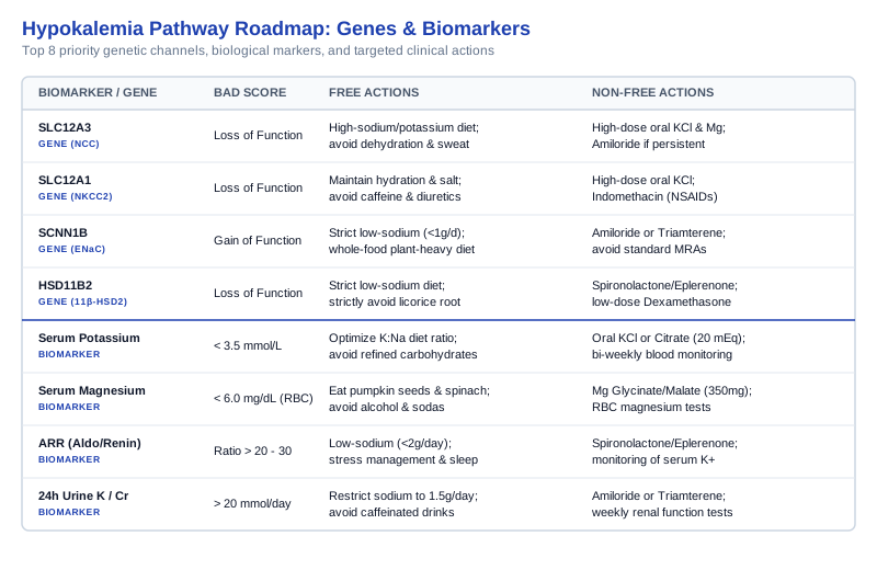
Grounded in clinical science and genetic research, this guide outlines two complementary pathways for reclaiming control. The primary strategy focuses on tracking six essential biomarkers that reveal how your kidneys and endocrine system manage electrolytes in real time. The bonus strategy explores seven key genes and epigenetic factors that set your baseline biological tolerance. Together, these frameworks offer a clear, actionable roadmap for reversing chronic hypokalemia and restoring cellular vitality.
Summary
This article provides an in-depth, clinically grounded guide to managing and reversing hypokalemia through systematic biomarker tracking and genomic analysis. By monitoring six core biomarkers—including serum potassium, magnesium, and the aldosterone-to-renin ratio—you can identify whether your low potassium levels stem from renal wasting, hormonal overactivity, or cellular shifts. Additionally, we analyze seven genetic variants, such as the SLC12A3 and SCNN1B genes, which dictate channel behaviors in the distal nephron. Combined with key takeaways from pioneering clinical literature on the sodium-potassium pump and five evidence-based complementary modalities, this guide empowers readers to build a personalized, data-driven plan to restore electrolyte balance.
Tracking the Essential Biomarkers for Potassium Balance
To understand why potassium levels remain chronically low, we must examine the markers that reflect renal function, hormone output, and acid-base balance. Potassium regulation is highly dynamic, and measuring blood levels alone provides only a snapshot of a highly complex system. By evaluating a comprehensive panel of biomarkers, you can determine whether your hypokalemia is driven by kidney wasting, hormonal signaling errors, or intracellular shifts. Below are the six most critical biomarkers to track, along with specific protocols for optimizing each score.
Biomarker 1: Serum Potassium
Serum potassium measures the concentration of potassium ions in the extracellular fluid. Under normal physiological conditions, the body maintains this level within a narrow margin, typically between 3.5 and 5.0 mmol/L, as outlined in the clinical guide on potassium disorders at StatPearls Hypokalemia. Even minor deviations can disrupt the electrical potentials of cardiac and skeletal muscle membranes. While serum potassium is the standard diagnostic tool, a low score simply confirms hypokalemia; it does not explain the mechanism. Chronically low serum potassium despite dietary sufficiency indicates that potassium is either being lost through the kidneys or gastrointestinal tract, or is shifting abnormally from the blood into the cells.
How to measure it
This is measured via a standard venous blood draw, which is widely available and typically costs between $15 and $30 out-of-pocket if not covered by insurance. It is crucial that the blood sample is processed quickly to prevent hemolysis (the rupturing of red blood cells), which can falsely elevate the potassium reading.
If the score is bad, the plan without supplements
If your serum potassium is mildly low, the first step is to optimize your dietary potassium-to-sodium ratio. Focus on consuming potassium-rich, whole foods such as avocados, wild-caught salmon, leafy greens, and organic potatoes, while reducing processed foods high in sodium. High sodium intake increases the delivery of sodium to the distal nephron of the kidney, which accelerates potassium excretion. Additionally, minimize intake of refined carbohydrates, as insulin surges cause transient shifts of potassium from the blood into skeletal muscle cells, worsening hypokalemia.
If the score is bad, the plan with supplements or equipment
For moderate hypokalemia, supplement with potassium chloride or potassium citrate. Potassium chloride is the preferred form for correcting hypokalemia associated with metabolic alkalosis, while potassium citrate is ideal if mild metabolic acidosis is present. A standard starting dose is 20 mEq (approximately 1,500 mg of elemental potassium) daily, taken in divided doses with large meals to prevent gastrointestinal irritation. Avoid taking large single doses, which can cause local mucosal damage in the gut or trigger cardiac arrhythmias. This protocol should be maintained continuously, with serum levels retested every two weeks until stable, and then quarterly. Side effects include abdominal cramps, nausea, and diarrhea. If you have chronic kidney disease, avoid potassium supplements unless under direct medical supervision.
Biomarker 2: Serum Magnesium
Serum magnesium is a critical cofactor for the Na+/K+-ATPase pump, which actively pumps potassium into cells and sodium out. Magnesium also regulates the Renal Outer Medullary Potassium (ROMK) channels in the distal convoluted tubule of the kidney. When intracellular magnesium levels are low, these ROMK channels lose their physical blockage, allowing potassium to leak freely into the urine, resulting in renal wasting. Clinical reviews such as the study on pediatric and adult hypokalemia at PMC9115714 emphasize that hypokalemia is often refractory to potassium supplementation unless co-existing magnesium deficiency is corrected first.
How to measure it
Serum magnesium is measured via a standard blood test, costing between $20 and $40. Because only 1% of the body's magnesium is stored in the blood, some integrative physicians recommend a red blood cell (RBC) magnesium test for a more accurate assessment of intracellular stores, which costs approximately $50 to $70. The target range for RBC magnesium is 6.0 to 6.5 mg/dL.
If the score is bad, the plan without supplements
Increase your consumption of magnesium-dense foods, including pumpkin seeds, dark chocolate (above 85% cacao), almonds, and spinach. Avoid consuming alcohol, carbonated sodas containing phosphoric acid, and refined sugars, as these substances increase renal magnesium excretion. Hydrate with mineral-rich spring water that naturally contains dissolved magnesium.
If the score is bad, the plan with supplements or equipment
Take 300 to 400 mg of elemental magnesium daily. Use highly bioavailable forms such as magnesium glycinate for overall cellular support, or magnesium malate to support muscle tissue. Avoid magnesium oxide due to poor absorption and laxative effects. Take magnesium in split doses, once in the morning and once before bed. Retest serum or RBC levels after six weeks. Possible side effects include loose stools and mild gastrointestinal cramping. Supplementation should be cycled off for 5 days every 8 weeks to assess baseline retention.
Biomarker 3: Aldosterone-to-Renin Ratio (ARR)
Aldosterone is a hormone produced by the adrenal glands that signals the kidneys to retain sodium and excrete potassium. Renin is an enzyme produced by the kidneys that controls aldosterone production. The Aldosterone-to-Renin Ratio (ARR) is the primary screening tool for primary aldosteronism, a condition characterized by autonomous, excessive aldosterone secretion. High aldosterone levels stimulate the epithelial sodium channels (ENaC) and ROMK channels in the distal nephron, driving continuous renal potassium wasting and sodium retention, which frequently presents as hypokalemia paired with hypertension.
How to measure it
ARR is measured via a morning blood draw after the patient has been standing or sitting upright for at least two hours. The test costs between $80 and $150. A ratio greater than 20 to 30, coupled with an elevated absolute aldosterone level (greater than 15 ng/dL), strongly suggests primary aldosteronism.
If the score is bad, the plan without supplements
Adhere to a strict low-sodium diet (less than 2,000 mg of sodium daily) to naturally lower blood pressure and reduce the kidney's stimulus for aldosterone excretion. Manage chronic psychological stress, as high cortisol levels can cross-react with mineralocorticoid receptors, mimicking the effects of aldosterone excess. Focus on maintaining consistent circadian sleep cycles to support balanced adrenal hormone output.
If the score is bad, the plan with supplements or equipment
If primary aldosteronism or aldosterone excess is confirmed, the standard protocol involves mineralocorticoid receptor antagonists (MRAs) such as spironolactone or eplerenone. Under medical supervision, initiate spironolactone at 25 mg daily. Eplerenone is a more selective alternative with fewer hormonal side effects, started at 25 to 50 mg daily. These medications block aldosterone from binding to receptors in the distal tubule, conserving potassium. Retest potassium and creatinine levels after one week of initiation, then monthly. Side effects of spironolactone include gynecomastia, menstrual irregularities, and hyperkalemia. No OTC supplements can replicate this action, though licorice root must be strictly avoided, as it contains glycyrrhizin, which inhibits the enzyme that deactivates cortisol, worsening aldosterone-like activity.
Biomarker 4: Serum Bicarbonate (HCO3-)
Serum bicarbonate is an indicator of the body's acid-base balance. Potassium regulation is closely tied to systemic pH. In states of metabolic alkalosis (high pH, elevated bicarbonate), hydrogen ions shift out of cells to balance the blood, forcing potassium ions to shift into the cells, resulting in hypokalemia. Conversely, metabolic acidosis (low pH, low bicarbonate) can also be associated with hypokalemia in specific renal disorders, such as Renal Tubular Acidosis (RTA). Measuring bicarbonate helps categorize the source of potassium loss.
How to measure it
Bicarbonate is routinely measured as part of a basic metabolic panel (BMP) or arterial blood gas (ABG) test. A BMP costs approximately $20 to $40. The normal reference range for serum bicarbonate is 22 to 29 mEq/L.
If the score is bad, the plan without supplements
If bicarbonate is elevated (alkalosis), reduce your intake of bicarbonate-producing foods and avoid alkaline water. Focus on deep, slow breathing exercises to normalize carbon dioxide retention, which naturally lowers blood pH. If bicarbonate is low (acidosis), increase your intake of organic vegetables, which yield alkaline ash, and reduce heavy consumption of animal proteins.
If the score is bad, the plan with supplements or equipment
For low bicarbonate (acidosis), use potassium citrate supplements. Citrate is metabolized in the liver to bicarbonate, helping to buffer excess blood acid while supplying potassium. Take 15 to 30 mEq of potassium citrate daily in divided doses with food. Monitor blood bicarbonate levels monthly. Side effects include mild diarrhea and stomach upset. For elevated bicarbonate (alkalosis), focus on replacing chloride ions using potassium chloride, which helps the kidneys excrete excess bicarbonate. Take 20 to 40 mEq of potassium chloride daily, monitored bi-weekly.
Biomarker 5: 24-Hour Urine Potassium-to-Creatinine Ratio
A 24-hour urine potassium test measures the total amount of potassium excreted by the kidneys over a full day. Comparing this to urine creatinine levels corrects for variations in urine concentration. This test distinguishes between renal potassium wasting and extrarenal potassium loss (such as poor dietary intake or gastrointestinal loss). If urinary potassium excretion is high (greater than 20 mmol/day) in the setting of hypokalemia, the kidneys are actively wasting potassium. If excretion is low (less than 15 mmol/day), the kidneys are attempting to conserve potassium, indicating that the loss is occurring elsewhere.
How to measure it
This test requires collecting all urine excreted over a 24-hour period into a specialized container. The sample is analyzed by a laboratory, with costs ranging from $40 to $90.
If the score is bad, the plan without supplements
If the kidneys are wasting potassium, reduce your dietary sodium intake significantly. High sodium delivery to the collecting duct increases the negative electrical charge in the tubule lumen, which pulls potassium out of the cells and into the urine. Restrict sodium to 1,500 mg daily while keeping hydration levels stable and avoiding caffeinated beverages, which act as weak diuretics.
If the score is bad, the plan with supplements or equipment
If renal wasting is confirmed, potassium-sparing diuretics that block ENaC channels are often utilized. Amiloride (5 to 10 mg daily) or triamterene (50 to 100 mg daily) are prescribed under medical supervision. These drugs reduce the negative electrical charge in the renal tubule, preventing potassium from being drawn into the urine. Monitor renal function and potassium levels weekly during the initial phase. Side effects include headache, dizziness, and hyperkalemia. Cycle these medications only under direct medical advice.
Biomarker 6: Serum Cortisol
Cortisol is the primary glucocorticoid hormone produced by the adrenal glands in response to stress. Cortisol has a structural similarity to aldosterone. At high physiological levels, cortisol can overwhelm the enzyme 11-beta-hydroxysteroid dehydrogenase type 2 (11β-HSD2), which normally deactivates cortisol in the kidneys. When this enzyme is overwhelmed, cortisol binds directly to mineralocorticoid receptors, acting as an aldosterone mimic. This triggers sodium reabsorption and severe renal potassium wasting, leading to stress-induced hypokalemia.
How to measure it
Serum cortisol is measured via a morning blood draw, typically around 8:00 AM when levels peak. The cost ranges from $35 to $60. A 4-point salivary cortisol test can also be used to evaluate cortisol rhythm throughout the day, costing approximately $100 to $150.
If the score is bad, the plan without supplements
Implement a strict sleep hygiene routine, keeping a consistent bedtime and waking schedule to align cortisol levels with the natural circadian rhythm. Avoid high-intensity exercise late in the evening, which raises night-time cortisol. Practice daily stress-reduction techniques such as progressive muscle relaxation, cold exposure therapy, and limiting blue light exposure after sunset.
If the score is bad, the plan with supplements or equipment
Use adaptogenic herbs to help regulate the hypothalamic-pituitary-adrenal (HPA) axis. Take 300 to 600 mg of standardized Ashwagandha extract (KSM-66) daily, split between morning and afternoon. Ashwagandha has been clinically shown to reduce serum cortisol levels. Additionally, take 300 mg of Phosphatidylserine in the evening to blunt excess nocturnal cortisol production. Cycle Ashwagandha by taking it for 8 weeks followed by a 2-week break. Side effects are rare but can include mild gastrointestinal upset or drowsiness.
Having detailed the dynamic biomarkers that reflect real-time potassium status, we must also consider the genetic architecture that sets your baseline susceptibility to electrolyte depletion.
Genetics and Epigenetics of Potassium Regulation: What Recent Research Suggests
While biomarkers reveal your current physiological state, your genetic blueprint determines how your kidney tubules, ion channels, and transport proteins are structured. Research led by geneticists like Ali Torkamani at Scripps Research and health commentators like Gary Brecka highlight how inherited single-nucleotide polymorphisms (SNPs) can predispose individuals to chronic electrolyte imbalances. Epigenetic modifications—such as DNA methylation changes driven by environmental stressors, diet, and toxins—further influence how these genes are expressed. By understanding your genetic profile, you can bypass standard protocols to address specific channel deficiencies.
Gene: SLC12A3
The SLC12A3 gene encodes the thiazide-sensitive sodium-chloride cotransporter (NCC) located in the distal convoluted tubule of the nephron. Pathogenic variants or loss-of-function mutations in this gene cause Gitelman syndrome, a salt-wasting tubulopathy. Because the NCC cotransporter is impaired, the kidney fails to reabsorb sodium and chloride, leading to increased sodium delivery to the collecting duct. This triggers secondary aldosterone activation, resulting in renal potassium and magnesium wasting, metabolic alkalosis, and low calcium in the urine. Clinical research, detailed in the clinical guide at StatPearls Gitelman Syndrome, indicates that patients with SLC12A3 mutations suffer from persistent, refractory hypokalemia.
If the gene is bad, the plan without supplements
Adhere to a high-sodium, high-potassium diet. Unlike individuals with primary hypertension, Gitelman syndrome patients need higher sodium intake to compensate for renal salt wasting and to suppress aldosterone activation. Consume high-quality sea salt and consume mineral-dense foods. Avoid dehydration and intense sweating, which can trigger severe cramping and weakness.
If the gene is bad, the plan with supplements or equipment
High-dose oral magnesium and potassium supplementation is required. Take potassium chloride (40 to 80 mEq daily, split into 3-4 doses with food) along with magnesium lactate or aspartate (300 to 500 mg daily). Retest blood levels every two weeks during dose adjustment. If hypokalemia persists, a low-dose ENaC inhibitor like amiloride (5 mg daily) can be prescribed to reduce potassium excretion. Side effects include stomach pain and nausea. This protocol is typically lifelong due to the genetic nature of the condition.
Gene: SLC12A1
The SLC12A1 gene encodes the sodium-potassium-chloride cotransporter (NKCC2) located in the thick ascending limb of the loop of Henle. Impairment of this transporter causes Bartter Syndrome Type 1. NKCC2 is responsible for reabsorbing approximately 25% of filtered sodium, potassium, and chloride. When this transporter is dysfunctional, massive salt wasting occurs, driving downstream potassium secretion via the ROMK channels. This presents as severe hypokalemia, metabolic alkalosis, and hypercalciuria (excess calcium in the urine, which can lead to kidney stones).
If the gene is bad, the plan without supplements
Maintain consistent hydration and salt intake. Consume foods with a high mineral content and avoid any dietary components that act as natural loop diuretics, such as caffeine and dandelion extract. Monitor urine output and stay indoors during periods of high heat to prevent volume depletion.
If the gene is bad, the plan with supplements or equipment
Supplementation requires high doses of potassium chloride (up to 100 mEq daily, divided into multiple doses) to replace renal losses. Nonsteroidal anti-inflammatory drugs (NSAIDs) like indomethacin (50 to 150 mg daily in divided doses) are often prescribed because prostaglandins are elevated in Bartter syndrome and drive renal salt wasting. NSAIDs help reduce urine output and conserve potassium. Side effects include gastrointestinal ulcers and nephrotoxicity with long-term use. Periodic renal function testing (creatinine and GFR) is mandatory every three months.
Gene: SCNN1B
The SCNN1B gene encodes the beta subunit of the Epithelial Sodium Channel (ENaC) located in the collecting duct. Gain-of-function mutations in SCNN1B cause Liddle Syndrome. This genetic variant prevents the normal degradation of ENaC channels, leading to their accumulation on the cell membrane. The result is constitutive, unregulated sodium reabsorption, which creates a strong negative electrical potential in the tubule lumen. This potential pulls potassium and hydrogen ions out of the cells and into the urine, causing hypokalemia, metabolic alkalosis, and severe, early-onset hypertension. More details are available in the clinical monograph at StatPearls Liddle Syndrome.
If the gene is bad, the plan without supplements
A strict low-sodium diet is essential. Because the genetic mutation causes the kidneys to continuously reabsorb sodium, dietary sodium must be restricted to less than 1,000 mg daily. Focus on a whole-food, plant-heavy diet and avoid all processed foods. Standard lifestyle measures for hypertension, such as stress reduction and moderate aerobic exercise, are supportive but must be paired with sodium restriction.
If the gene is bad, the plan with supplements or equipment
Traditional anti-hypertensive drugs like ACE inhibitors, ARBs, and spironolactone are ineffective because Liddle syndrome bypasses normal aldosterone pathways. The primary medical therapy is amiloride (5 to 10 mg daily) or triamterene (50 to 100 mg daily), which directly block the ENaC channel. These medications lower blood pressure and correct hypokalemia. Supplementation with potassium is generally not required once amiloride therapy is established, as the drug effectively halts potassium wasting. Monitor blood pressure and serum potassium weekly during initial dosing. Side effects include hyperkalemia, headache, and muscle cramps.
Gene: CACNA1S
The CACNA1S gene encodes the alpha-1S subunit of the L-type calcium channel in skeletal muscle cells. Mutations in CACNA1S are the primary cause of Hypokalemic Periodic Paralysis (HypoPP) Type 1. Under specific triggers—such as high-carbohydrate meals or stress, which release insulin and adrenaline—mutated calcium channels cause an abnormal leakage of electrical current. This depolarizes the muscle membrane, causing potassium to shift rapidly from the blood into the intracellular space of the muscle. This sudden extracellular drop leads to acute hypokalemia and temporary muscle paralysis.
If the gene is bad, the plan without supplements
Avoid major triggers, particularly large, carbohydrate-rich meals that trigger insulin surges, as insulin stimulates the Na+/K+-ATPase pump to pull potassium into cells. Keep your carbohydrate intake low-glycemic and spread meals throughout the day. Avoid sudden exposure to cold and strenuous physical exertion followed by rest, which are classic triggers for paralytic episodes.
If the gene is bad, the plan with supplements or equipment
During an acute episode, oral potassium chloride (either in solution or powder form, 20 to 40 mEq) is administered to restore serum levels. For long-term prevention, carbonic anhydrase inhibitors such as acetazolamide (125 to 250 mg daily) are prescribed. Acetazolamide induces a mild metabolic acidosis, which promotes potassium release from cells into the blood, preventing hypokalemic paralysis. Side effects of acetazolamide include paresthesia (tingling in fingers and toes), fatigue, and altered taste. Renal function and electrolytes should be monitored quarterly.
Gene: SCN4A
The SCN4A gene encodes the alpha subunit of the voltage-gated sodium channel Nav1.4 in skeletal muscle. Mutations in SCN4A cause Hypokalemic Periodic Paralysis Type 2. Similar to CACNA1S mutations, these variants cause the skeletal muscle sodium channels to leak ions at rest. When triggered by insulin, epinephrine, or cold, the muscle cells depolarize, leading to a rapid uptake of potassium from the blood into the skeletal muscle. This results in acute hypokalemia, muscle weakness, or paralysis.
If the gene is bad, the plan without supplements
Implement a low-carbohydrate, high-fat diet (such as a modified ketogenic diet) to minimize insulin excursions. Avoid high-stress environments, as epinephrine increases Na+/K+-ATPase activity, pushing potassium into cells. Ensure proper physical warm-ups before exercise and cool downs afterward.
If the gene is bad, the plan with supplements or equipment
Use oral potassium chloride during acute paralytic episodes. For preventative therapy, acetazolamide or dichlorphenamide (50 mg twice daily) is used. Dichlorphenamide is a potent carbonic anhydrase inhibitor that reduces the frequency of paralytic attacks. Monitor for side effects such as cognitive slowing, paresthesias, and metabolic acidosis. Retest potassium levels monthly during titration.
Gene: KCNJ2
The KCNJ2 gene encodes the Kir2.1 inward rectifier potassium channel, which plays a key role in maintaining the resting membrane potential of cardiac and skeletal muscle cells. Mutations in KCNJ2 lead to Andersen-Tawil Syndrome, which presents with a triad of periodic paralysis, cardiac arrhythmias (specifically prolonged QT intervals and ventricular ectopy), and developmental features. Dysfunction of Kir2.1 prevents potassium from flowing normally out of cells, rendering muscle and cardiac tissues susceptible to electrical instability during periods of hypokalemia.
If the gene is bad, the plan without supplements
Avoid strenuous exercise, sudden starts, and stressful stimuli, which can trigger adrenaline surges and cardiac arrhythmias. Maintain consistent hydration and follow a balanced diet with a stable potassium content. Avoid medications that prolong the QT interval, such as certain antibiotics and anti-emetics.
If the gene is bad, the plan with supplements or equipment
Maintain serum potassium levels in the high-normal range (4.0 to 4.5 mmol/L) to stabilize the cardiac membrane. Supplement with potassium chloride (20 to 40 mEq daily). Carbonic anhydrase inhibitors like acetazolamide may be used to prevent paralytic episodes, but must be paired with beta-blockers like flecainide or nadolol under cardiology supervision to manage arrhythmia risks. Retest ECGs and potassium levels monthly.
Gene: HSD11B2
The HSD11B2 gene encodes the enzyme 11-beta-hydroxysteroid dehydrogenase type 2. This enzyme is expressed in the kidneys, where it converts active cortisol into inactive cortisone. Because cortisol has a high affinity for the mineralocorticoid receptor (MR), HSD11B2 acts as a gatekeeper, preventing cortisol from binding to and activating the MR. A loss-of-function mutation or epigenetic downregulation (often caused by chronic stress, inflammation, or licorice consumption) causes Apparent Mineralocorticoid Excess (AME). In AME, cortisol continuously binds to renal MRs, triggering sodium retention, volume expansion, hypertension, and severe hypokalemia.
If the gene is bad, the plan without supplements
Implement a strict low-sodium diet and avoid licorice root, which contains glycyrrhizin, a natural inhibitor of 11β-HSD2. Manage chronic stress through mindfulness and vagal nerve stimulation to reduce circulating cortisol levels, which reduces the load on the remaining enzyme.
If the gene is bad, the plan with supplements or equipment
Treatment involves mineralocorticoid receptor blockers, specifically spironolactone (50 to 100 mg daily) or eplerenone. In some cases, low-dose dexamethasone (0.125 to 0.25 mg daily) is used to suppress pituitary ACTH secretion, which reduces endogenous cortisol production. Retest blood pressure, cortisol, and potassium weekly during titration. Side effects of dexamethasone include weight gain, immune suppression, and adrenal suppression.
By understanding how these genes shape your physiology, we can look at the overall cellular pump mechanism and how dietary adjustments can restore balance.
Optimizing the Cellular Pump: Key Insights from 'The High Blood Pressure Solution'
To understand the relationship between sodium, potassium, and cellular health, we can look to the book The High Blood Pressure Solution: A Scientific Approach to Preventing and Treating Hypertension with the Potassium-to-Sodium Ratio by Dr. Richard D. Moore. Dr. Moore presents a clinical framework centered on the Na+/K+-ATPase pump, illustrating how maintaining a high potassium-to-sodium ratio (the K-Factor) is essential for preventing chronic disease. Below is a summary of the ten most impactful concepts from his work, translated into practical steps for managing hypokalemia.
1. The Primacy of the Na+/K+-ATPase Pump
Dr. Moore explains that the sodium-potassium pump is the most energy-consuming mechanism in the human body, utilizing up to one-third of all cellular ATP. This pump actively moves three sodium ions out of the cell for every two potassium ions it pulls in. This action maintains the negative electrical charge inside the cell, which is necessary for muscle contraction, nerve conduction, and nutrient transport. When this pump lacks potassium, cellular voltage drops, leading to muscle weakness and cardiovascular dysfunction.
2. The Concept of the K-Factor
The K-Factor is the ratio of potassium to sodium in the diet and within cells. Dr. Moore argues that modern diets have inverted this ratio, delivering high sodium and low potassium, which stresses the cellular pumps. A high dietary K-Factor (aiming for a 4:1 ratio of potassium to sodium) optimizes pump function, allowing the body to maintain the high intracellular potassium concentrations needed for cellular metabolic processes.
3. How Sodium Excess Deactivates the Pump
High sodium levels in the blood increase the workload on the Na+/K+-ATPase pump. If intracellular sodium rises due to a low K-Factor diet, the cell swells with water, diluting intracellular potassium. Over time, this fluid retention and pump fatigue lead to vascular resistance, elevated blood pressure, and renal potassium wasting, as the kidneys attempt to excrete sodium along with potassium.
4. Intracellular Potassium Depletion and Vascular Tone
A decrease in intracellular potassium changes the resting membrane potential of vascular smooth muscle cells. This depolarization opens voltage-gated calcium channels, allowing calcium to enter the cells. The resulting calcium accumulation causes the smooth muscles surrounding arteries to constrict, increasing vascular resistance and blood pressure. Maintaining high intracellular potassium keeps these calcium channels closed, promoting vasodilation.
5. The Synergistic Role of Magnesium
Dr. Moore notes that magnesium acts as the chemical spark plug for the Na+/K+-ATPase pump. Because ATP must bind to magnesium to become biologically active, magnesium deficiency prevents the pump from utilizing energy. As a result, potassium cannot be pulled into cells, and sodium cannot be pumped out, leading to cellular potassium depletion. Magnesium supplementation is therefore necessary to support pump function.
6. Insulin Sensitivity and Pump Regulation
The sodium-potassium pump is regulated by insulin. When insulin binds to its receptor, it stimulates pump activity, causing potassium to shift into cells. Dr. Moore explains that insulin resistance impairs this pathway, leading to reduced pump activity, cellular potassium depletion, and elevated blood pressure. Improving insulin sensitivity helps maintain normal cellular potassium uptake.
7. The Renal Potassium Leak
When systemic potassium is low, the kidneys attempt to conserve it, but this process is less efficient if dietary sodium is high. High sodium intake increases sodium delivery to the distal nephron, which creates a negative electrical gradient that draws potassium out of cells and into the urine. Restricting sodium is therefore a key step in reducing renal potassium loss.
8. The Fallacy of Isolated Electrolyte Management
Dr. Moore critiques the clinical practice of treating blood pressure and electrolyte status in isolation. He argues that supplementing potassium without reducing sodium, or supplementing magnesium without addressing insulin resistance, is ineffective. A successful strategy requires addressing the entire pump system, balancing dietary intake with hormone levels and cellular health.
9. Target K-Factor Ratios for Prevention
To support cellular function, Dr. Moore recommends a daily intake of 4,700 mg of potassium and no more than 1,200 mg of sodium. This achieves a K-Factor ratio of approximately 4:1. For individuals with genetic renal wasting syndromes, this ratio may need to be adjusted, but the principle of prioritizing potassium over sodium remains key.
10. Reversing Hypertension via Pump Re-activation
By restoring the K-Factor, the Na+/K+-ATPase pump can lower intracellular sodium, reduce cell swelling, and normalize vascular smooth muscle tone. This approach treats hypertension at the cellular level, addressing a root cause rather than simply suppressing symptoms with medication.
While dietary and genetic strategies focus on physical intake and transport, complementary modalities can help regulate the nervous system to support potassium conservation.
Complementary and Alternative Modalities for Electrolyte Harmony
Complementary therapies can support potassium homeostasis by regulating the nervous system. Chronic stress activates the sympathetic nervous system and the HPA axis, increasing cortisol and aldosterone production, which promote renal potassium wasting. By utilizing mind-body practices, you can help reduce autonomic stress and lower these hormone levels. Below are five evidence-based modalities, along with specific protocols for supporting potassium conservation.
Mindfulness Meditation / MBSR
Mindfulness-Based Stress Reduction (MBSR) is a structured program that uses mindfulness practices to reduce physiological stress responses. Chronic HPA-axis activation elevates cortisol, which can bind to mineralocorticoid receptors and drive renal potassium excretion. Reducing stress through mindfulness helps lower cortisol levels, reducing this pathway of potassium wasting.
A standardized protocol involves practicing daily mindfulness meditation for 20 to 30 minutes, focusing on breath awareness and non-judgmental observation of bodily sensations. Clinical research, such as the review on stress and cortisol at PMC4662693, indicates that MBSR programs can significantly reduce circulating cortisol levels in stressed populations.
To apply this practice, dedicate 20 minutes each morning to sitting in a quiet space, closing your eyes, and focusing on the sensation of your breath. If your mind wanders, gently redirect your attention to your breathing. Practice this consistently for eight weeks to help establish autonomic balance, and monitor your serum cortisol and potassium levels to track progress.
Breathing-Based Therapies
Slow, paced breathing techniques help modulate autonomic nervous system activity. Slowing the respiratory rate stimulates the vagus nerve, increasing parasympathetic activity and dampening sympathetic output. This reduction in sympathetic tone helps lower renin and aldosterone secretion, reducing the drive for renal potassium excretion.
The protocol involves practicing paced diaphragmatic breathing at a rate of 6 breaths per minute (a 5-second inhalation and a 5-second exhalation) for 15 minutes twice daily. Research on slow breathing and autonomic function, such as the review at PMC9115714, shows that regular slow breathing improves baroreflex sensitivity and reduces sympathetic overactivity.
To implement this, sit comfortably with one hand on your chest and the other on your abdomen. Inhale deeply through your nose for 5 seconds, allowing your abdomen to expand, then exhale slowly through pursed lips for 5 seconds. Use a visual pacer app to maintain the rhythm. Practice this daily, especially during periods of stress, to help support electrolyte balance.
Biofeedback
Heart Rate Variability (HRV) biofeedback training teaches individuals to regulate their autonomic nervous system. By monitoring heart rate variations in real time, users learn to align their breathing with their cardiovascular rhythm, which helps reduce sympathetic activity and the secretion of epinephrine, a hormone that can trigger intracellular potassium shifts.
The protocol involves using an HRV monitor and practicing resonance frequency breathing (typically between 5.5 and 6 breaths per minute) for 20 minutes daily. Clinical trials have demonstrated that HRV biofeedback training improves autonomic balance, reduces anxiety, and lowers blood pressure.
To use this modality, acquire a consumer-grade HRV finger or ear sensor that connects to a biofeedback app. Practice breathing in sync with the app's visual guide, aiming to maximize your HRV coherence score. Perform this training in a quiet environment, and check your serum potassium levels after six weeks of daily practice.
Yoga
Yoga integrates physical postures, breath control, and meditation to help lower systemic stress. By downregulating the HPA axis and reducing sympathetic nervous system activity, yoga helps lower blood pressure and reduce aldosterone levels, which supports potassium retention in the kidneys.
The protocol involves a 60-minute Hatha or Restoration yoga session, practiced three times per week, incorporating gentle stretching, pranayama (breathing exercises), and a final relaxation period (Savasana). Clinical studies indicate that regular yoga practice leads to reductions in systolic blood pressure and serum cortisol.
To apply this, join a beginner-friendly restoration yoga class or follow a structured home video program. Focus on slow movements and coordinated breathing, avoiding hot yoga formats that cause heavy sweating and potential potassium loss. Retest your blood pressure and electrolytes after eight weeks of consistent practice.
Microbiome-Directed Therapies
The gut microbiome plays a role in mineral absorption, including magnesium and potassium. High-fiber diets that support the production of short-chain fatty acids (SCFAs) like butyrate help maintain a healthy colonic environment, which can improve mineral absorption and support overall electrolyte status.
The protocol involves daily supplementation with prebiotic fibers (such as inulin or acacia gum at 10 grams daily) alongside a diverse diet rich in fermented foods. Clinical studies show that prebiotic supplementation can improve intestinal absorption of minerals like calcium and magnesium, which supports potassium balance.
To apply this, start with 3 grams of inulin daily to assess tolerance, gradually increasing to 10 grams as tolerated. Pair this with a daily serving of unsweetened kefir or lacto-fermented vegetables. Monitor your digestive comfort and serum magnesium levels, as improving magnesium status is key to resolving renal potassium wasting.
Conclusion
Managing chronic hypokalemia requires moving beyond simple dietary fixes to address the renal, hormonal, and genetic factors that regulate potassium balance. By monitoring a comprehensive panel of biomarkers—including serum potassium, magnesium, and the aldosterone-to-renin ratio—you can identify the specific mechanisms driving your electrolyte depletion. Pairing these insights with genetic analysis of key transport channels allows for a more personalized, targeted approach to stabilization.
With this information, you can collaborate more effectively with your healthcare team to address the root causes of your symptoms. The next step is to request the necessary laboratory tests from your doctor, review your dietary potassium-to-sodium ratio, and implement the physiological protocols outlined in this guide. Taking these steps can help restore cellular energy, normalize blood pressure, and support overall vitality.