This article contains AI generated content.
· UpdatedHypoparathyroidism: 6 Genes And 7 Biomarkers To Track
Living With Hypoparathyroidism: Why Tracking the Right Numbers Changes Everything
Hypoparathyroidism is not a condition most people have heard of until they are living it. The parathyroid glands, four tiny structures resting against the thyroid, quietly regulate calcium and phosphorus every minute of your life. When they fail — whether from surgery, autoimmunity, genetic mutation, or unknown causes — the resulting chemical imbalance touches nearly everything: your muscles, your nerves, your kidneys, your mood, and your sleep. Yet many patients spend years managing symptoms without a clear picture of what specific numbers are driving their problems.
Generic advice — "take your calcium and vitamin D" — is a starting point, not a strategy. The reality is that hypoparathyroidism involves a cascade of interacting deficiencies and compensatory changes, and the same prescription can work well for one person and create new complications for another. Urinary calcium that creeps too high can deposit in kidneys silently. Magnesium quietly undermines the effectiveness of every other supplement. Active vitamin D behaves completely differently from the standard vitamin D most clinicians measure by default. The gap between feeling managed and feeling genuinely well often comes down to which numbers you are watching and how precisely.
This article takes a more targeted approach. The first and most developed section focuses on the 7 key biomarkers most relevant to hypoparathyroidism — what each one reveals, how to measure it, and what actions actually move the needle, with and without supplementation. A second, supporting section covers the 6 most relevant genes identified in hypoparathyroidism research, which matter most for people who have had genetic testing or want to understand the deeper drivers of their condition. An additional section summarizes the most insightful book on mineral physiology for this condition, and a final section reviews complementary approaches with real clinical evidence.
Better information does not replace medical care. But it gives you and your doctor something specific to act on, which is far more useful than waiting for symptoms to worsen before adjusting a protocol.
7 Biomarkers to Track in Hypoparathyroidism
The following seven biomarkers form the core monitoring panel recommended in clinical practice and increasingly in precision medicine approaches to hypoparathyroidism. They are not equal in importance: intact PTH and ionized calcium are the most fundamental, while urinary calcium and calcitriol are the most frequently overlooked — and often the most clinically meaningful.
1. Intact Parathyroid Hormone (iPTH)
Why it matters: Intact PTH is the defining biomarker of this condition. In a healthy person, PTH rises within seconds when calcium drops, stimulating bone resorption, increasing renal calcium reabsorption, and activating vitamin D conversion. In hypoparathyroidism, this signal is absent or severely blunted. Monitoring iPTH tells you the degree of residual gland function — which matters because even small amounts of residual PTH secretion reduce the risk of hypercalciuria and improve treatment tolerance significantly.
A level consistently below 10 pg/mL in the context of low calcium is diagnostic. But many patients on treatment drift between 10 and 20 pg/mL, and understanding where you sit shapes how aggressively you need to supplement and how carefully you need to monitor kidneys. Research on PTH monitoring in hypoparathyroidism — PubMed
How to Measure It
Blood draw, fasting preferred. The assay is called "intact PTH" or "PTH 1-84." Cost ranges from $30 to $90 depending on the lab and whether it is bundled with a calcium panel. Most primary care labs offer it. Aim to test every 3 to 6 months when stable, more frequently during dosage adjustments.
If the Score Is Low — The Plan Without Supplements
If iPTH is undetectable or near-zero, lifestyle levers are limited but not irrelevant. Dietary phosphorus restriction matters because high phosphorus intake further suppresses the calcium-PTH axis and accelerates soft tissue calcification. Reducing processed foods, sodas (especially colas), and processed cheese — the main dietary phosphorus sources — can improve the calcium-phosphorus balance even without medication changes. Resistance exercise has a modest stimulatory effect on bone metabolism and supports calcium retention independent of PTH. Frequency: 3 sessions per week minimum.
If the Score Is Low — The Plan With Supplements or Equipment
The gold standard pharmacological support is now rhPTH 1-34 (teriparatide) or rhPTH 1-84 (natpara/palopegteriparatide) — the latter specifically approved for hypoparathyroidism in some countries. These are injectable hormone replacement therapies that mimic endogenous PTH. For those not eligible or not yet prescribed PTH replacement, optimizing calcium citrate dosing (preferring citrate over carbonate for absorption), ensuring magnesium sufficiency, and using activated vitamin D analogs (calcitriol or alfacalcidol) remain the backbone. Side effects of PTH analogs include injection site reactions, hypercalcemia risk, and theoretical concern about osteosarcoma with very long-term use (primarily a rat-model finding; human surveillance has not confirmed this risk at standard clinical doses).
2. Ionized Calcium
Why it matters: Total serum calcium is the biomarker most commonly ordered, but it is the ionized fraction — roughly 45% of total calcium — that is biologically active and drives symptoms. Total calcium is strongly affected by albumin levels, and patients with low albumin (common in illness or malnutrition) will have falsely reassuring total calcium results. Ionized calcium correlates directly with symptom burden: numbness, tingling, muscle cramps, tetany, and cardiac arrhythmias all track more precisely with ionized calcium than total calcium. Ionized calcium and symptom correlation in hypoparathyroidism — PubMed
The target range in treated hypoparathyroidism is typically the low-normal end (1.12 to 1.20 mmol/L), deliberately kept below the upper-normal range to limit hypercalciuria risk.
How to Measure It
Requires a separate blood draw with special handling — the sample must be processed quickly on ice to prevent equilibration shifts. Not all labs offer it routinely; you may need to request it specifically. Cost: $40 to $120. For most routine monitoring, total calcium corrected for albumin is an acceptable proxy, but ionized calcium is superior when symptoms are difficult to interpret or when albumin is abnormal.
If the Score Is Low — The Plan Without Supplements
Timing and spacing of calcium intake is underappreciated. The gut absorbs calcium most efficiently in doses of 500 mg or less, and absorption is dramatically enhanced by stomach acid (favoring calcium citrate for those on acid-suppressing medications). Distributing calcium intake across the day rather than taking it in one dose meaningfully improves steady-state ionized calcium levels without increasing total daily dose. Reducing high-oxalate foods (spinach, almonds in excess, beet greens) that bind dietary calcium also helps, though this is less critical than spacing.
If the Score Is Low — The Plan With Supplements or Equipment
Calcium citrate (preferred over carbonate, especially in low-acid environments) at doses guided by labs — typically 1000 to 2500 mg elemental calcium per day in divided doses. Calcitriol (1,25-OH vitamin D) is essential as the activating step that enables intestinal calcium transport. Without calcitriol or alfacalcidol, calcium supplements are far less effective. Frequency of dose adjustments should be guided by labs every 4 to 8 weeks during titration. Monitor urinary calcium simultaneously — over-supplementation drives nephrocalcinosis.
3. Serum Phosphorus
Why it matters: PTH is the primary hormone that signals the kidneys to excrete phosphorus. Without adequate PTH, phosphorus accumulates in the blood — a defining feature of hypoparathyroidism that is sometimes undertreated. Elevated phosphorus directly inhibits the renal conversion of 25-OH vitamin D to its active calcitriol form, creating a vicious cycle that worsens hypocalcemia. High phosphorus also binds calcium in the gut and bloodstream, reducing its bioavailability, and over time contributes to calcification of soft tissues and blood vessels. Hyperphosphatemia management in hypoparathyroidism — PubMed
Target range in treated hypoparathyroidism: ideally 2.5 to 4.5 mg/dL.
How to Measure It
Standard blood panel, often included in a comprehensive metabolic panel (CMP). Cost is typically bundled at $15 to $50. Test fasting in the morning for most reliable results — phosphorus has mild diurnal variation. Frequency: every 3 to 6 months when stable.
If the Score Is High — The Plan Without Supplements
Dietary phosphorus reduction is the most direct lever. Prioritize reducing phosphate additives (E numbers in processed foods), not just naturally occurring phosphorus from whole foods, as the former is absorbed nearly completely while the latter is absorbed at roughly 40 to 60%. Avoid phosphoric acid in cola drinks — even small amounts raise serum phosphorus measurably. Increase water intake to support renal phosphorus excretion.
If the Score Is High — The Plan With Supplements or Equipment
Calcium carbonate taken with meals acts as a phosphate binder in the gut — one of the reasons it is preferred over calcium citrate in some protocols despite its lower absorption efficiency. If phosphorus remains elevated despite dietary changes, prescription phosphate binders may be considered; sevelamer is commonly used when calcium-based binders contribute to hypercalcemia. These are prescription medications with side effects including gastrointestinal discomfort and fat-soluble vitamin malabsorption with long-term use.
4. Serum Magnesium
Why it matters: This is the most consistently underestimated biomarker in hypoparathyroidism. Magnesium has a dual critical role: first, it is required for PTH secretion from residual parathyroid tissue — even modest magnesium deficiency suppresses PTH output; second, it is required for PTH receptor signaling at target tissues. A patient with low-normal magnesium may have blunted response to their own residual PTH secretion and reduced sensitivity to exogenous PTH analogs. Standard serum magnesium can be deceptively normal when intracellular stores are depleted — only about 1% of total body magnesium circulates in the blood. Magnesium deficiency and PTH suppression — PubMed
Target: serum magnesium above 0.85 mmol/L (2.0 mg/dL), ideally in the upper third of the normal range.
How to Measure It
Standard blood draw. Cost: $15 to $50, often included in CMPs. For a more sensitive assessment of intracellular magnesium status, RBC magnesium (red blood cell magnesium) is more accurate than serum magnesium and costs $50 to $150. The serum test will miss up to 40% of cases of true magnesium deficiency.
If the Score Is Low — The Plan Without Supplements
Dietary magnesium optimization is underrated. Dark leafy greens, pumpkin seeds, dark chocolate (85%+), and legumes are high-density sources. Reducing alcohol (which dramatically increases renal magnesium wasting) and caffeine (mild urinary loss) helps preserve stores. Avoid high-dose zinc supplementation, which competes with magnesium absorption.
If the Score Is Low — The Plan With Supplements or Equipment
Magnesium glycinate or magnesium malate are the preferred forms for oral supplementation — better absorbed and far less likely to cause the diarrhea associated with magnesium oxide or citrate at higher doses. Typical supplemental doses: 200 to 400 mg elemental magnesium per day in split doses (morning and evening). Start low and titrate up. Transdermal magnesium (magnesium chloride oil applied to skin) is advocated as a way to bypass gut absorption limits, though evidence for its superiority over oral supplementation is limited. Cycling is generally not necessary — this is a continuous daily support nutrient. Monitor serum or RBC magnesium at 4 to 6 weeks after starting.
5. 25-OH Vitamin D (Calcidiol)
Why it matters: 25-OH vitamin D is the storage form and the standard test ordered to assess vitamin D status. In hypoparathyroidism, it serves as the raw substrate for conversion to active calcitriol (1,25-OH vitamin D). Without adequate substrate, even the best-dosed calcitriol analogs may underperform. More critically, 25-OH vitamin D at adequate levels provides a buffer — modest amounts are converted non-enzymatically and support immune regulation, muscle function, and cardiovascular health independently of the PTH-calcitriol pathway. Vitamin D status and treatment outcomes in hypoparathyroidism — PubMed
Target in hypoparathyroidism: 40 to 60 ng/mL (100 to 150 nmol/L) — somewhat higher than the general population minimum, given the impaired conversion pathway.
How to Measure It
Standard blood test. Cost: $30 to $100. The 25-OH D3 form is the one to request (not D2 separately unless you are specifically supplementing D2). Test in late winter for a true baseline; supplementation decisions should be guided by labs, not season or symptom guess.
If the Score Is Low — The Plan Without Supplements
Sensible sun exposure — 15 to 30 minutes of midday sun to the arms and legs, several times weekly during summer months — can meaningfully raise 25-OH D in lighter skin types. For darker skin, this threshold increases substantially. Dietary sources (fatty fish, egg yolks, fortified foods) contribute but rarely achieve adequate levels alone.
If the Score Is Low — The Plan With Supplements or Equipment
Vitamin D3 (cholecalciferol) supplementation is safe and effective. Doses of 2000 to 5000 IU daily are commonly needed to maintain levels above 40 ng/mL in most adults. Important note for hypoparathyroidism: regular vitamin D3 is not a substitute for calcitriol or alfacalcidol, as the final conversion step requires functional PTH signaling. Both are needed — vitamin D3 fills the storage pool; calcitriol fills the active slot. Vitamin K2 (MK-7, 100 to 200 mcg daily) is commonly co-supplemented to direct calcium into bone and away from arteries. Side effects at standard doses are rare; toxicity begins above prolonged 10,000+ IU daily with elevated serum levels.
6. 1,25-OH Vitamin D (Calcitriol)
Why it matters: This is the active form of vitamin D — the form that actually opens intestinal calcium channels, stimulates renal calcium reabsorption, and modulates bone metabolism. In healthy individuals, PTH drives its production in the kidneys. In hypoparathyroidism, this conversion is severely impaired, creating a functional deficiency of calcitriol even when 25-OH D is perfectly adequate. This is why patients can have "normal" vitamin D levels on a standard test yet have profoundly impaired calcium absorption. Calcitriol supplementation in hypoparathyroidism — PubMed
Target: within the normal range, adjusted to maintain serum calcium at goal without causing hypercalciuria.
How to Measure It
Blood test labeled "1,25-dihydroxyvitamin D" or "1,25-OH D." Not routinely included in standard panels — must be specifically requested. Cost: $80 to $200. This test is particularly valuable when treatment response is poor despite adequate 25-OH D levels, as it confirms whether the conversion step is the limiting factor.
If the Score Is Low — The Plan Without Supplements
There is no reliable dietary source of active calcitriol. Sun exposure does not improve the conversion in the absence of PTH. The only lifestyle lever is ensuring the substrate (25-OH D) is not limiting by maintaining adequate vitamin D3 intake, reducing factors that accelerate vitamin D catabolism (obesity, inflammation, certain medications including anticonvulsants and corticosteroids).
If the Score Is Low — The Plan With Supplements or Equipment
Calcitriol (0.25 to 1 mcg twice daily) or alfacalcidol (slightly more forgiving dosing due to its longer half-life) are the standard pharmacological corrections. These are prescription medications requiring careful dose titration. Hypercalcemia and hypercalciuria are the primary risks — this is why concurrent urinary calcium monitoring (next biomarker) is essential whenever calcitriol is being adjusted. Never adjust calcitriol dose without checking labs, as the therapeutic window is narrow.
7. 24-Hour Urinary Calcium Excretion
Why it matters: This is the safeguard biomarker — the one that tells you whether your treatment is silently damaging your kidneys. In healthy physiology, PTH instructs the kidneys to reabsorb most filtered calcium, keeping urinary calcium low. Without PTH, this reabsorption is impaired, and the calcium you supplement — even to achieve target serum levels — tends to spill into the urine at higher-than-normal rates. Over years, this causes nephrocalcinosis (calcium deposits in kidney tissue), nephrolithiasis (kidney stones), and eventually chronic kidney disease. Studies show renal complications in up to 40% of hypoparathyroidism patients over time, primarily driven by unchecked hypercalciuria. Urinary calcium and renal risk in hypoparathyroidism — PubMed
Target: ideally below 250 mg per day in women, below 300 mg per day in men.
How to Measure It
24-hour urine collection — the patient collects all urine over a full day into a provided container, which is then analyzed. Cost: $50 to $150. A spot urine calcium-to-creatinine ratio (uCa/Cr) can provide a quick estimate between formal collections. This test should be done every 6 to 12 months in all treated hypoparathyroidism patients — more frequently during treatment adjustments. Many clinicians skip it; patients should specifically request it.
If the Score Is High — The Plan Without Supplements
Increasing fluid intake to at least 2 to 2.5 liters per day dilutes urine and significantly reduces crystallization risk. Dietary sodium restriction is highly effective — sodium and calcium share a renal transport mechanism, and reducing sodium intake by 2 to 3 g per day can meaningfully lower urinary calcium excretion without changing calcium supplementation. Moderate dietary protein (avoid very high-protein diets) reduces the acid load that drives calciuria.
If the Score Is High — The Plan With Supplements or Equipment
Thiazide diuretics (hydrochlorothiazide, chlorthalidone) are a clinically proven strategy to reduce urinary calcium excretion by 30 to 50% — they work by enhancing calcium reabsorption in the distal tubule, a PTH-independent mechanism. This is often the first medication added when hypercalciuria is identified on treatment. Side effects include electrolyte shifts (watch potassium and sodium), mild glucose effects, and blood pressure lowering. Amiloride is sometimes added to potassium-spare the thiazide effect. Dose adjustments of calcitriol or calcium supplements are frequently required in parallel when starting thiazides.
Moving from the biomarker panel, understanding the genetic architecture of hypoparathyroidism can add a meaningful layer to treatment decisions — particularly for patients with a family history, younger onset, or additional features suggesting a syndromic form.
The Genetic Underpinnings: 6 Key Genes in Hypoparathyroidism
Genetics does not replace biomarker monitoring, but it contextualizes why a person developed hypoparathyroidism, predicts associated risks in other organ systems, and increasingly guides treatment decisions. The six genes below account for the majority of identified genetic hypoparathyroidism cases.
GCM2 (Glial Cells Missing 2)
GCM2 is the master transcription factor for parathyroid gland development. Without functioning GCM2, parathyroid glands fail to form or are severely hypoplastic. Mutations in GCM2 are the most commonly identified genetic cause of isolated hypoparathyroidism, with both dominant loss-of-function mutations (haploinsufficiency) and recessive mutations documented. GCM2 mutations in isolated hypoparathyroidism — PubMed
If the Gene Is Impaired — Plan Without Supplements
GCM2 mutations mean the parathyroid tissue itself is structurally deficient or absent — this is not correctable with lifestyle or diet. However, ensuring no additional suppressors of residual parathyroid function exist (adequate magnesium, avoiding prolonged alkalosis, reducing glucocorticoid exposure) maximizes output from any remaining tissue.
If the Gene Is Impaired — Plan With Supplements or Equipment
Standard hypoparathyroidism management applies: calcitriol, calcium supplementation, thiazide diuretics for hypercalciuria. GCM2-related hypoparathyroidism is one of the clearest indications for PTH replacement therapy (rhPTH 1-84), which bypasses the absent gland by providing the hormone exogenously. Genetic counseling for family members is advisable given the heritable nature.
CASR (Calcium-Sensing Receptor)
Activating mutations in CASR cause the calcium-sensing receptor to be constitutively switched on — the receptor behaves as if calcium is always high, suppressing PTH secretion and promoting renal calcium excretion even when actual calcium is low. This is called autosomal dominant hypocalcemia type 1 (ADH1) and is an important diagnostic distinction because standard treatment (calcitriol + calcium supplementation) carries particularly high hypercalciuria risk in these patients due to the concurrent renal calcium-wasting mutation. CASR activating mutations and autosomal dominant hypocalcemia — PubMed
If the Gene Is Impaired — Plan Without Supplements
Dietary calcium should be modest — not restricted severely, but not aggressively high. Sodium restriction and high fluid intake are even more important in CASR-mutation carriers than in other hypoparathyroidism patients, given the dual mechanism of hypercalciuria.
If the Gene Is Impaired — Plan With Supplements or Equipment
Standard calcitriol and calcium still apply but require exceptionally careful urinary calcium monitoring (every 3 to 6 months). Emerging pharmacology: calcilytic drugs (which block the hyperactive CASR) are in clinical trials specifically for ADH1 — they represent a potentially mechanism-correcting treatment for this genetic subtype. Thiazide diuretics are frequently essential in this group.
PTH Gene
Direct mutations in the PTH gene itself — encoding the preproPTH protein — cause autosomal dominant or recessive hypoparathyroidism by impairing hormone production or processing. These are rarer than CASR and GCM2 mutations and typically present in infancy or early childhood. The condition is biochemically indistinguishable from other causes; genetics are needed for diagnosis. PTH gene mutations in familial hypoparathyroidism — PubMed
If the Gene Is Impaired — Plans
Management is the same as standard isolated hypoparathyroidism: calcitriol, calcium, magnesium optimization, urinary calcium monitoring. Because these mutations impair hormone synthesis rather than gland structure, there is theoretical possibility that residual secretion exists from normal alleles in heterozygous cases, making magnesium optimization particularly relevant (magnesium is required as a cofactor in PTH secretion). PTH replacement therapy can be considered in refractory cases.
GATA3
GATA3 mutations cause HDR syndrome — hypoparathyroidism, sensorineural deafness, and renal dysplasia. In patients where hypoparathyroidism is accompanied by any hearing impairment or kidney structural abnormality, GATA3 should be considered. The hypoparathyroidism component is often the presenting feature in adulthood, while renal anomalies may be subtle and missed without targeted imaging. GATA3 and HDR syndrome — PubMed
If the Gene Is Impaired — Plans
Standard hypoparathyroidism management with additional monitoring: annual renal ultrasound (for nephrocalcinosis and structural abnormalities), audiometry, and urinalysis. The renal component means urinary calcium thresholds should be held conservatively — even mildly elevated calciuria is more dangerous when there is pre-existing renal structural anomaly. Nephrology co-management is advisable.
TBX1
TBX1 haploinsufficiency is the primary molecular driver of 22q11.2 deletion syndrome (DiGeorge syndrome), in which hypoparathyroidism occurs due to failure of pharyngeal pouch development — the same embryological origin as the parathyroid glands and thymus. Hypoparathyroidism in DiGeorge is often neonatal and may improve partially with age, though adults frequently retain subclinical parathyroid insufficiency. The syndrome also involves congenital cardiac defects, immune deficiency (from thymic aplasia), and palatal anomalies. TBX1 and DiGeorge hypoparathyroidism — PubMed
If the Gene Is Impaired — Plans
Immunological status should be co-assessed (T-cell levels, thymic function). Cardiac surveillance as indicated. Hypoparathyroidism management follows standard protocols, with awareness that the condition may partially recover — monitoring at 6-month intervals is advisable rather than assuming permanent maximal deficiency.
AIRE (Autoimmune Regulator)
AIRE mutations cause autoimmune polyendocrinopathy-candidiasis-ectodermal dystrophy (APECED / APS-1), in which hypoparathyroidism results from autoimmune destruction of parathyroid tissue. This is the most common cause of autoimmune hypoparathyroidism and is associated with adrenal insufficiency and chronic mucocutaneous candidiasis — a triad that, when present, is pathognomonic. Measuring anti-NALP5 and anti-CaSR antibodies can confirm autoimmune etiology even without genetic testing. AIRE and autoimmune hypoparathyroidism — PubMed
If the Gene Is Impaired — Plans
Screen for adrenal insufficiency before initiating aggressive calcium/calcitriol treatment — treating hypocalcemia without correcting untreated adrenal insufficiency can precipitate adrenal crisis. Annual monitoring for additional autoimmune endocrinopathies. Consider measuring anti-parathyroid antibodies. Management of hypoparathyroidism is otherwise standard. Immunomodulatory treatments are investigational for the autoimmune mechanism specifically.
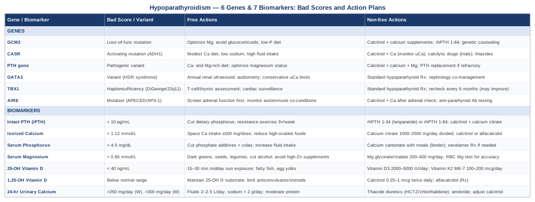
A Summary Table of Genes and Biomarkers to Track
The Mineral Fix: The Book That Reframes Electrolyte Deficiency
One of the most practically useful books for anyone with hypoparathyroidism or mineral imbalance is The Mineral Fix by Dr. James DiNicolantonio, published in 2021. DiNicolantonio is a cardiovascular research scientist known for challenging mainstream nutritional orthodoxy with large reference sets of human studies. This book is particularly relevant because it systematically reviews how modern diet creates widespread electrolyte depletion — exactly the terrain hypoparathyroidism patients navigate every day.
Below are ten of the most impactful insights from the book, each with direct relevance to hypoparathyroidism management.
1. Magnesium Deficiency Is Far More Prevalent Than Serum Tests Suggest
DiNicolantonio documents that standard serum magnesium tests detect only about 1% of total body stores. The vast majority of people with genuine tissue magnesium deficiency have normal serum results. He argues for RBC magnesium as the minimal standard for assessing true status — a distinction that is particularly critical for hypoparathyroidism patients, where even subtle magnesium insufficiency can suppress residual PTH secretion and blunt treatment response.
2. Calcium Supplements Are Often Counterproductive Without Co-Factors
Calcium supplementation without adequate vitamin K2 and magnesium diverts calcium into arteries rather than bone, contributing to vascular calcification. DiNicolantonio extensively reviews trials showing that K2 (particularly MK-7) directs calcium to bone matrix — a concern directly applicable to hypoparathyroidism patients who often take substantial amounts of supplemental calcium long-term.
3. Sodium Restriction Reduces Urinary Calcium Losses
One of the book's most clinically useful takeaways: sodium and calcium share the same renal cotransporter in the proximal tubule. For every gram of sodium excreted, approximately 26 mg of calcium follows. Reducing dietary sodium from a typical 3500 mg per day to below 2000 mg can reduce urinary calcium by 70 to 100 mg per day — equivalent in impact to a meaningful dose adjustment in some patients.
4. Most Processed Foods Are Designed to Deplete Minerals
Phosphate additives in processed foods — used as preservatives, emulsifiers, and acidulants — are absorbed at nearly 100% efficiency compared to 40 to 60% for organic phosphates in whole foods. DiNicolantonio's review of food additive research shows that the average processed-food consumer is absorbing substantially more inorganic phosphorus per day than nutritional databases suggest, directly relevant to the hyperphosphatemia challenge in hypoparathyroidism.
5. Potassium Is the Forgotten Mineral in Calcium Balance
Adequate potassium intake reduces urinary calcium excretion independently of sodium — through alkalinization of urine, which reduces calcium mobilization from bone and increases renal calcium reabsorption. DiNicolantonio points to several trials showing that potassium citrate supplementation meaningfully reduces urinary calcium — a particularly relevant point for hypoparathyroidism patients with borderline hypercalciuria who want to avoid starting thiazides.
6. Vitamin D Toxicity Is Real But Rare, and Most People Are Far Too Conservative
The book reviews trial data suggesting that optimal 25-OH D levels of 40 to 60 ng/mL provide substantially different physiological outcomes than the minimum of 20 ng/mL that many guidelines define as sufficient. In the context of hypoparathyroidism, this matters because higher substrate levels give more buffer for the impaired conversion pathway to work with.
7. Zinc and Magnesium Compete — Most Zinc Supplements Interfere With Magnesium Absorption
This is a commonly overlooked interaction. The book notes that zinc supplementation above 25 mg per day significantly impairs intestinal magnesium absorption. For hypoparathyroidism patients taking high-dose zinc (for immune support or wound healing), this interaction can quietly worsen magnesium status — and through it, undermine PTH secretion from residual tissue.
8. The Gut Microbiome Directly Regulates Calcium and Magnesium Absorption
DiNicolantonio reviews emerging evidence that short-chain fatty acids produced by gut bacteria — especially butyrate — directly upregulate calcium and magnesium transport proteins in the colon. A depleted microbiome results in measurably reduced mineral absorption. This supports paying attention to gut health not as a wellness trend but as a direct lever on mineral balance.
9. Muscle Cramps Are a Mineral Problem First
The book challenges the conventional view that exercise-associated muscle cramps are primarily dehydration. DiNicolantonio's review of electrolyte trial data implicates magnesium, sodium, and potassium deficiencies as the primary drivers in most populations. For hypoparathyroidism patients for whom cramps are a daily reality, this supports comprehensive electrolyte assessment rather than adjusting calcium alone.
10. Mineral Balance Must Be Assessed as a System, Not in Isolation
Perhaps the most important meta-message of the book: individual minerals do not operate in isolation. Calcium needs magnesium for metabolism, magnesium needs B6 for cellular uptake, vitamin D needs K2 for safe routing of calcium, and potassium balances sodium's effect on calcium excretion. Treating hypoparathyroidism as purely a "calcium and calcitriol problem" misses this systemic web — and it is why tracking the full biomarker panel matters more than optimizing any one number.
Complementary Approaches With Relevant Evidence
No complementary approach replaces calcitriol or calcium supplementation in hypoparathyroidism. The four approaches below are selected for having meaningful human evidence relevant to the symptom burden and physiological challenges of this specific condition.
Mindfulness-Based Stress Reduction (MBSR)
Hypoparathyroidism is a chronic condition with unpredictable symptom episodes — tetany, brain fog, fatigue, and anxiety — that take a documented toll on quality of life. Psychological stress independently increases urinary mineral excretion and can precipitate episodes of relative hypocalcemia in borderline-controlled patients through mechanisms involving cortisol and catecholamines. MBSR was specifically designed for chronic conditions to interrupt this stress-symptom cycle.
A systematic review of MBSR in chronic endocrine and metabolic conditions found significant reductions in disease-related anxiety and improved quality of life outcomes. The standard 8-week MBSR program — PubMed evidence involves two weekly sessions (typically 45 to 90 minutes each) with daily home practice. The Jon Kabat-Zinn protocol remains the most studied format.
For practical application: begin with 10 minutes of daily body-scan meditation focusing on detecting early tetany symptoms before they escalate — this type of somatic awareness gives patients more time to intervene (hydrate, correct posture, reduce triggers) before a full episode. Evidence for MBSR specifically in hypoparathyroidism is limited to case series; the strongest evidence is extrapolated from chronic disease management broadly.
Progressive Muscle Relaxation (PMR)
Muscle spasms, carpopedal spasm, and generalized cramping are among the most disruptive symptoms of suboptimal calcium control. Progressive muscle relaxation is a systematic technique of sequentially tensing and releasing muscle groups, which has documented efficacy in reducing involuntary muscle tension and spasm frequency across several conditions including nocturnal leg cramps and spastic disorders.
A randomized trial in patients with musculoskeletal pain and involuntary muscle tension found that twice-daily PMR practice over 4 weeks — PubMed significantly reduced both frequency and perceived intensity of muscle cramping events. The mechanism involves reducing baseline neuromuscular excitability — which is already elevated in hypocalcemia.
Practical application: 15 to 20 minutes, twice daily, working from feet to face in sequence. Most useful as a nightly routine before bed, when hypocalcemia symptoms often worsen. PMR does not address the underlying mineral deficiency but can meaningfully reduce the symptom burden and sleep disruption that accompany it.
Microbiome-Directed Therapies
Emerging research has linked gut microbiome composition to the efficiency of mineral absorption — particularly for calcium and magnesium. Butyrate-producing bacteria (notably Bifidobacterium and Lactobacillus species) upregulate the expression of calcium transport proteins TRPV6 and calbindin-D9k in the intestinal epithelium. In a setting where every milligram of absorbed calcium matters, gut microbiome quality becomes a potentially modifiable factor in calcium bioavailability. Gut microbiome and calcium absorption — PubMed
A human intervention study examining fermented dairy and prebiotic supplementation found improved calcium and magnesium retention at 12 weeks in participants with microbiome-targeted intervention versus control. The protocol used prebiotic fiber (inulin-type fructans, 8 to 10 g daily) combined with a multi-strain probiotic (including Lactobacillus acidophilus and Bifidobacterium longum).
Practical application: start with dietary prebiotic sources — chicory root, garlic, onion, asparagus, slightly unripe bananas — before moving to supplemental inulin, which can cause gas and bloating if introduced too quickly. Reduce dose if GI symptoms appear. This is a long-term strategy with a minimum 8 to 12 weeks before expecting measurable mineral retention changes. Evidence specifically for hypoparathyroidism is limited; the mechanism is plausible and the risk profile is low.
Breathing-Based Therapies — With an Important Caution
Slow diaphragmatic breathing activates the parasympathetic nervous system, reduces cortisol, and improves autonomic regulation — all relevant to managing the anxiety and autonomic instability that often accompanies hypocalcemia. Critical caveat: hyperventilation dramatically drops partial pressure of CO2, causes respiratory alkalosis, and reduces ionized calcium — this can precipitate or worsen tetany in hypoparathyroidism patients. Breathing techniques must focus exclusively on slow, controlled breathing (not rapid or high-volume techniques).
The 4-7-8 breathing method (inhale 4 counts, hold 7, exhale 8) and paced respiration at 5 to 6 breaths per minute have documented evidence for parasympathetic activation in chronic disease. Slow breathing and autonomic regulation — PubMed
Practical application: 5 to 10 minutes of paced breathing (6 breaths per minute) during anticipated stressful periods. Absolutely avoid Wim Hof method, breath-of-fire yoga breathing, or any technique that involves rapid cycles — these can directly precipitate tetany in this population. If any tingling or perioral numbness begins during a breathing session, return to normal breathing rate immediately.
Conclusion
Hypoparathyroidism management rarely goes wrong because of the wrong medication — it goes wrong because of incomplete monitoring. Patients who track all seven biomarkers and adjust their protocols accordingly have a far better chance of staying in the narrow therapeutic window where calcium is adequate, kidneys are protected, and quality of life is meaningful. The genetic picture adds depth: knowing which gene is driving your condition shapes how aggressively you protect your kidneys, whether you screen family members, and whether emerging therapies like calcilytics or PTH analogs are specifically relevant for you.
The next smart step is not a new supplement — it is a conversation with your endocrinologist specifically about 24-hour urinary calcium and ionized calcium if these are not already in your regular panel. Bring the list. Ask for the numbers. The difference between "treated" and "well-managed" almost always lives in the results you have not been checking yet.
Endocrine & Metabolic Autoimmune
Musculoskeletal: Bone Conditions Muscle Conditions
Endocrine & Metabolic: Adrenal Conditions
Urological: Kidney Conditions
Women's Health: Hormonal Conditions