This article contains AI generated content.
· UpdatedHypophosphatasia - 3 Genes And 7 Biomarkers To Track
Introduction
If you've been told your alkaline phosphatase is "a bit low" and sent home without further investigation, you're not alone. For most clinicians, a low ALP value gets dismissed as a statistical outlier or ignored in favor of more familiar abnormalities. But for people living with hypophosphatasia — a rare inherited metabolic disease — that single number, persistently low when it should not be, is the biochemical fingerprint of a condition that can affect bones, teeth, muscles, and even the nervous system across an entire lifetime.
Hypophosphatasia (HPP) is caused by deficient activity of tissue-nonspecific alkaline phosphatase (TNAP), an enzyme encoded by the ALPL gene. Because it presents so variably — from fatal perinatal forms with unmineralized bone to mild adult cases where the only clue is early tooth loss in childhood — it is frequently misdiagnosed or simply missed for years. The result is that people with HPP often receive treatment protocols designed for osteoporosis or chronic pain, without anyone addressing the underlying enzymatic failure. In the most dangerous scenario, they are prescribed bisphosphonates — the standard of care for low bone density — which are actively harmful in HPP.
Generic wellness advice is not wrong, but it's incomplete when the real issue is a genetic defect in how the body mineralizes bone and metabolizes key substrates like vitamin B6 and inorganic pyrophosphate. Knowing which specific biomarkers are running out of range and understanding whether a gene variant is involved gives you and your medical team a much sharper picture of what is actually happening. Sharper information leads to safer, more targeted decisions.
This article takes a practical approach to HPP from two complementary angles. The first, and most actionable, is a detailed breakdown of the 7 most useful biomarkers to track — what each one reveals, how to measure it, and what to do when it falls outside the optimal range. The second is a focused look at the 3 genes most relevant to this condition, what different variants mean clinically, and how to work around genetic findings both with and without supplementation. There is also a section drawing from Peter Attia's longevity medicine framework, which is one of the more useful public resources for understanding why HPP is so consistently overlooked. Better information genuinely can change the trajectory of this condition.
Summary
This article maps out the 7 most clinically relevant biomarkers for hypophosphatasia and the 3 genes that drive or modify it. You will learn why persistently low alkaline phosphatase is never a trivial finding, what it means when vitamin B6 metabolites accumulate in your blood despite no deficiency, and how a urine test for phosphoethanolamine can confirm what a standard blood panel cannot. Beyond diagnosis, each biomarker section explains what to do when scores fall out of range — including actionable protocols for nutrition, physical activity, targeted supplementation, and equipment, with guidance on frequency, cycling, and side effects. The genetics section covers the primary ALPL gene in plain language, plus two modifier genes that can shift severity in ways most genetic reports overlook. You will find a synthesis of the most practically useful insights from Peter Attia's bone health framework, followed by four complementary modalities — photobiomodulation, mindfulness, massage, and breathwork — that have real clinical rationale for HPP-related pain and musculoskeletal support. If you have been living with unexplained bone pain, early tooth loss, or a low ALP that no one has taken seriously, this article is written for you.
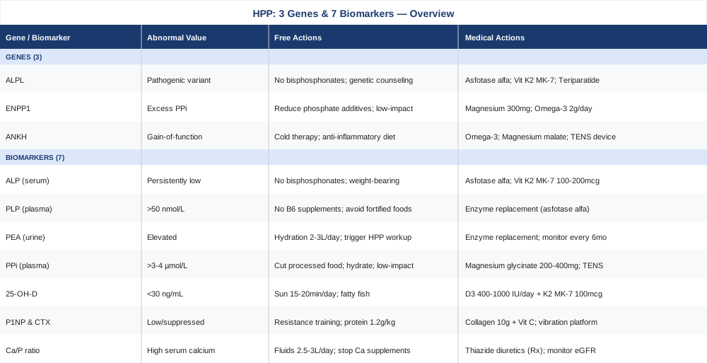
7 Biomarkers to Track in Hypophosphatasia
Tracking the right biomarkers in HPP serves a purpose that goes well beyond confirming a diagnosis. It reveals disease activity, monitors progression or stability, evaluates treatment response, and flags complications — like hypercalcemia or joint calcification — before they become severe. The seven markers below represent the most informative and actionable panel available for this condition.
1. Serum Alkaline Phosphatase (ALP) — The Primary Signal
Why it matters: ALP is the enzyme directly encoded by the ALPL gene. When TNAP is dysfunctional due to gene mutations, serum ALP activity is persistently low. This is the defining biochemical feature of HPP. While elevated ALP reliably prompts investigation for liver or bone pathology, persistently low ALP is systematically under-investigated — most automated lab systems do not flag low values with the same urgency as high ones, and most reference ranges were not built to capture HPP.
What it reveals: ALP activity reflects the functional output of the ALPL gene in real time. Lower activity correlates broadly with more severe disease phenotype, though the relationship is not strictly linear — some individuals with very low ALP have mild clinical presentations, while others with moderate reductions experience significant bone fragility and dental disease. Age- and sex-adjusted interpretation is essential: ALP naturally declines with age, and what reads as "low normal" in a 55-year-old may represent clinically significant enzyme deficiency if that person has bone pain or a history of early tooth loss.
How to measure it: Standard serum chemistry panel, included in most comprehensive metabolic panels (CMP). Cost: $10–40 as part of routine bloodwork, or $20–80 standalone. Requires age- and sex-specific reference interpretation. HPP referral centers use condition-specific thresholds rather than general population ranges. The test should be repeated on at least two separate occasions to confirm persistence.
If the score is low — the plan without supplements: The first and most critical intervention is avoiding bisphosphonates entirely — alendronate, risedronate, zoledronate, and related drugs are contraindicated in HPP and can dramatically worsen bone disease by suppressing bone remodeling without correcting the mineralization defect. Beyond that, prioritize weight-bearing physical activity: walking and resistance training 3–4 times per week support bone remodeling through mechanical loading, which functions partially independently of TNAP activity. Adequate dietary phosphorus from whole foods — fish, legumes, dairy — is important; avoid phosphate-depleting diets. Prolonged immobilization worsens HPP bone disease and should be minimized even after fractures where possible.
If the score is low — the plan with supplements or equipment: Asfotase alfa (brand name Strensiq) is the only FDA-approved treatment for HPP and works by replacing the deficient TNAP enzyme. It is a prescription injectable: standard dosing is 2 mg/kg subcutaneously three times per week, or 1 mg/kg six times per week. Side effects include injection site reactions (lipodystrophy with repeated use at the same site), rare hypersensitivity, and nephrocalcinosis at high doses — monitoring renal function is standard. Teriparatide (PTH 1-34 analog) has been used off-label in adult HPP with recalcitrant stress fractures; evidence consists of case series and small cohorts, so this requires specialist evaluation. Vitamin K2 in the MK-7 form at 100–200 mcg per day supports bone mineralization through TNAP-independent activation of osteocalcin and matrix Gla protein — it can be taken continuously with a low side effect profile. Avoid calcium supplementation beyond dietary amounts, as HPP already predisposes to hypercalcemia.
PubMed: Low ALP and Hypophosphatasia Diagnosis
2. Pyridoxal-5-Phosphate (PLP) — The Vitamin B6 Accumulation Marker
Why it matters: PLP — the biologically active form of vitamin B6 — is a natural substrate of TNAP. In healthy individuals, TNAP dephosphorylates PLP to pyridoxal, which crosses the blood-brain barrier to support neurotransmitter synthesis. When TNAP is deficient, this dephosphorylation fails and PLP accumulates in plasma. Elevated plasma PLP is one of the most specific biochemical markers of HPP, used both diagnostically and to track treatment response. In neonates and infants with severe HPP, this PLP accumulation causes pyridoxine-responsive seizures — a neurological emergency with a direct enzymatic cause.
What it reveals: Plasma PLP above roughly 50 nmol/L (normal reference: approximately 5–50 nmol/L; in HPP, values can reach 200–1,000 nmol/L or higher) confirms that TNAP substrates are accumulating. This is particularly valuable when ALP is borderline — PLP elevation provides independent biochemical evidence of enzyme dysfunction. It also serves as a treatment monitoring tool: when asfotase alfa therapy normalizes TNAP activity, PLP levels drop toward the reference range, providing a quantitative indicator of therapeutic response.
How to measure it: Plasma PLP assay, available through specialty reference labs (ARUP Laboratories, Quest Diagnostics). Cost: $50–120. Requires fasting and a light-protected collection tube — light degrades B6 compounds rapidly, making sample handling critical. This test is not part of standard panels and must be explicitly ordered, ideally by a metabolic or endocrine specialist familiar with HPP.
If the score is elevated — the plan without supplements: Do not supplement vitamin B6 in any form — this is counterintuitive but important. Adding pyridoxine to an already elevated PLP pool further increases substrate accumulation and provides no benefit. Reduce dietary sources of high-dose synthetic B6: fortified protein powders, B-complex supplements, and processed cereals with added vitamins. Prioritize natural food sources of B6 (poultry, fish, potatoes) which do not bypass the regulatory mechanisms that limit PLP elevation from dietary intake. Track neurological symptoms longitudinally: numbness, tingling, proprioceptive loss, and gait instability can signal peripheral neuropathy from chronic PLP dysregulation.
If the score is elevated — the plan with supplements or equipment: No supplement directly lowers plasma PLP in HPP — the mechanism driving accumulation is enzyme deficiency, not dietary excess. The therapeutic lever is enzyme replacement: asfotase alfa restores TNAP activity and normalizes PLP catabolism within weeks of initiation. PLP should be rechecked every 3–6 months in stable disease and every 4–8 weeks when starting or adjusting enzyme replacement therapy to confirm the expected normalization.
PubMed: Plasma PLP in Hypophosphatasia
3. Phosphoethanolamine (PEA) in Urine — The Classic Substrate Accumulation Marker
Why it matters: PEA is a second natural substrate of TNAP. When the enzyme is deficient, PEA accumulates and is excreted in urine in elevated amounts. Elevated urinary PEA was one of the first biochemical abnormalities identified in HPP, described in early foundational research on the condition, and it remains clinically useful today — particularly when ALP is only mildly reduced and PLP testing is unavailable or borderline.
What it reveals: Elevated urine PEA, expressed as a ratio to creatinine to correct for hydration status, confirms substrate accumulation and provides independent support for an HPP diagnosis. It is particularly useful in adult-onset or odontohypophosphatasia cases where ALP may be only slightly below the normal range. While PEA is generally considered less sensitive than PLP in distinguishing HPP from carriers or healthy controls in some studies, it adds diagnostic specificity when used alongside the other markers.
How to measure it: 24-hour urine collection or spot urine with creatinine correction, analyzed via amino acid or organic acid profile at a metabolic reference lab. Cost: $80–180. Not routinely ordered — you will typically need to specifically request it through a metabolic specialist or a physician familiar with HPP. Results should be interpreted against age-matched reference ranges as PEA excretion varies across the lifespan.
If the score is elevated — the plan without supplements: Adequate hydration — 2 to 3 liters per day — supports optimal renal clearance of accumulated substrates, though it will not normalize PEA in HPP. There is no direct dietary modification that reduces PEA accumulation because the driver is TNAP deficiency rather than dietary PEA intake. Use an elevated PEA result as a clinical trigger: pursue full HPP evaluation including serum ALP, plasma PLP, and consideration of ALPL genetic testing if not already completed.
If the score is elevated — the plan with supplements or equipment: As with PLP, normalization of urine PEA requires restoration of TNAP function through enzyme replacement. PEA is a useful longitudinal monitoring marker — tracking it every 6 months in diagnosed HPP provides a secondary signal of disease activity and treatment response that complements serum ALP and plasma PLP.
4. Inorganic Pyrophosphate (PPi) in Plasma — The Calcification Risk Marker
Why it matters: Inorganic pyrophosphate is a potent natural inhibitor of hydroxyapatite crystal formation — that is, it normally prevents unwanted calcification of soft tissues and joints. In healthy bone, TNAP hydrolyzes PPi at the mineralization front to allow controlled bone mineral deposition. In HPP, TNAP deficiency means PPi accumulates in the extracellular space, which paradoxically does two opposing things: it blocks normal bone mineralization (causing the rickets and osteomalacia characteristic of HPP) while simultaneously predisposing to ectopic crystallization in joints and soft tissues — a mechanism behind the calcium pyrophosphate deposition (CPPD) disease and chondrocalcinosis seen in many adult HPP patients.
What it reveals: Plasma PPi above roughly 3–4 µmol/L (reference: approximately 1–3 µmol/L; in HPP, values can reach 5–15 µmol/L) confirms excess substrate accumulation central to the HPP mechanism. Elevated PPi helps explain the otherwise puzzling clinical picture of a patient who simultaneously has poor bone mineralization and joint calcification — both are downstream consequences of the same enzyme deficiency.
How to measure it: Plasma inorganic pyrophosphate assay — a specialized test available at metabolic research centers and select academic reference labs. Cost: $100–250. Not widely available commercially; may require referral to an HPP specialist or a metabolic bone disease center. Sample handling is critical: the assay is enzymatic and requires immediate processing on ice-cold plasma to prevent ex vivo PPi degradation or generation.
If the score is elevated — the plan without supplements: Reduce dietary ultra-processed foods and foods high in inorganic phosphate additives (found in fast food, processed meats, soft drinks, packaged bakery products) — these elevate the total phosphate burden and indirectly increase the substrate pool for PPi generation. Stay well hydrated, as dehydration concentrates PPi in plasma. Low-impact exercise — swimming, cycling, elliptical — maintains joint mobility without high-impact loading on already calcified cartilaginous structures. Avoid prolonged NSAIDs for joint symptoms without addressing the underlying PPi excess.
If the score is elevated — the plan with supplements or equipment: Magnesium glycinate or malate at 200–400 mg per day: magnesium competes with calcium in crystallization reactions and may modulate PPi-driven ectopic calcification. This can be taken continuously; monitor kidney function with long-term use, particularly in HPP where nephrocalcinosis is already a risk. TENS (transcutaneous electrical nerve stimulation) or low-level laser therapy over affected joints addresses pain from CPPD but does not reduce PPi itself. Colchicine is the standard medical intervention for acute CPPD flares — prescription-only, not self-managed. Probenecid has been studied for PPi reduction in research contexts but is not standard clinical practice.
PubMed: PPi Accumulation and Calcification in HPP
5. 25-Hydroxyvitamin D (25-OH-D) — The Bone Context Marker
Why it matters: Vitamin D deficiency independently impairs bone mineralization and can compound the skeletal disease already present in HPP. At the same time, aggressive high-dose vitamin D supplementation — the standard recommendation in general bone health protocols — carries a real and specific risk in HPP. HPP patients are prone to hypercalcemia and hypercalciuria because the failure of bone mineralization leaves calcium that would otherwise be deposited in bone circulating in the blood. Excessive vitamin D amplifies this calcium mobilization and can precipitate kidney stones and nephrocalcinosis.
What it reveals: Serum 25-OH-D reflects total vitamin D stores. In HPP, the pragmatic target is generally 30–50 ng/mL — sufficient to support calcium homeostasis without pushing hypercalcemia risk. Values below 20 ng/mL may worsen osteomalacia; values above 60 ng/mL increase toxicity risk in HPP more than in the general population because of the underlying impaired calcium handling.
How to measure it: Standard serum test, widely available in virtually every laboratory. Cost: $30–80 standalone; included in many wellness and bone health panels. Should be measured at baseline and every 6 months when supplementing or during seasonal variation.
If the score is below 30 ng/mL — the plan without supplements: Sensible sun exposure — 15 to 20 minutes of midday sun on arms and legs, three to five days per week — supports endogenous vitamin D synthesis at levels that are difficult to overdose from. Dietary sources — fatty fish (salmon, mackerel, sardines), egg yolks, and naturally fortified foods in moderation — provide additional input. Recheck at 3 months.
If the score is below 30 ng/mL — the plan with supplements or equipment: In HPP, start at 400–1,000 IU of vitamin D3 per day — not the 4,000–10,000 IU used in general longevity protocols. Always pair with vitamin K2 (MK-7 form, 100–200 mcg per day) to direct calcium toward bone via osteocalcin and away from soft tissue via matrix Gla protein. Recheck serum 25-OH-D, serum calcium, and urine calcium at 6–8 weeks. Any sign of hypercalcemia — fatigue, increased thirst, nausea, kidney stones — requires immediate dose reduction and specialist review. Never megadose vitamin D in HPP without metabolic bone specialist oversight.
6. Bone Turnover Markers (P1NP and CTX) — The Remodeling Snapshot
Why it matters: Bone formation and resorption operate in a tightly coupled cycle. P1NP (procollagen type 1 N-terminal propeptide) reflects the rate of new bone matrix production by osteoblasts; CTX (C-terminal telopeptide of type I collagen) reflects the rate of bone matrix breakdown by osteoclasts. In HPP, both markers are often suppressed or abnormally uncoupled, reflecting the failure of normal mineralization-coupled remodeling. Tracking them longitudinally distinguishes stable disease from active deterioration and reveals whether any intervention — physical, nutritional, or pharmaceutical — is actually moving bone metabolism in the right direction.
What it reveals: Low P1NP with suppressed CTX, in the context of low ALP, points to profoundly impaired bone formation consistent with moderate-to-severe HPP. Monitoring the CTX-to-P1NP ratio can also identify whether a patient has been inadvertently treated with anti-resorptive agents, which further suppress CTX while leaving the formation deficit untouched.
How to measure it: Serum P1NP and CTX — available through most endocrinology-affiliated or metabolic bone labs; request specifically as they are not in standard panels. Cost: $80–160 for the pair. For CTX, a fasting morning sample is optimal as values fluctuate with feeding; P1NP is more stable. Recheck every 6–12 months when disease is stable; every 3 months when adjusting treatment.
If scores are abnormal — the plan without supplements: Resistance training three times per week is one of the few non-pharmacological stimuli that can upregulate bone formation signaling through mechanical loading, partially independent of TNAP function. Adequate dietary protein at 1.2–1.6 g per kg of body weight provides the collagen precursors needed for P1NP production. Sleep optimization — 7–9 hours consistently — matters because bone remodeling peaks during slow-wave sleep and disruption measurably suppresses bone formation markers. Avoid bisphosphonates unconditionally — they suppress CTX even further and are harmful in HPP regardless of what the DXA scan shows.
If scores are abnormal — the plan with supplements or equipment: Hydrolyzed collagen peptides at 10 g per day, taken with vitamin C (500 mg), have shown support for bone matrix quality in clinical trials of connective tissue conditions. Take daily on an ongoing basis; side effects are minimal. Whole-body vibration platforms (low-magnitude, high-frequency: approximately 30 Hz at 0.3g) have shown modest but consistent benefit for bone density in low bone turnover conditions in several small trials — 10 minutes per day, five days per week. Evidence in HPP specifically is lacking, but the mechanism is condition-relevant. Teriparatide under specialist supervision remains the most potent anabolic agent for refractory cases with healing-impaired stress fractures.
PubMed: Bone Turnover Markers in Metabolic Bone Disease
7. Serum Calcium and Phosphorus Ratio — The Metabolic Balance Check
Why it matters: Calcium and phosphorus are the two primary mineral constituents of hydroxyapatite, the crystalline matrix of bone. In HPP, their handling is disrupted not because they are absent from the diet but because the machinery that incorporates them into bone is defective. The result is that calcium intended for bone remains in circulation or is excreted in urine, creating a risk of hypercalcemia, hypercalciuria, kidney stones, and nephrocalcinosis — particularly in infants, children, and any patient receiving more calcium or vitamin D than their kidneys can manage.
What it reveals: Serum calcium (target: 8.5–10.2 mg/dL), serum phosphorus (target: 2.5–4.5 mg/dL), and the urine calcium-to-creatinine ratio together provide a metabolic safety snapshot. Elevated serum calcium with low ALP in HPP is a red flag requiring immediate reassessment of all supplementation. Elevated urine calcium signals kidney stone risk that may need active management.
How to measure it: Basic metabolic panel (BMP) or CMP covers serum calcium and phosphorus — widely available, typically $15–50. Urine calcium can be assessed as a spot ratio to creatinine (cost: $30–60) or as a 24-hour collection (cost: $60–100). Both should be measured at HPP diagnosis and rechecked every 6–12 months, or more frequently when adjusting vitamin D or calcium intake.
If calcium is elevated — the plan without supplements: Increase fluid intake to 2.5–3 liters per day — this is the single most effective non-pharmacological intervention for reducing urinary calcium concentration and kidney stone risk. Stop all calcium supplements; derive calcium entirely from dietary sources at approximately 700–1,000 mg per day from food. A low-calcium diet is not appropriate — the goal is adequate, not excessive, calcium intake from real food. Reduce sun exposure temporarily if vitamin D is already sufficient. Avoid thiazide diuretics without specialist direction (they reduce urinary calcium but require careful management in HPP).
If calcium is elevated — the plan with supplements or equipment: Nothing should be added when calcium is already elevated. Thiazide diuretics are used medically to reduce urinary calcium excretion in HPP hypercalciuria — prescription only. Track kidney function (creatinine, eGFR, urine protein) alongside calcium at every recheck — nephrocalcinosis is a real long-term risk in HPP and requires nephrology input if early signs appear on ultrasound.
PubMed: Calcium Complications in Hypophosphatasia
With a clear picture of the biomarker landscape, the natural next step is understanding where the dysfunction originates — at the genetic level. The three genes below explain why HPP varies so dramatically between individuals and what that means for long-term management.
The Genetic Architecture of Hypophosphatasia: 3 Key Genes
Understanding the genetics of HPP is not an academic exercise reserved for researchers. It clarifies why two people carrying an HPP diagnosis can present so differently, it guides family screening decisions, it identifies treatment eligibility for enzyme replacement therapy, and it increasingly informs a more precise approach to management. These three genes form the core of what current evidence supports.
Gene 1: ALPL — The Causal Gene
The ALPL gene encodes tissue-nonspecific alkaline phosphatase (TNAP), expressed in bone, liver, kidney, and neural tissue. Over 400 distinct pathogenic variants have been identified in ALPL, ranging from missense mutations — which alter a single amino acid and may retain partial enzyme function — to frameshift and nonsense mutations that abolish enzyme activity entirely. The ALPL database maintained at the University of Versailles (SESEP) catalogs these variants and their phenotypic associations.
Inheritance pattern: The severe perinatal lethal and infantile forms of HPP are typically autosomal recessive — requiring two mutated alleles, one from each parent. Milder forms, including childhood HPP, adult HPP, and odontohypophosphatasia (the form where early tooth loss is the main feature), can be autosomal dominant — a single mutated allele is sufficient to produce clinical disease. This is why a parent of a child with HPP may themselves have a history of unexplained early tooth loss or stress fractures that was never investigated.
Dominant-negative mechanism: Some ALPL variants produce a mutant TNAP protein that physically interferes with the function of the normal protein produced by the second allele. These dominant-negative mutations can cause surprisingly significant disease in heterozygous carriers — worse than would be predicted from having only one functional copy.
If the ALPL variant is pathogenic — the plan without supplements: - Document your specific variant in medical records — this directly affects treatment eligibility and determines whether enzyme replacement therapy is indicated. - Avoid all bisphosphonates permanently, regardless of DXA results — this point cannot be overstated and has been the source of significant clinical harm in undiagnosed HPP. - Pursue genetic counseling for first-degree relatives: parents, siblings, and children each have a 25–50% probability of carrying variants depending on the inheritance mode. - Dental management: use a dentist experienced in HPP; premature exfoliation of primary teeth with intact roots is pathognomonic and warrants immediate pediatric dental and metabolic evaluation. - Weight-bearing exercise as tolerated, structured around fracture risk — a physiotherapist experienced in metabolic bone disease is valuable.
If the ALPL variant is pathogenic — the plan with supplements or equipment: - Asfotase alfa (Strensiq): currently FDA-approved for perinatal-, infantile-, and juvenile-onset HPP; adult indications are being evaluated with growing evidence. Injection-site rotation is mandatory to prevent lipodystrophy with long-term use. - Vitamin K2 (MK-7): 100–200 mcg per day continuously. Activates osteocalcin and matrix Gla protein through a pathway independent of TNAP activity — providing partial mineralization support that does not require normal enzyme function. - Teriparatide (PTH 1-34): prescription only, off-label in HPP; evidence consists of case series showing fracture healing improvement, particularly for stress fractures of the metatarsals and tibiae. Requires specialist oversight and monitoring for hypercalcemia. - Collagen peptides (10 g/day with vitamin C): support bone matrix substrate production, which is partially independent of the mineralization step and benefits from adequate collagen precursor availability.
PubMed: ALPL Pathogenic Variants in HPP
Gene 2: ENPP1 — The Pyrophosphate Generator
ENPP1 encodes ectonucleotide pyrophosphatase/phosphodiesterase 1, the enzyme responsible for producing the majority of extracellular inorganic pyrophosphate (PPi) in bone and other mineralized tissues. ENPP1 and TNAP have opposing roles in PPi regulation: ENPP1 generates PPi from ATP to inhibit ectopic mineralization; TNAP hydrolyzes PPi at the growth plate and bone mineralization front to allow controlled hydroxyapatite deposition. Together they maintain the mineralization balance. Loss-of-function ENPP1 mutations cause generalized arterial calcification of infancy (GACI) and autosomal recessive hypophosphatemic rickets type 2 (ARHR2). Gain-of-function variants or elevated ENPP1 expression can contribute to excess PPi accumulation — overlapping mechanistically with HPP.
Why it matters in the context of HPP: A patient carrying an ALPL mutation and also harboring ENPP1 variants that increase PPi production may have disproportionately elevated plasma PPi relative to the severity of their ALPL genotype. This can translate to more prominent joint calcification, more severe CPPD disease, and a worse-than-expected articular phenotype. Research into ENPP1 inhibitors is active in academic settings as a potential therapeutic strategy for conditions of pathological PPi excess.
If ENPP1 activity is a concern — the plan without supplements: - Monitor plasma PPi longitudinally — if it is disproportionately elevated relative to ALP severity, consider ENPP1 sequencing or functional activity testing. - Substantially reduce dietary inorganic phosphate additives (E-numbers 338–341 on food labels, present in processed meats, fast food, cola drinks, and packaged bakery goods) — these directly increase the phosphate load and the substrate available for PPi generation. - Increase dietary magnesium via green vegetables, nuts, and seeds — magnesium competes with calcium in crystallization reactions and may reduce PPi-driven mineralization at ectopic sites. - Low-impact joint-protective exercise: swimming and cycling maintain range of motion without overloading calcified cartilage.
If ENPP1 activity is a concern — the plan with supplements or equipment: - Magnesium glycinate or malate: 300–400 mg per day. Ongoing use; monitor kidney function at 6-month intervals. This is one of the most consistently useful supplements in the PPi excess context. - Omega-3 fatty acids (EPA + DHA): 2–3 g per day from fish oil or algae-based sources. Three months on, one month off cycling is reasonable to prevent any platelet effect accumulation. Anti-inflammatory benefit for joint symptoms; some evidence for reducing PPi-associated joint inflammation. - Discuss with a specialist whether ENPP1 sequencing adds information to your existing genetic workup — particularly if joint calcification is prominent relative to bone disease severity.
PubMed: ENPP1, PPi, and Bone Mineralization
Gene 3: ANKH — The Pyrophosphate Channel
ANKH (ankyrin repeat homolog, encoded by the ANKH gene) is a transmembrane transporter protein that regulates the export of intracellular PPi to the extracellular matrix. Gain-of-function mutations in ANKH increase extracellular PPi and produce a syndrome phenotypically overlapping with HPP: joint calcification (particularly chondrocalcinosis and CPPD), progressive arthropathy, bone pain, and elevated plasma PPi with near-normal ALP in some cases. Loss-of-function ANKH mutations cause premature fusion of cranial sutures (craniosynostosis) and a different pattern of skeletal abnormality.
Why it matters in the context of HPP: Some adults presenting with the joint calcification phenotype of HPP — CPPD, unexplained arthritis, and mildly low ALP — may have concurrent or primary ANKH gain-of-function variants rather than, or in addition to, ALPL mutations. Testing ANKH alongside ALPL in adults with unexplained joint calcification, bone pain, and low-normal ALP is increasingly recommended at specialist metabolic bone centers, particularly when the ALP is only borderline low and PPi is prominently elevated.
If ANKH is implicated — the plan without supplements: - Joint-protective low-impact activity: swimming, cycling, and water aerobics maintain cartilage nutrition without high-impact loading on calcified structures. - Cold therapy (10–15 minutes with an ice pack over acutely inflamed joints): reduces CPPD flare intensity without the gastrointestinal risk of prolonged NSAIDs. - Anti-inflammatory dietary pattern: Mediterranean-style diet emphasizing whole grains, olive oil, oily fish, legumes, and abundant vegetables has the strongest evidence base for chronic musculoskeletal inflammation. - Adequate sleep hygiene: joint inflammation has a strong reciprocal relationship with sleep disruption; improving sleep quality demonstrably reduces perceived joint pain severity.
If ANKH is implicated — the plan with supplements or equipment: - Omega-3 fatty acids (EPA + DHA, 2–3 g/day): meaningful evidence for reducing synovial inflammation. Cycle 3 months on, 1 month off. - Magnesium malate or threonate: 300–400 mg per day. The threonate form has the additional advantage of crossing the blood-brain barrier, which may reduce the central sensitization component of chronic joint pain. Ongoing use with periodic kidney function checks. - Colchicine for acute CPPD flares: prescription-only, not self-managed, but worth discussing proactively with a rheumatologist before the next acute episode. - TENS device: 20–30 minute sessions over affected joints, used as needed for pain management during flares. No interaction with HPP pharmacotherapy; available over-the-counter.
PubMed: ANKH Variants and Pyrophosphate Metabolism
The biomarker and genetic frameworks above describe the mechanistic landscape of HPP in considerable detail. Stepping back to the broader context of bone health and metabolic medicine, the work of clinician-researchers like Peter Attia provides a useful lens for understanding why these signals are consistently overlooked in standard care — and what a more rigorous approach looks like.
10 Insights That Change How You Think About HPP
Peter Attia's 2023 book Outlive: The Science and Art of Longevity and his podcast The Drive represent one of the most rigorously evidence-referenced public resources on metabolic bone health, alkaline phosphatase, and the failure of conventional medicine to interpret standard lab values with adequate precision. While Attia does not focus specifically on HPP, the framework he constructs — treating biomarkers as continuous variables interpreted in physiological context rather than as binary pass/fail flags — is exactly the framework that catches HPP when standard reference-range thinking does not.
1. Low ALP is not reassuring — it is a clinical question
Attia argues consistently that any deviation from physiologically optimal, not merely from the population reference range, deserves investigation. Persistently low ALP does not mean "healthy." In the HPP context, it means TNAP output is reduced — and the downstream consequences of that reduction are real regardless of where the value sits relative to the printed lab range.
2. Age-adjusted interpretation changes everything
ALP declines naturally with age, which means population-derived reference ranges effectively shift the "acceptable" threshold downward for older adults — masking HPP that would be obvious against a physiologically appropriate comparator. Attia's emphasis on using the right comparator for the right individual is directly applicable here.
3. Bone pain without elevated inflammatory markers should trigger enzyme evaluation
Many HPP patients with chronic bone pain, fatigue, or recurrent stress fractures have completely normal CRP, ESR, and inflammatory markers — which leads clinicians to dismiss the complaint or label it functional. ALP is not routinely ordered in this context. It should be.
4. Bisphosphonates cause iatrogenic harm in HPP — and it is preventable
Attia discusses extensively how the reflex prescription of bisphosphonates for low DXA scores without understanding the underlying mechanism represents a failure of diagnostic precision. In HPP, this reflex causes direct harm. Atypical femoral fractures, jaw osteonecrosis, and accelerated bone disease have been documented in HPP patients who received bisphosphonates before diagnosis. Mechanism-first prescribing would prevent this entirely.
5. Vitamin D dosing is not one-size-fits-all
Attia acknowledges that the aggressive vitamin D supplementation common in longevity medicine — often 4,000–8,000 IU/day — is based on population-level evidence that does not account for metabolic variation. In HPP, where hypercalcemia risk is structural, conservative dosing with calcium and kidney monitoring is mandatory. The standard protocol is the wrong starting point.
6. Muscle weakness is a clue, not a complaint
TNAP substrates affect skeletal muscle energy metabolism as well as bone. Proximal muscle weakness — difficulty climbing stairs, rising from a low chair — in the absence of an obvious cause should prompt ALP evaluation, particularly in adults with any history of early dental abnormalities.
7. Vitamin B6 status is not simple in metabolic bone disease
Attia's nuanced approach to B-vitamin status — recognizing that plasma PLP can be elevated for mechanistic reasons unrelated to intake — mirrors the HPP-specific finding that high plasma PLP is a marker of enzyme deficiency, not supplementation excess. Treating an elevated PLP with B6 supplementation in HPP makes the problem worse.
8. The teeth encode diagnostic information
Attia's integrative approach to organ systems as interacting variables extends to oral health. In HPP, the early loss of primary teeth — specifically, teeth that fall out with roots intact before age 5 — is a diagnostic signal that typically precedes bone symptoms by years. Dentists see this; most do not know what it means. This is information that should flow directly to a metabolic evaluation.
9. Genetic testing is underutilized in adults with bone fragility
Attia's framework strongly supports genetic evaluation as a tool for understanding individual metabolic variation — not just for hereditary cancer syndromes. In bone medicine, ALPL sequencing in adults with low ALP, bone fragility, and early dental disease remains far less common than the prevalence of undiagnosed adult HPP would warrant.
10. Enzyme replacement changes the natural history of severe HPP
The clinical trials establishing asfotase alfa as the standard treatment for perinatal and infantile HPP represent one of the more striking examples of mechanistic therapy successfully reversing a metabolic disease course. Early identification — before irreversible neurological or skeletal damage occurs — is what makes treatment maximally effective. In longevity medicine terms: earlier intervention, better outcome. The biomarker and genetic framework in this article is the mechanism for earlier identification.
PubMed: Asfotase Alfa Clinical Trials in HPP
Alongside the biomarker-guided and genetically-informed approaches described above, several evidence-supported complementary modalities can meaningfully improve quality of life in HPP — particularly around the chronic pain, fatigue, and musculoskeletal limitations that medical treatment alone often does not fully resolve.
Complementary Approaches Worth Considering
For HPP specifically, complementary modalities serve a realistic and well-defined role: they address the symptom burden — chronic bone pain, muscle weakness, fatigue, anxiety about disease progression — that standard medical management does not fully reach. They do not treat the underlying ALPL dysfunction, but used appropriately they can substantially improve daily function and wellbeing. Evidence in HPP specifically is limited; the selections below represent modalities with the strongest mechanistic and clinical rationale for this condition's symptom profile.
Low-Level Laser Therapy and Photobiomodulation
Photobiomodulation (PBM) uses specific wavelengths of red to near-infrared light (630–1,000 nm) to stimulate cytochrome c oxidase in mitochondria, reduce local inflammation, and promote tissue repair. For HPP, its relevance lies in two areas: the established evidence base for musculoskeletal pain management, and the emerging evidence for bone healing support — both directly relevant to the bone pain, stress fractures, and CPPD-associated joint inflammation that characterize adult HPP.
A randomized controlled trial published in PubMed (Chow et al.) found that low-level laser therapy applied to the neck and back produced clinically meaningful reductions in chronic musculoskeletal pain compared to sham treatment, with effects sustained at 12-week follow-up. Animal studies and small human trials have shown PBM can upregulate osteoblast activity and accelerate fracture healing — an effect that would be mechanistically beneficial in HPP, though direct HPP-specific trials have not yet been conducted.
Practical application in HPP: Use a clinical class 3B or class 4 laser, or a quality near-infrared home panel, targeting pain sites — most commonly the tibiae, metatarsals, femoral necks, and acromion, which are the stress fracture-prone regions in adult HPP. Protocol: 10–20 minutes per session, 3–5 times per week for an initial 8-week period, then reassess. Begin at lower fluence settings and increase only if well-tolerated. Available through physiotherapy clinics; home devices exist but evidence quality varies significantly by device power. Avoid direct application over active CPPD flares in acutely inflamed joints.
PubMed: Photobiomodulation for Musculoskeletal Pain
Mindfulness Meditation and MBSR
Mindfulness-Based Stress Reduction is an 8-week structured program developed by Jon Kabat-Zinn that trains participants to observe pain, physical sensation, and anxiety without reactive amplification. For HPP, two distinct but overlapping symptom areas make this modality particularly relevant: chronic bone pain, which in HPP often has a central sensitization component that amplifies nociceptive signals beyond what the underlying tissue damage alone would generate; and the psychological burden of living with a rare, frequently misdiagnosed condition — anxiety, vigilance, and diagnostic frustration are common and have measurable negative effects on pain perception and immune regulation.
A large systematic review and meta-analysis published in JAMA Internal Medicine (Goyal et al., 2014) examined 47 randomized trials and found moderate-quality evidence that mindfulness meditation programs improved pain, depression, and anxiety in chronic disease populations, with effect sizes in the small-to-moderate range across conditions. While HPP-specific trials are absent, the central sensitization mechanism is condition-independent — it operates through prefrontal-limbic regulation of pain signaling and is directly modifiable by consistent mindfulness practice.
Practical application in HPP: The standard MBSR program is delivered over 8 weeks with 2.5-hour weekly group sessions plus daily home practice. Hospital-based programs, university health centers, and fully online versions (University of Massachusetts Medical School offers an online MBSR program) are available. For patients unable to commit immediately to a full program, daily body scan practice — 10 to 20 minutes of systematic attention to physical sensation without judgment — provides meaningful entry-level benefit. No contraindications; can be combined with physical therapy or PBM without interaction. Consistency matters far more than duration: 10 minutes daily outperforms 90 minutes once per week.
PubMed: MBSR and Chronic Pain Meta-Analysis
Massage Therapy
Therapeutic massage — particularly gentle Swedish massage, myofascial release, and trigger point work — addresses the secondary musculofascial consequences of HPP-related skeletal changes. Abnormal gait mechanics from bone deformity, compensatory muscle loading around fracture sites, and the direct myopathic involvement seen in some HPP patients all create patterns of sustained muscle tension and trigger point formation that massage can directly and reliably relieve. Massage also has consistent evidence for improving sleep quality and reducing anxiety — both of which have downstream effects on pain perception and bone remodeling regulation.
A Cochrane systematic review on massage therapy for chronic musculoskeletal pain found short-term benefit for pain intensity and physical function across multiple musculoskeletal conditions, with the strongest effect sizes for back and neck pain. Evidence for HPP specifically does not exist, but the musculoskeletal symptom profile of adult HPP — bone pain, proximal muscle weakness, gait abnormality, fatigue — maps closely to the populations where benefit has been demonstrated.
Practical application in HPP: Monthly sessions with a licensed massage therapist who has experience working with medical conditions. Briefing the therapist on HPP before the first session is essential — specifically communicating the fracture risk, locations of known bone fragility, and the presence of any joint calcification. Deep tissue work and high-pressure techniques are contraindicated over areas of bone fragility or known stress fractures. Gentle myofascial release of the lumbar paraspinals, hip flexors, and calf muscles — common sites of compensatory tension in HPP — is safe and well-tolerated. Can be usefully combined with PBM in the session.
Breathing-Based Therapies
Slow, regulated breathing at approximately 5–6 breaths per minute (resonance-frequency breathing, box breathing, or physiological sighing) activates parasympathetic nervous system tone, reduces cortisol, and measurably modulates the inflammatory cytokine environment. For HPP, the relevance is mechanistic: chronic pain activates the hypothalamic-pituitary-adrenal axis and sustains sympathetic dominance, which elevates cortisol — and elevated cortisol is a catabolic signal for bone that further suppresses bone formation markers like P1NP. Interrupting this cycle through regular breathwork practice has direct relevance to bone health, not just stress management.
Research by Vaschillo et al. and Mark Russo et al. published in peer-reviewed journals have demonstrated that resonance-frequency breathing training over 4–6 weeks significantly increases heart rate variability, reduces perceived stress scores, and reduces salivary cortisol levels. Heart rate variability is a reliable proxy for autonomic regulation and has been associated with lower inflammatory burden in chronic disease populations.
Practical application in HPP: Box breathing (4 seconds inhale, 4-second hold, 4-second exhale, 4-second hold) or resonance breathing at 5–6 cycles per minute for 5–10 minutes daily. An HRV biofeedback app (Elite HRV, Welltory) can guide pacing and provide objective feedback on autonomic improvement over time. The physiological sigh (double inhale through the nose followed by a long, slow exhale) can be used acutely during pain spikes for rapid parasympathetic activation. No equipment required; no contraindications; can be practiced lying down during pain flares when other activity is not possible.
PubMed: Breathing Regulation and Autonomic Modulation
Conclusion
Hypophosphatasia is a condition where the difference between years of mismanagement and appropriate care often comes down to a single number — serum ALP — being correctly interpreted rather than dismissed. Persistently low ALP is not reassuring. Elevated plasma PLP is not a vitamin supplementation excess. The simultaneous presence of poor bone mineralization and joint calcification is not contradictory — it is the expected consequence of TNAP deficiency. These are not subtle findings; they are mechanistically coherent signals that point to a treatable enzyme defect.
The seven biomarkers in this article give you and your medical team a coherent, actionable picture of disease activity in HPP. The three genes explain why severity varies so widely and what the genetic findings mean for you and your family. The complementary approaches offer real tools for managing the symptom burden that standard treatment cannot fully address.
The most useful next step is a targeted, well-interpreted metabolic bone panel: serum ALP, plasma PLP, 25-OH-D, serum calcium, serum phosphorus, P1NP, and CTX. If ALP is consistently low with any relevant symptoms — bone pain, stress fractures, early tooth loss, muscle weakness — pursue ALPL genetic testing through a metabolic bone or endocrinology specialist, and explicitly confirm that bisphosphonates have been avoided. If you already have a diagnosis, the biomarker monitoring cadence outlined above gives you the structure to track what matters most. Better information, systematically gathered and correctly interpreted, genuinely changes the trajectory of this condition.
Urological Endocrine & Metabolic
Musculoskeletal: Bone Conditions Joint Conditions
Neurological: Nerve Conditions Epilepsy & Seizures