This article contains AI generated content.
· UpdatedIgA Vasculitis Genes and Biomarkers — 6 Genes and 7 Biomarkers to Track
Introduction
IgA vasculitis does not behave the same way in every person it touches. For one child it arrives as a dramatic rash across the legs and disappears within weeks without a trace. For an adult, or for someone carrying a particular biological vulnerability, the same diagnosis can mean months of joint pain, recurrent abdominal crises, and a kidney quietly accumulating damage in the background. If your experience has felt impossible to predict — flaring without obvious cause, resolving and returning — that unpredictability is not imagined. It is biological, and it is largely driven by individual differences in immune architecture that standard workups rarely capture.
Standard medical management is built for the average presentation. That is necessary — guidelines have to work across populations — but it means the molecular profile driving your specific disease often goes unmeasured. The degree to which your immune cells produce abnormally glycosylated IgA1, whether your complement system is chronically activated, whether you carry a genetic variant that amplifies your inflammatory response: these details can determine whether you recover fully, relapse repeatedly, or develop nephritis — yet they are rarely part of a first-line workup.
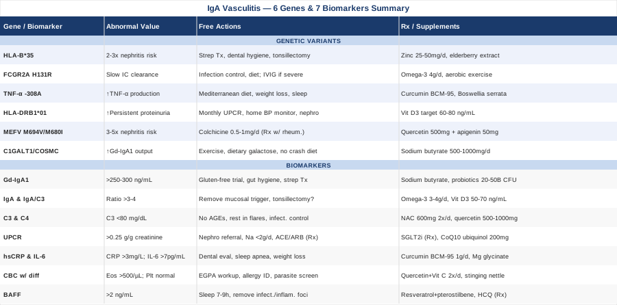
This article takes a more targeted approach. Rather than repeating broad advice about symptom monitoring and rest, it focuses on the seven most informative biomarkers for IgA vasculitis and six genetic variants that current research links to susceptibility, severity, and renal outcomes. Each section is designed to be practical: what to measure, what an abnormal result means, and what evidence-based steps — with or without supplements — may help if results come back concerning.
None of this replaces specialist care. But better information leads to more targeted conversations. When you understand what galactose-deficient IgA1 means, why complement C3 matters as much as CRP, or how a specific MEFV variant can multiply kidney risk three to fivefold, you can ask better questions and advocate more effectively. The sections ahead build that picture systematically — starting with the seven biomarkers you can begin tracking today, then moving into the genetic layer that shapes the underlying risk, then the gut-immune science that is reshaping how experts think about this condition entirely.
Summary
Most patients with IgA vasculitis are never told about galactose-deficient IgA1 — a molecular marker at the center of the disease mechanism that can predict kidney damage before it becomes irreversible. They are rarely told that a single variant in the MEFV gene can multiply nephritis risk three to five times, or that their complement C3 level reveals something their CRP cannot.
This article maps out 7 measurable biomarkers that track IgA vasculitis with genuine precision — what each reveals, how to measure it with cost estimates, and what targeted steps with or without supplements may help when results come back abnormal. It then covers 6 genetic variants with documented links to disease risk, inflammatory severity, and renal outcomes, including what to do differently if you carry them.
Beyond the clinical layer, you will find a synthesis of the gut-immune research that is quietly overturning assumptions about what drives abnormal IgA production — plus five evidence-supported complementary approaches with real human trial data, including the Autoimmune Protocol from Dr. Sarah Ballantyne whose framework is directly relevant to any condition rooted in mucosal immune dysregulation.
If you have wondered why your case follows its own script, the answers are often written in these markers and variants. This article helps you read them.
7 Biomarkers to Track in IgA Vasculitis
Understanding IgA vasculitis in real time requires moving beyond a standard blood count or single CRP result. The seven biomarkers below form a practical, progressive monitoring panel — some available at any routine clinic visit, others requiring specialty labs — that together reveal inflammatory intensity, immune complex burden, and kidney trajectory with far more resolution than any single test provides.
Biomarker 1: Galactose-Deficient IgA1 (Gd-IgA1)
Why it matters
Gd-IgA1 sits at the mechanistic center of IgA vasculitis. Normal IgA1 molecules carry O-linked sugar chains — specifically galactose residues — attached at their hinge region. In IgA vasculitis and its renal-predominant sibling IgA nephropathy, a proportion of these sugar chains lose their galactose, leaving exposed N-acetylgalactosamine (GalNAc) residues that the immune system reads as foreign. This triggers immune complex formation: autoantibodies bind these aberrant IgA1 molecules, producing macromolecular aggregates that deposit in small vessel walls and glomerular mesangium, activating complement and driving vascular injury. The higher the circulating Gd-IgA1 level, the greater the supply of this immune complex-forming substrate — and the higher the risk of kidney involvement. Multiple cohort studies in IgA nephropathy have confirmed Gd-IgA1 as an independent predictor of disease activity, proteinuria, and long-term renal function decline.
How to measure it
Gd-IgA1 is measured via a lectin-based ELISA assay using Helix aspersa agglutinin (HAA), which selectively binds exposed GalNAc residues. This test is not part of standard clinical panels at most hospitals but is available through academic nephrology centers and specialty reference laboratories. Approximate cost: $150–$400. Levels above 250–300 ng/mL have been associated with active disease and nephritis risk in published cohorts, though formal clinical cutoffs are still being standardized. A serum sample is sufficient.
If the score is high: the plan without supplements
Elevated Gd-IgA1 reflects a glycosylation defect in IgA-producing B cells in the gut and tonsils that is partly reversible through mucosal immune normalization. The most evidence-supported approach without supplements is reducing mucosal immune overactivation: eliminating recurring streptococcal infections through prompt treatment and hygiene, conducting a minimum 3-month gluten-free diet trial (particularly if any gluten sensitivity exists — small IgA nephropathy trials show IgA reductions in sensitive patients), and addressing coexisting gut dysbiosis, since the gut-associated lymphoid tissue is the primary site of both normal and abnormal IgA production.
If the score is high: the plan with supplements or equipment
Sodium butyrate (500–1000 mg/day): Butyrate — a short-chain fatty acid produced by gut bacteria — has shown in preclinical studies the capacity to upregulate COSMC, the molecular chaperone required for C1GALT1 to correctly galactosylate IgA1. Begin with dietary butyrate through resistant starch (green banana flour, cooked and cooled potato, 15–20 g/day) before supplementing directly. Continuous use; reassess at 3 months. Side effects: occasional bloating in week one.
Multi-strain probiotics (20–50 billion CFU/day): Strains including Lactobacillus rhamnosus GG and Bifidobacterium longum BB536 have demonstrated modulation of gut mucosal IgA secretion and may reduce the B-cell stimulation driving abnormal IgA1 production over time. Side effects: rare, minimal.
Biomarker 2: Serum IgA Level and the IgA/C3 Ratio
Why it matters
Total serum IgA is elevated in approximately 50–70% of adults with IgA vasculitis during active disease. While non-specific in isolation, in context it serves as a practical correlate of disease activity and mucosal immune activation. More informative is the IgA/C3 ratio: when IgA is elevated alongside low-normal or reduced C3, the pattern reflects active immune complex formation with ongoing complement consumption — a profile associated with more aggressive disease and higher nephritis risk. Persistently elevated IgA after clinical remission suggests the immune stimulus driving abnormal IgA production has not been eliminated, and that recurrence probability remains higher than it appears.
How to measure it
Standard serum immunoglobulin panel (IgA, IgG, IgM) at any commercial laboratory. Cost: $30–$80. Normal adult IgA: 70–400 mg/dL. Calculate the ratio by dividing IgA (mg/dL) by C3 (mg/dL); values above 3–4 during active disease suggest significant immune complex load. Repeat at 3-month intervals during active phases and every 6 months in remission.
If the score is high: the plan without supplements
The most effective step is identifying and removing the mucosal IgA trigger. The three most commonly overlooked sources are recurrent streptococcal pharyngitis (track any sore throat episodes against IgA flares), gastrointestinal dysbiosis or food antigen sensitivity (a structured 4-week elimination trial can identify this), and chronic dental or tonsillar infection. Tonsillectomy has shown meaningful IgA reductions in Japanese cohorts of IgA nephropathy patients with documented tonsillar infection foci — a finding worth discussing with your specialist if repeated streptococcal pharyngitis is clearly triggering flares.
If the score is high: the plan with supplements or equipment
Omega-3 fatty acids (EPA + DHA combined, 3–4 g/day): The landmark controlled trial by Donadio and colleagues in the New England Journal of Medicine (1994) established that fish oil supplementation significantly slowed kidney function decline in IgA nephropathy, with subsequent analyses confirming IgA-lowering effects. Use enteric-coated formulations to minimize GI side effects. Monitor for increased bleeding time above 4 g/day.
Vitamin D3 targeting 50–70 ng/mL (25-OH-D3): Vitamin D suppresses IgA-promoting B-cell differentiation and Tfh cell activity. Deficiency below 30 ng/mL is common in this population and independently associated with faster IgA nephropathy progression. Supplement with D3 and K2-MK7 (100 mcg) at 2,000–5,000 IU/day depending on baseline. Recheck at 3 months.
Biomarker 3: Complement C3 and C4
Why it matters
IgA vasculitis activates complement predominantly through the lectin pathway — MASP-1 and MASP-2 bind galactose-deficient IgA1 immune complexes and initiate the cascade. As complement is consumed in this process, circulating C3 falls. Low C3 during an active flare is a strong indicator of immune complex load and has been associated in observational studies with more severe organ involvement, particularly nephritis. A C3 below 80 mg/dL during a flare should prompt nephrology referral even if urinalysis appears clean — subclinical glomerular injury may already be occurring. C4 is less consistently reduced (the lectin pathway partially bypasses C4 cleavage), but low C4 alongside low C3 can indicate co-activation through the classical pathway, a pattern seen in more inflammatory adult presentations.
How to measure it
Complement panel (C3, C4, CH50) at any clinical laboratory. Cost: $30–$100 for the full panel. Normal C3: 90–180 mg/dL; Normal C4: 16–47 mg/dL. Serial measurements every 4–6 weeks during active disease and every 3–6 months in remission are more informative than a single-point reading.
If the score is low: the plan without supplements
Low complement during active disease reflects consumption by circulating immune complexes. The primary non-supplement intervention is reducing immune complex-forming substrate: aggressive infection management, dietary reduction of advanced glycation end products (AGEs from heavily grilled or fried foods, which independently activate the lectin pathway), and avoiding strenuous exercise during flares, which can transiently redistribute immune complexes into small vessel walls.
If the score is low: the plan with supplements or equipment
N-acetylcysteine (NAC, 600 mg twice daily): As a glutathione precursor and anti-inflammatory agent, NAC has shown complement-modulating effects in several inflammatory conditions. Evidence in IgA vasculitis is indirect. Low-risk adjunct; discuss with physician before adding. Cycling: 3 months on, reassess.
Quercetin (500–1000 mg/day with meals): In vitro studies have shown quercetin inhibits lectin pathway complement activation — the dominant complement route in IgA vasculitis. Avoid concurrent use with fluoroquinolone antibiotics. Cycling: 8 weeks on, 2 weeks off. Side effects: mild GI upset occasionally; generally well tolerated.
Biomarker 4: Urine Protein-to-Creatinine Ratio (UPCR)
Why it matters
Renal involvement is the most serious complication of IgA vasculitis, occurring in 20–60% of adults during the initial episode. UPCR is the most sensitive early warning signal for glomerular injury — consistently more reliable than dipstick urinalysis, which routinely misses low-level proteinuria in the 100–200 mg/day range. In adults, even modest but rising proteinuria (above 0.1 g/g creatinine) warrants close follow-up, because trajectory is as informative as absolute value. Microhematuria combined with proteinuria is particularly concerning and should never be attributed to incidental causes without investigation — this combination on consecutive urine samples is a hallmark of glomerulonephritis and a strong indication for nephrology referral.
How to measure it
A single random urine sample (first morning void preferred) submitted for protein and creatinine at any clinical laboratory. Cost: $10–$30. Threshold for adult concern: above 0.25 g/g (25 mg/mmol). Recommended monitoring schedule: at 1, 3, 6, and 12 months after the initial episode, then annually in remission. Automated home dipstick systems such as Minuteful Kidney offer semi-quantitative screening between clinic visits for approximately $30–$50 per month — a practical option for high-risk or recently diagnosed patients.
If the score is high: the plan without supplements
A high UPCR is a medical urgency. Immediate steps are nephrology referral and kidney biopsy consideration if proteinuria exceeds 0.5 g/day or is rising. From a supportive standpoint, sodium restriction below 2 g/day reduces intraglomerular pressure and slows proteinuria progression — the most evidence-backed dietary intervention across all forms of glomerulonephritis. Blood pressure control below 130/80 mmHg using an ACE inhibitor or ARB provides renoprotection beyond blood pressure lowering through modulation of glomerular filtration pressure and TGF-β signaling. Vigorous exercise should be paused during episodes of heavy proteinuria.
If the score is high: the plan with supplements or equipment
SGLT2 inhibitors (prescription — discuss with nephrologist): Dapagliflozin and empagliflozin demonstrated renoprotective effects in the DAPA-CKD and EMPA-KIDNEY trials, including specific benefit in IgA nephropathy subgroups. These are increasingly used in IgA vasculitis nephritis off-label and are worth discussing explicitly with your nephrology team if UPCR remains elevated on standard treatment.
Coenzyme Q10 (ubiquinol form, 200–300 mg/day): Mitochondrial protectant with proteinuria-reducing effects in small glomerulonephritis trials, through podocyte energy metabolism support. Ubiquinol has superior bioavailability to standard CoQ10. Side effects: minimal. Daily use; reassess at 3 months.
Biomarker 5: High-Sensitivity CRP (hsCRP) and Interleukin-6 (IL-6)
Why it matters
CRP is an acute-phase reactant produced by the liver in response to IL-6, itself secreted by macrophages in inflamed vessel walls. In IgA vasculitis, hsCRP provides a real-time readout of systemic vasculitic inflammation. Its trajectory during and after a flare — how high it rises, how quickly it returns to baseline, and whether true baseline is actually below 1 mg/L — is more clinically useful than any single measurement. IL-6 is the upstream signal most worth monitoring in adults with frequent recurrence or refractory disease. A persistently elevated IL-6 (above 7 pg/mL) between clinical flares may indicate smoldering vasculitis or occult infection-driven inflammation — a pattern that can precede a major flare by weeks and that is increasingly targeted pharmacologically with IL-6 inhibitors in refractory adult cases.
How to measure it
hsCRP: $20–$50 at any clinical lab. Optimal chronic disease target: below 1 mg/L. A level above 3 mg/L between flares consistently warrants upstream investigation. IL-6: $80–$200 at specialty reference labs. Most useful in adults with recurrent or atypical presentations.
If the score is high: the plan without supplements
Elevated hsCRP between flares almost always indicates a persistent inflammatory driver. The three most commonly overlooked sources before turning to supplements:
Chronic periodontal disease — among the most potent sustained IL-6 sources in the body; dental evaluation and treatment can reduce hsCRP by 0.5–1.5 mg/L in controlled trials. Sleep apnea — drives repetitive nighttime IL-6 surges through intermittent hypoxia; if snoring and daytime fatigue coexist with elevated hsCRP, a sleep study should precede any supplement protocol. Visceral adiposity — adipose tissue is a major IL-6 source independent of diet; a 5–7% reduction in body weight reduces hsCRP proportionally.
If the score is high: the plan with supplements or equipment
Curcumin in bioavailable form (BCM-95 or phospholipid complex, 500–1000 mg/day): Multiple meta-analyses confirm clinically significant reductions in CRP and IL-6 with high-absorption curcumin formulations. Standard curcumin powder has negligible bioavailability — formulation is critical. Cycling: 8 weeks on, 2 weeks off. Avoid with anticoagulants at high doses.
Magnesium glycinate (300–400 mg elemental magnesium/day): Magnesium deficiency is prevalent and independently associated with elevated CRP. This form has high bioavailability and GI tolerability. Side effects: loose stools above 500 mg/day elemental dose.
Biomarker 6: Complete Blood Count — Platelet Count and Eosinophils
Why it matters
The CBC in IgA vasculitis serves two specific and clinically critical purposes. First, a normal platelet count is a defining feature of IgA vasculitis and the primary test that distinguishes it from immune thrombocytopenic purpura — both conditions cause purpura, but IgA vasculitis occurs with a normal platelet count. Thrombocytopenia in the presence of purpura demands a completely different diagnostic and treatment path; this distinction cannot be missed.
Second, peripheral eosinophilia (eosinophils above 500/μL) requires urgent attention. It can indicate a parasitic infection triggering vasculitis, an atopic immune component, or — critically — eosinophilic granulomatosis with polyangiitis (EGPA), a distinct vasculitis that can mimic IgA vasculitis but requires entirely different immunosuppression. Missing EGPA in a workup labeled as IgA vasculitis is a clinically consequential error.
How to measure it
Standard CBC with differential: $15–$40 at any clinical lab. No preparation required. Repeat at each flare and routinely in patients on immunosuppression.
If the eosinophil count is high: the plan without supplements
Elevated eosinophils require systematic cause investigation before any supplement protocol: stool ova and parasites, serology for Toxocara and Ascaris, IgE panel for environmental and food allergens, and EGPA workup (p-ANCA, chest HRCT) if eosinophilia exceeds 1500/μL. If atopy is identified as the driver, structured elimination of the relevant allergen normalizes eosinophils within 3–6 weeks in most cases.
If the eosinophil count is high: the plan with supplements or equipment
Quercetin (500 mg) + vitamin C (1000 mg) twice daily: This combination stabilizes mast cells and inhibits eosinophil chemotaxis in allergic inflammation. Evidence is strongest in allergic rhinitis but the mechanism is directly relevant to eosinophilia of allergic origin. Side effects: minimal.
Freeze-dried stinging nettle leaf (300–600 mg twice daily): Has shown antihistamine and anti-eosinophilic effects in RCTs for allergic conditions. Use during symptomatic periods. Cycling: 6–8 weeks. Side effects: minimal.
Biomarker 7: BAFF (B-Cell Activating Factor)
Why it matters
BAFF — also known as BLyS — is a cytokine that promotes the survival, proliferation, and differentiation of B cells into IgA-secreting plasma cells. In IgA vasculitis and IgA nephropathy, elevated serum BAFF has been associated with higher total IgA production, greater Gd-IgA1 formation, and a more active, treatment-resistant disease trajectory. BAFF is now an active therapeutic target: belimumab (an anti-BAFF monoclonal antibody) is under active investigation in IgA nephropathy trials, with early signals of proteinuria reduction and IgA normalization. From a monitoring standpoint, a high BAFF level during apparent clinical remission suggests B-cell activation is still running at elevated baseline — and that relapse probability is higher than it appears from clinical presentation alone.
How to measure it
BAFF is measurable via ELISA at specialty immunology and rheumatology reference laboratories. Cost: $100–$300. Normal serum BAFF: approximately 0.3–1.0 ng/mL; levels above 2 ng/mL are associated with active B-cell driven inflammation. Not yet a standard clinical panel, but available through academic medical centers and selected commercial reference labs.
If the score is high: the plan without supplements
High BAFF indicates sustained upstream B-cell stimulation that supplements alone cannot reliably normalize. The most important step is removing the activating signal — typically mucosal infections, gut dysbiosis, or chronic inflammatory foci. Sleep optimization is also directly relevant: BAFF secretion is partially regulated by circadian signals, and chronic sleep deprivation elevates BAFF in ways that mimic low-grade autoimmune activation. Targeting consistent 7–9 hours of quality sleep is not a generic health tip in this context — it is a BAFF-modulating intervention.
If the score is high: the plan with supplements or equipment
Resveratrol + pterostilbene (500 mg + 50 mg daily): Both polyphenols have shown in vitro modulation of BAFF pathway signaling in B cells, and resveratrol has demonstrated BAFF-driven proliferation reduction in lupus models. Cycling: 12 weeks on, 4 weeks off. Side effects: resveratrol at high doses has mild estrogenic activity — avoid during pregnancy or in hormone-sensitive conditions.
Hydroxychloroquine (prescription — discuss with rheumatologist): This antimalarial has documented BAFF-lowering properties and is used off-label in IgA nephropathy and some autoimmune vasculitides at 200–400 mg/day. Worth raising explicitly with a specialist if BAFF remains elevated alongside disease activity markers.
The seven biomarkers above form the most actionable monitoring framework currently available for IgA vasculitis. Understanding the genetic layer beneath them helps explain why those numbers look the way they do — and which interventions are likely to have the greatest leverage in your specific case.
The Genetic Layer: 6 Variants That Shape Risk and Severity
IgA vasculitis has a clear genetic component: first-degree relatives of affected individuals carry elevated risk, identical twin concordance significantly exceeds fraternal, and certain ethnic groups show markedly higher disease prevalence. Genome-wide association studies and candidate gene analyses have identified a growing list of variants that influence IgA glycosylation, immune complex handling, and inflammatory signaling. None of these variants predicts disease with certainty — they shift probability, not destiny. But knowing which variants you carry can identify the specific biological pathways that deserve the most targeted attention, and most can be accessed through clinical HLA typing, rheumatology-ordered genetic panels, or direct-to-consumer sequencing platforms combined with raw-data interpretation tools.
Gene 1: HLA-B*35
What it is and why it matters
HLA-B*35 is among the most consistently replicated genetic associations in IgA vasculitis across multiple ethnic populations. The HLA-B gene encodes a class I MHC molecule that presents intracellular peptides to CD8+ T cells. The *35 allele appears to bias the immune response to common mucosal pathogens — particularly streptococcal antigens — in a way that amplifies IgA immune complex formation. Studies in European, Turkish, and Asian cohorts have found a 2–3-fold higher frequency of HLA-B*35 in IgA vasculitis patients compared to healthy controls. The association is stronger in patients with concurrent renal involvement, suggesting this allele amplifies nephritis risk above and beyond susceptibility to the initial vasculitic episode.
If the gene is bad: the plan without supplements
HLA-B*35 carriers benefit most from systematic streptococcal trigger management: throat cultures at every pharyngitis episode with prompt antibiotic treatment when culture-positive, discussion of prophylactic penicillin with a physician if streptococcal pharyngitis is a documented flare trigger, and optimization of dental hygiene since periodontitis can harbor streptococcal species that maintain low-grade IgA stimulation. Tonsillectomy merits explicit discussion when tonsils serve as a recurring streptococcal focus, given the strong evidence from Japanese IgA nephropathy cohorts.
If the gene is bad: the plan with supplements or equipment
Zinc gluconate or picolinate (25–50 mg/day): Zinc supports regulatory T-cell function, directly inhibits streptococcal mucosal adhesion, and has documented antimicrobial effects against common upper respiratory pathogens. Pair with copper (2 mg/day) to prevent zinc-induced copper deficiency at higher doses. Cycling: 3 months on, 1 month off.
Elderberry extract standardized (300–600 mg/day): Multiple RCTs confirm reduced upper respiratory infection duration and severity. Use seasonally (autumn and winter) rather than year-round. Avoid in patients on immunosuppressants requiring preserved immune clearance.
Gene 2: FCGR2A — H131R Polymorphism (rs1801274)
What it is and why it matters
FcγRIIa (encoded by FCGR2A) is a receptor on macrophages, neutrophils, and platelets that facilitates phagocytic clearance of IgG-coated immune complexes from circulation. The H131R variant (rs1801274) produces a receptor with reduced affinity for IgG2 — a subclass involved in opsonizing IgA immune complexes for macrophage removal. Individuals homozygous for the R131 allele have measurably slower immune complex clearance, resulting in prolonged circulation and greater deposition in vessel walls and glomeruli. In IgA vasculitis, the R131/R131 genotype has been associated in several cohort studies with higher nephritis risk and more severe clinical presentations. This variant can be identified through direct-to-consumer genetic testing and raw-data interpretation platforms.
If the gene is bad: the plan without supplements
Impaired immune complex clearance means that whatever substrate is generated, it circulates longer and deposits more extensively. The most effective counterstrategy is reducing total immune complex production — addressing the same upstream levers as Gd-IgA1 and serum IgA: infection control, dietary trigger elimination, and mucosal immune normalization. During active and severe flares, discussing IVIG with a specialist is worth considering: at doses used for autoimmune conditions, IVIG can accelerate Fc receptor-mediated immune complex clearance, the same mechanism exploited in Kawasaki disease treatment.
If the gene is bad: the plan with supplements or equipment
High-dose omega-3 (EPA + DHA, 4 g/day): Reduces immune complex formation by modulating IgA synthesis and complement activation, directly reducing the substrate burden that FcγRIIa must clear. Use during remission phases; monitor for bleeding time.
Moderate aerobic exercise (30 minutes, 5 days/week) outside of active flares: Upregulates macrophage Fc receptor expression and phagocytic capacity, partially compensating for the reduced FcγRIIa binding affinity of the R131 variant. This benefit is lost during active vasculitic episodes — rest during flares.
Gene 3: TNF-α -308G/A Promoter Variant (rs1800629)
What it is and why it matters
TNF-alpha is a master inflammatory cytokine that amplifies vasculitic tissue injury once immune complex deposition has initiated the inflammatory cascade. The -308G/A variant in the TNF gene promoter region increases transcription, resulting in higher TNF-α production in response to immune activation. Carriers of the A allele — whether G/A heterozygotes or A/A homozygotes — show elevated TNF-α both at baseline and during inflammatory challenges. Several IgA vasculitis cohort studies have found higher -308A allele frequency specifically in patients who develop nephritis compared to those without renal involvement, suggesting this variant amplifies the inflammatory response to immune complex deposition in the glomerulus.
If the gene is bad: the plan without supplements
A TNF-α-amplifying genotype means your inflammatory response, once engaged, burns hotter and likely longer than average. Reducing background inflammatory load carries proportionally greater benefit in high-TNF genotypes than in the general population. A high-glycemic diet independently amplifies TNF-α production and should be replaced with a Mediterranean-style pattern (the most rigorously studied dietary anti-inflammatory intervention). Visceral adiposity is a major autonomous TNF-α source — even modest weight loss of 5–7% reduces TNF-α levels significantly. Sleep deprivation of even a single night transiently doubles TNF-α production in controlled studies; sleep quality is not a secondary consideration for this genotype.
If the gene is bad: the plan with supplements or equipment
Curcumin (BCM-95 or nanoparticle form, 1 g/day): Among the best-studied natural TNF-α inhibitors; multiple meta-analyses confirm significant TNF-α reductions with bioavailable formulations. Standard curcumin powder is inadequate here — formulation determines efficacy. Cycling: 8 weeks on, 2 weeks off.
Boswellia serrata (65% AKBA standardized, 300 mg/day): Specifically inhibits 5-lipoxygenase with secondary TNF-α modulating effects. Well-tolerated at standard doses; no cycling required. Evidence is strongest in arthritis and inflammatory bowel conditions with shared inflammatory pathways.
Gene 4: HLA-DRB1*01
What it is and why it matters
HLA-DRB1 encodes class II MHC molecules involved in activating CD4+ T helper cells, which in turn direct B-cell class switching and antibody production. Certain HLA-DRB1 alleles have been linked in European cohort studies to nephritis risk in IgA vasculitis, with the *01 group associated with higher rates of persistent proteinuria after the initial episode. The proposed mechanism involves a Tfh (follicular helper T cell) differentiation bias that supports IgA class-switching and promotes the prolonged survival of Gd-IgA1-producing plasma cells in the bone marrow. Carriers may therefore have both more Gd-IgA1 production and a more sustained nephritis trajectory once kidney involvement begins. HLA typing is clinically available through rheumatology and transplant immunology services.
If the gene is bad: the plan without supplements
HLA-DRB1*01 carriers should adopt a proactive renal monitoring protocol from diagnosis: UPCR and urinalysis at 1, 2, 3, 6, and 12 months after the initial episode rather than the standard 3 and 6-month schedule. Weekly home blood pressure tracking using a validated upper-arm automated monitor ($40–$80) enables early detection of hypertension — the single most modifiable renal risk factor — before clinic visits would catch it. Nephrology co-management from the outset rather than waiting for proteinuria thresholds to be crossed is appropriate in this genotype context.
If the gene is bad: the plan with supplements or equipment
Vitamin D3 targeting 60–80 ng/mL serum 25-OH-D3: Vitamin D actively suppresses Tfh differentiation and IgA class-switching — the specific immune pathway amplified by HLA-DRB1*01. Carriers of this allele may have proportionally more to gain from vitamin D optimization than the average IgA vasculitis patient. Supplement D3 with K2-MK7 (100 mcg) at 3,000–5,000 IU/day depending on baseline. Recheck at 3 months.
Validated home blood pressure monitor: As above — an equipment recommendation, not a supplement. At-home BP monitoring enables early detection and prompt treatment of the most consequential modifiable renal risk factor for this genotype.
Gene 5: MEFV Variants — M694V and M680I
What it is and why it matters
The MEFV gene encodes pyrin, a protein that regulates IL-1β secretion through the NLRP3-related inflammasome. Biallelic pathogenic variants in MEFV cause familial Mediterranean fever (FMF). However, heterozygous carriers of the M694V or M680I variants — who do not have FMF — have been found in multiple independent studies to have significantly worse outcomes in IgA vasculitis. The M694V variant has been associated with a 3–5-fold increase in nephritis risk among IgA vasculitis patients in Turkish and Israeli cohorts. The mechanism is a lowered inflammasome threshold: heightened IL-1β secretion amplifies the inflammatory response to IgA immune complex deposition in vessel walls, transforming what would be a contained vasculitic episode into a more destructive one.
MEFV testing is clinically available through rheumatology and genetic testing services. In populations with higher FMF carrier rates — Mediterranean, Middle Eastern, Armenian, and Sephardic Jewish ancestries — this gene should be part of the standard IgA vasculitis workup.
If the gene is bad: the plan without supplements
MEFV heterozygous carriers with IgA vasculitis should discuss colchicine prophylaxis directly with their rheumatologist. Colchicine — the standard FMF treatment — has shown in controlled trials to reduce flare frequency and IL-1β-driven inflammation in MEFV carriers even without full FMF. It is inexpensive, well-characterized, and has a favorable long-term safety profile at standard doses. This is one of the clearest and most evidence-backed genotype-directed therapeutic recommendations available in IgA vasculitis management.
If the gene is bad: the plan with supplements or equipment
Colchicine (0.5–1 mg/day, prescription): Warrants specific mention despite crossing into medication territory given the strength of evidence in this genotype. Side effects: diarrhea at higher doses, resolving with dose adjustment. Avoid in significant renal impairment.
Quercetin + apigenin (500 mg + 50 mg daily): Both flavonoids have demonstrated NLRP3 inflammasome inhibitory activity in preclinical studies — directly relevant to the pyrin dysregulation caused by MEFV variants. Clinical evidence in MEFV carriers is still emerging, but the mechanism is on-target and the side effect profile is favorable. Daily use; no cycling required.
Gene 6: C1GALT1 and COSMC (Galactosylation Enzyme Variants)
What it is and why it matters
C1GALT1 encodes the enzyme that adds galactose to the O-linked sugar chains of IgA1 — the exact enzymatic step that fails in patients with elevated Gd-IgA1. COSMC (C1GALT1C1) encodes its obligate molecular chaperone; without functional COSMC, C1GALT1 misfolds and cannot attach galactose correctly. Variants that reduce C1GALT1 or COSMC activity directly increase the fraction of Gd-IgA1 produced — making these genes the most mechanistically proximal genetic risk factors in the disease. A genome-wide association study confirmed C1GALT1 as a genome-wide significant susceptibility locus for IgA nephropathy (Gharavi AG et al., Nat Genet, 2011), with the shared pathophysiology of IgA vasculitis making this finding directly relevant. Carriers of reduced-function C1GALT1 or COSMC variants are essentially biochemically primed to produce the IgA1 that drives disease.
If the gene is bad: the plan without supplements
Reduced galactosylation capacity can be partially compensated by supporting the biochemical environment in which B cells operate. Moderate aerobic exercise upregulates AMPK-dependent glycosylation enzyme expression in B cells — exercise during remission is a direct biological intervention, not generic advice. Avoiding prolonged caloric restriction matters here: aggressive dieting downregulates glycosyltransferase activity. Ensuring adequate dietary galactose from dairy, legumes, and certain vegetables provides the enzymatic reaction's substrate.
If the gene is bad: the plan with supplements or equipment
Sodium butyrate (500–1000 mg/day): Butyrate upregulates COSMC expression in IgA-producing B cells in preclinical models, directly compensating for the chaperone deficiency caused by COSMC gene variants. This is the most mechanistically grounded nutritional strategy currently available for targeting the galactosylation defect at its genetic source.
Dietary resistant starch (15–20 g/day): The preferred whole-food route to butyrate production — increases butyrate-producing bacteria (Faecalibacterium prausnitzii, Roseburia intestinalis) without supplementation. Green banana flour, cooked and cooled potato, and slightly undercooked whole oats are practical daily sources.
What the genetic and biomarker data share is a common thread: the gut, its microbial composition, and the mucosal immune responses it drives. The research on this connection has grown rapidly enough that it deserves its own focus — starting with what a rigorous, study-backed body of work has revealed about the gut-IgA axis.
What the Gut-Immune Research Reveals About IgA Dysregulation
The gut is not just where IgA vasculitis may be triggered — it is where IgA is produced. Up to 70–80% of the body's total IgA is generated in the gut-associated lymphoid tissue in response to the microbial environment there. The composition of the gut microbiome directly determines how much IgA the immune system secretes, what quality it produces, and how well the galactosylation process functions in IgA-producing plasma cells. The Huberman Lab has covered this connection in depth in a long-form episode featuring Dr. Justin Sonnenburg from Stanford's microbiome research group, synthesizing his landmark research including a 2021 randomized trial published in Cell. The ten most important and frequently overlooked findings from that work follow below — each has direct implications for IgA vasculitis management.
1. Fermented Foods, Not Just Fiber, Were the Real Game-Changer
Sonnenburg's 2021 Cell trial found that a diet high in fermented foods — kimchi, kefir, yogurt, kombucha, fermented vegetables — reduced 19 key inflammatory markers including IL-6, IL-17, and CXCL10 within 10 weeks. A high-fiber diet alone did not reduce inflammation in individuals with low baseline microbiome diversity. For IgA vasculitis management, this means fermented food intervention may be more immediately impactful than fiber supplementation alone for most patients, particularly those who have undergone antibiotics or whose diet has historically been low in fiber.
2. The Gut Produces More IgA Than Any Other Tissue in the Body
Secretory IgA in the gut lumen coats commensal bacteria and pathogens, shaping microbial community composition. Gut dysbiosis disrupts the coating pattern, and the resulting IgA misdirection — coating harmless commensal species rather than true pathogens — feeds a cycle of abnormal IgA stimulation that spills into systemic circulation. The gut is not peripheral to IgA vasculitis biology; it is central to it.
3. Microbiome Diversity Is an Immune Regulator, Not Just a Gut Metric
Higher microbiome diversity correlates with more balanced Th1/Th2/Th17 ratios, lower BAFF, and better B-cell regulatory control. Every factor that reduces diversity — antibiotics, ultra-processed food, chronic psychological stress, sleep deprivation — moves the immune system toward the kind of dysregulation that characterizes IgA vasculitis. Microbiome diversity is a tracking variable as meaningful as serum IgA.
4. Antibiotic Exposure Can Reshape IgA Production for Years
A single course of broad-spectrum antibiotics can reduce populations of butyrate-producing bacteria by up to 90%, with recovery taking months to years. The downstream effect includes reduced COSMC expression in gut IgA-producing B cells — directly increasing the fraction of Gd-IgA1 produced during the recovery period. Mapping antibiotic history relative to flare timing is a clinically informative exercise that most patients have never been asked to do.
5. Dietary Changes Shift the Microbiome Faster Than Most People Expect
The microbiome can shift measurably within 3–4 days of significant dietary change. This is an encouraging finding: meaningful immune modulation through diet is not a multi-year project. Even short-term dietary interventions during remission — increased fermented foods, diverse plant fiber — can reduce the mucosal immune stimulation driving IgA abnormality within weeks.
6. Mucin Layer Health Depends on Fiber Diversity, Not Fiber Quantity
The mucin layer separating gut bacteria from the underlying immune tissue depends on diverse polysaccharide inputs to remain thick and intact. A diet diverse in plant fiber types — not just total grams — prevents mucin layer degradation that allows bacterial antigens to reach and abnormally stimulate IgA-producing cells in the lamina propria. Fiber variety matters more than fiber quantity.
7. Short-Chain Fatty Acids Are the Primary Gut-Immune Regulatory Signal
Butyrate, propionate, and acetate produced by gut bacteria act directly on immune cells in the intestinal lamina propria, regulating IgA production, B-cell class-switching, and regulatory T-cell differentiation. Declining butyrate production — from dysbiosis, antibiotics, or low-fiber diet — removes the primary physiological brake on excessive IgA secretion. This makes butyrate production capacity a central modifiable factor in IgA vasculitis biology.
8. Stress Hormones Directly Alter Mucosal IgA Secretion
Cortisol and catecholamines have direct receptors on IgA-secreting plasma cells in the gut lining. Acute psychological stress can reduce secretory IgA within 20 minutes while simultaneously increasing serum IgA — a shift that moves IgA distribution from protective mucosal coating toward systemic immune complex formation. Chronic stress is not a background variable in IgA vasculitis; it is a direct and measurable disease driver.
9. Sleep Is the Single Most Impactful Restoration Tool for Gut Immune Health
During deep sleep, the gut undergoes mucin layer repair, butyrate-producing bacteria peak in activity, and regulatory T cells in the gut-associated lymphoid tissue undergo phenotypic reset. Even a single night of poor sleep measurably reduces gut IgA regulatory capacity. In a condition where flares are often preceded by infection and stress — themselves sleep disruptors — sleep quality is both a monitoring signal and a high-leverage intervention target.
10. Individual Variation in Response to Dietary Interventions Is High
Sonnenburg's research consistently showed that identical dietary changes produce widely variable microbiome responses between individuals, shaped by existing microbiome composition, genetics (including HLA type), and antibiotic history. This is not discouraging — it is an argument for personalization. Stool microbiome testing through services such as Viome or Genova Diagnostics GI Effects can identify specific population gaps (low butyrate producers, absent Akkermansia, high Proteobacteria) that prebiotic, probiotic, or dietary interventions can address with far more precision than a generic protocol.
The gut-immune connection established above points toward several complementary approaches that work by modulating the same biological systems — inflammation load, mucosal immune balance, and autonomic nervous system tone. The most evidence-supported of these for conditions sharing IgA vasculitis's immune mechanisms are reviewed below.
Complementary Approaches with Human Evidence
The five modalities below have human clinical evidence — ranging from robust to preliminary — specifically for autoimmunity, small vessel inflammation, and mucosal immune dysregulation. Each addresses a different leverage point in the disease biology, and several are synergistic with the biomarker and genetic strategies described above.
The Autoimmune Protocol (AIP)
Developed by Dr. Sarah Ballantyne and detailed in The Paleo Approach and its companion cookbook, the Autoimmune Protocol is a structured dietary and lifestyle elimination framework designed specifically for autoimmune and immune-mediated conditions. It begins with a 30–90-day elimination phase removing grains, legumes, nightshades, eggs, dairy, nuts, seeds, alcohol, and refined sugar — all identified as potential sources of molecular mimicry antigens, gut permeability agents, or mucosal immune triggers. The elimination phase is followed by systematic reintroduction to identify individual-specific drivers.
The rationale for IgA vasculitis is direct. The AIP specifically targets gut epithelial integrity and mucosal immune activation — the same biological pathway feeding abnormal IgA production and Gd-IgA1 elevation. A 2017 randomized trial published in Inflammatory Bowel Diseases found that the Autoimmune Protocol achieved clinical remission in 73% of participants with active inflammatory bowel disease, a condition driven by the same mucosal IgA dysregulation central to IgA vasculitis. Ballantyne's framework explicitly addresses the role of increased intestinal permeability in perpetuating autoimmune cycles and provides specific tools to interrupt this pattern. The protocol has been used widely in IgA nephropathy and vasculitis communities, with substantial anecdotal reports of reduced flare frequency and improved laboratory markers.
Start the elimination phase during remission — not during a flare, when nutritional needs are higher and caloric restriction increases risk. Working with a registered dietitian familiar with elimination protocols significantly improves adherence and reintroduction accuracy. Reintroduce one food category per 3 days, monitoring UPCR, serum IgA, and symptom score before advancing. Most people identify their key dietary triggers within 60 days of systematic reintroduction. Full protocols and research are available through Dr. Ballantyne's published books and website.
Mindfulness-Based Stress Reduction (MBSR)
MBSR is a structured 8-week program developed by Jon Kabat-Zinn that combines mindfulness meditation, body scan practices, and gentle yoga. Its relevance to IgA vasculitis extends beyond general stress management: chronic psychological stress drives the cortisol-mediated shift in mucosal IgA distribution described in the gut section above, and MBSR's documented capacity to reduce IL-6, CRP, and HPA axis dysregulation means it operates on biomarkers directly relevant to disease monitoring and management.
A 2016 meta-analysis in Brain, Behavior, and Immunity covering 45 controlled studies found that mindfulness-based interventions significantly reduced circulating IL-6 and CRP — effect sizes directly relevant to the inflammatory biomarkers tracked in IgA vasculitis. A separate RCT in systemic lupus erythematosus showed that MBSR reduced both disease activity scores and IL-17 levels over 8 weeks, with effects maintained at 6-month follow-up. Given the shared autoimmune inflammatory architecture between lupus and IgA vasculitis, these findings are meaningful rather than merely analogical.
The full MBSR program is available in-person through certified instructors (find practitioners at the UMass Center for Mindfulness website) and online through Palouse Mindfulness at no cost. The core practice requires 20–45 minutes of daily formal meditation. If that commitment feels too large at the outset, start with 10 minutes daily of slow diaphragmatic breathing — studies show measurable cortisol reduction within 2 weeks. Build toward the full program over 4–6 weeks.
Microbiome-Directed Therapies
The gut microbiome's role in regulating IgA production and galactosylation quality is now well-established in translational research. Microbiome-directed therapies — targeted probiotic use, prebiotic dietary interventions, and precision supplementation — represent one of the most mechanistically grounded non-pharmacological approaches available for IgA vasculitis, directly addressing the gut-immune axis that feeds the disease process.
A 2022 systematic review in Frontiers in Immunology documented that IgA nephropathy patients — sharing IgA vasculitis's core immune mechanism — show consistent reductions in butyrate-producing bacteria (Faecalibacterium prausnitzii and Roseburia intestinalis) and elevated inflammatory Proteobacteria compared to healthy controls. Targeted interventions restoring these populations showed modest but consistent reductions in serum IgA and proteinuria markers in small clinical trials. Fecal microbiome transfer (FMT) for IgA conditions is investigational and not yet appropriate outside of controlled research settings.
For practical application, begin with feeding existing butyrate-producing bacteria through dietary resistant starch (10–20 g/day from green banana flour, cooked and cooled potatoes, whole legumes) before considering capsule supplementation. A 4–6-week targeted probiotic trial with strains including Lactobacillus rhamnosus GG, Bifidobacterium longum BB536, and encapsulated Akkermansia muciniphila is a reasonable second step. Stool microbiome analysis (Viome or Genova Diagnostics GI Effects) can identify which bacterial populations need the most support and make subsequent interventions meaningfully more precise. Retest at 3 months to assess microbiome trajectory.
Chinese Herbal Medicine
Chinese herbal medicine has a substantial clinical trial database for kidney and vascular inflammatory conditions, driven by the high prevalence of IgA nephropathy in East Asian populations and decades of parallel TCM and conventional treatment research. The most studied formula relevant to IgA vasculitis is Huangqi (Astragalus membranaceus) granules, which have shown in multiple Chinese RCTs the capacity to reduce proteinuria, serum IgA, and inflammatory markers in IgA nephropathy alongside conventional treatment. Compound formulations including Tripterygium glycoside tablets have also been evaluated as adjuncts to corticosteroid therapy in IgA nephropathy, with some trials reporting significant proteinuria reduction.
A meta-analysis of Chinese herbal medicine in IgA nephropathy published in Evidence-Based Complementary and Alternative Medicine found that herbal formulas added to conventional treatment significantly reduced 24-hour proteinuria and improved renal function markers compared to conventional treatment alone across included trials. It is important to note that most trials were conducted in China with preparations standardized in that context, and product quality varies significantly in Western markets. Evidence strength is moderate rather than definitive, and most trials have methodological limitations.
Practical application requires working with a licensed TCM practitioner experienced in autoimmune kidney conditions. Self-prescribing from online vendors is explicitly not recommended — several Tripterygium preparations carry hepatotoxicity and reproductive toxicity risk without professional guidance and dose titration. For any formula used, request a certificate of analysis confirming testing for heavy metals and microbial contamination — a legitimate concern with some imported herbal products.
Breathing-Based Therapies
Slow diaphragmatic breathing at approximately 5–6 breaths per minute activates the vagal-parasympathetic nervous system, suppresses cortisol, reduces TNF-α and IL-6, and improves heart rate variability (HRV). In IgA vasculitis, where psychological stress directly amplifies mucosal IgA dysregulation through the HPA axis and catecholamine-driven shifts in gut IgA secretion, improving vagal tone through breathing is a direct immunological intervention — not a relaxation technique.
A 2023 RCT published in Cell Reports Medicine found that structured breathwork, including cyclic sighing (double inhale through the nose, extended exhale through the mouth) practiced for 5 minutes daily, produced greater reductions in anxiety, physiological arousal, and resting respiratory rate compared to mindfulness meditation alone. Reductions in cortisol and improvements in HRV were observed within the first 4 weeks, with benefits sustained through the trial period.
The practical daily protocol for IgA vasculitis management: 5 minutes of 5-breath-per-minute diaphragmatic breathing (4-second inhale, 6-second exhale) at the most stressful point of the day and before sleep. Biofeedback-assisted breathing using a wearable device such as the Garmin HRV4Training app or a dedicated HRV monitor can quantify vagal tone improvement over time, making the intervention measurable and the progress concrete. Begin with unguided practice before investing in devices — most consistent practitioners achieve measurable HRV improvement within 2–3 weeks.
Conclusion
IgA vasculitis is a condition with real biological complexity beneath its clinical label. The path to better outcomes — fewer flares, preserved kidney function, or simply understanding why your case follows its own trajectory — runs through that complexity rather than around it. The seven biomarkers in this article represent the most actionable layer: measurable signals that can guide specialist conversations, inform intervention timing, and confirm whether the steps you are taking are actually working. The six genetic variants add the context that explains individual differences in risk and severity — and points toward the specific pathways most worth targeting in your case.
The smartest next step is usually the simplest one available today. Get a complement panel, a UPCR, and an hsCRP if you have not had them recently. Ask your specialist whether Gd-IgA1 or BAFF testing is available through their network. Review your antibiotic and infection history against the genetic variants most common in your ancestry. If you are working with a rheumatologist or nephrologist, bring specific questions about MEFV testing, SGLT2 inhibitor eligibility, or targeted probiotic protocols — those conversations are far more productive when you arrive with precise, informed questions.
Better information is not a cure. But it is the foundation on which better decisions are built, and better decisions, compounded over months, are where real outcomes are made.
Musculoskeletal: Joint Conditions
Cardiovascular: Vascular Conditions
Skin: Inflammatory Skin Conditions
Autoimmune: Inflammatory Conditions
Urological: Kidney Conditions