This article contains AI generated content.
Iliotibial Band Calcification: 6 Genes And 7 Biomarkers To Track
Introduction
If you're dealing with iliotibial band calcification, you've probably already heard the standard script: rest, ice, foam roll, stretch the hip, strengthen the glutes. That advice isn't wrong, but it's built for garden-variety IT band friction, not for a chalky deposit that shows up on imaging and doesn't budge no matter how disciplined your mobility routine is. When calcium is actually depositing in soft tissue, the mechanical explanation only tells part of the story.
Generic advice tends to stop at the level of the tendon itself — tightness, friction, overuse. It rarely asks the upstream question: why is calcium ending up there in the first place, and what is regulating where calcium goes in your body at all. That's a systemic, biochemical question, not a stretching question, and it requires looking at the blood work and, in some cases, the genetics that govern mineral metabolism and connective tissue quality.
This article takes that deeper route. Instead of repeating stretch protocols, it walks through the biomarkers that reveal how your body is handling calcium, phosphate, inflammation, and the vitamin K-dependent proteins that act as calcification's traffic control system — plus the genetic variants that can predispose certain people to laying down calcium where it doesn't belong.
None of this promises a cure, and nobody should expect a single blood test or gene panel to explain a calcified deposit on its own. But better information changes the decisions you can make. This piece covers the biomarkers worth tracking, the genetic variants worth understanding, a book that reframes how calcium and vitamin K interact, and a handful of complementary approaches with real supporting evidence — each one narrowing the gap between "just rest it" and actually understanding what's going on.
Summary
Iliotibial band calcification sits at an odd crossroads: it looks like a mechanical overuse problem, but underneath it may involve the same calcium-regulation machinery that governs arterial and cartilage calcification elsewhere in the body. Below, you'll find seven blood biomarkers — from basic serum calcium to a specialized vitamin K-dependent protein most doctors never order — that reveal whether your body is managing mineral deposition well or poorly, along with concrete, cost-aware plans for each one. After that, six genes tied to collagen structure and calcification inhibition show why some people seem more prone to this than others, and what — realistically — can be done about it. A book that upends conventional calcium-and-vitamin-D advice, plus a short review of complementary approaches with actual evidence behind them, round out the picture. If you've been told to just "give it time," the numbers below are a more precise place to start.
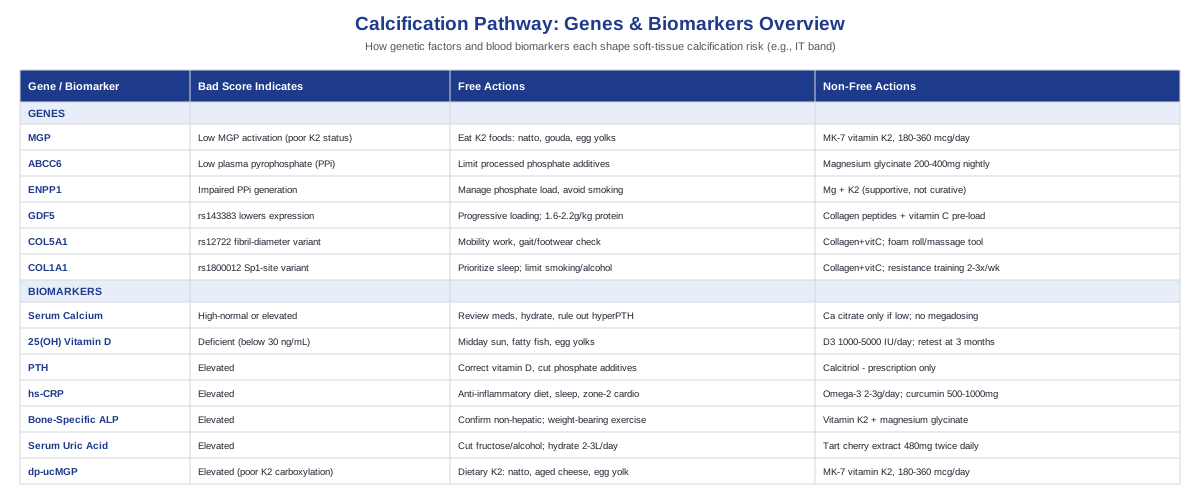
7 Biomarkers That Reveal Your Calcification Risk
Before looking at genes, it's worth looking at what's happening right now in your blood. Genes describe predisposition; biomarkers describe current state — and current state is what you can actually act on. The approach below borrows from the tracking philosophy popularized by physicians like Peter Attia: don't guess about a mechanism, measure it, then re-measure after an intervention to see if it actually moved. That's especially relevant here, because calcification is downstream of a handful of measurable, modifiable systems: calcium handling, vitamin D and parathyroid signaling, systemic inflammation, bone turnover, and — the piece most people have never heard of — vitamin K-dependent calcification inhibitors.
Serum Calcium (Total and Ionized)
Total and ionized calcium reflect how much calcium is circulating and available for deposition. This isn't a direct measure of what's happening inside the IT band, but chronically elevated calcium — even mild, "high-normal" elevation — increases the overall mineral load your body has to manage, and secondary causes of high calcium (mild hyperparathyroidism, excess supplementation, certain medications) are worth ruling out when calcification shows up somewhere it shouldn't.
How to measure it: a basic metabolic panel includes total calcium and costs roughly 10 to 20 dollars out of pocket if not already covered by insurance; ionized calcium is a more accurate but slightly pricier add-on, typically 20 to 40 dollars, and is more reliable if you have abnormal protein levels.
If the score is bad, the plan without supplements: review medications (thiazide diuretics, lithium, excess calcium-fortified foods), improve hydration, and ask your physician to rule out primary or secondary hyperparathyroidism. Retest in 4 to 6 weeks after any change.
If the score is bad, the plan with supplements or equipment: this is one of the few markers where the fix is rarely "add a supplement" — high calcium calls for identifying the source, not adding more. If calcium is low rather than high and contributing to secondary hyperparathyroidism, calcium citrate 500 to 600 mg with meals, retested every 8 to 12 weeks, is reasonable; megadosing calcium on your own is a bad idea and can itself raise ectopic calcification risk.
25-Hydroxyvitamin D
Vitamin D status governs calcium absorption and parathyroid hormone output. Deficiency is common, and research on calcific tendinopathy has proposed that low vitamin D can trigger transient secondary hyperparathyroidism, which may itself be an aetiopathogenetic factor in heterotopic (ectopic) calcification around tendons — see this discussion of calcific tendinopathy and vitamin D status. Vitamin D deficiency has also been linked to worse tendon-healing outcomes after rotator cuff repair, a related tendon-and-bone-interface tissue, in a retrospective cohort study.
How to measure it: a standard 25(OH)D blood test runs about 40 to 100 dollars out of pocket; at-home fingerstick kits are available for around 60 dollars if you want to track it without a clinic visit.
If the score is bad, the plan without supplements: 15 to 20 minutes of midday sun exposure several times a week (adjusted for skin tone and latitude), and more dietary fatty fish, egg yolks, and fortified foods.
If the score is bad, the plan with supplements or equipment: vitamin D3, 1000 to 2000 IU daily for maintenance, or up to 4000 to 5000 IU daily short-term if you're significantly deficient, under physician guidance. Retest at 3 months and taper down once replete — don't stay on a high maintenance dose indefinitely. Side effects at appropriate doses are rare, but excessive, unsupervised high-dose D3 without adequate vitamin K2 can theoretically push more calcium into circulation than your body can direct properly, which is exactly the dynamic explored later in this article.
Parathyroid Hormone (PTH)
PTH is the master regulator connecting vitamin D, calcium, and bone. Elevated PTH — whether from vitamin D deficiency, kidney disease, or a primary parathyroid problem — increases bone calcium turnover and shifts the body's mineral-handling equilibrium, which is part of why it's flagged as a plausible contributor to heterotopic calcification in the tendon research cited above.
How to measure it: intact PTH is typically ordered alongside calcium and vitamin D, adding roughly 50 to 100 dollars if paid out of pocket.
If the score is bad, the plan without supplements: correct the underlying driver first — usually vitamin D repletion — and reduce excess dietary phosphate from processed foods and phosphate-containing additives, which compete with calcium regulation. Retest in 6 to 8 weeks.
If the score is bad, the plan with supplements or equipment: there's no supplement that lowers PTH directly; correcting vitamin D is the main lever available without a prescription. In cases of secondary hyperparathyroidism from kidney disease, active vitamin D analogs (calcitriol) exist but are prescription-only and require close monitoring for hypercalcemia — this is not a self-directed intervention.
High-Sensitivity CRP (hs-CRP)
hs-CRP is a general marker of low-grade systemic inflammation. Chronic low-grade inflammation is a known driver of tendon degeneration, and research in osteoarthritis has shown that systemic hs-CRP levels correlate with local joint inflammation, likely via synovial IL-6 signaling — see this study on hs-CRP and local inflammatory findings. Tendon and connective tissue that stay in a low-grade inflamed state for a long time are the same tissues where degenerative calcific deposits tend to form.
How to measure it: an hs-CRP test is inexpensive, typically 15 to 30 dollars, and widely available through standard labs or direct-to-consumer testing services.
If the score is bad, the plan without supplements: prioritize an anti-inflammatory eating pattern (less refined carbohydrate and processed food, more fatty fish and fiber), consistent sleep of 7 to 9 hours, weight management, and 150 minutes weekly of moderate (zone 2) cardio.
If the score is bad, the plan with supplements or equipment: omega-3 fish oil, 2 to 3 grams combined EPA/DHA daily, taken continuously with periodic reassessment; curcumin with piperine, 500 to 1000 mg daily for 8 to 12 weeks, then reassess hs-CRP. Side effects: fish oil can mildly thin the blood, so use caution if you're on anticoagulants; curcumin can cause GI upset and should be avoided with active gallstone disease.
Bone-Specific Alkaline Phosphatase (ALP)
Bone-specific ALP reflects osteoblastic activity — how actively your body is building and remodeling mineralized tissue. Elevated levels can indicate an active mineralization process, which is relevant when soft tissue is starting to lay down calcium in a bone-like pattern, as happens in calcific tendon deposits.
How to measure it: a bone-specific ALP isoenzyme panel costs about 40 to 80 dollars; it's worth pairing with GGT to rule out a liver source of elevated total ALP first.
If the score is bad, the plan without supplements: confirm the source isn't hepatic, add regular weight-bearing exercise to support healthy bone remodeling signaling, and retest in 8 to 12 weeks.
If the score is bad, the plan with supplements or equipment: vitamin K2 (detailed below) to help direct mineralization appropriately, and magnesium glycinate, 200 to 400 mg nightly, which supports enzymatic cofactor function in bone metabolism. Cycle for 3 months, then reassess dietary sufficiency before continuing. Side effect: loose stools at higher magnesium doses — reduce if this occurs.
Serum Uric Acid
Uric acid matters here for two reasons: it helps distinguish calcium pyrophosphate deposition disease (CPPD, sometimes called pseudogout) from gout when a joint or peri-tendinous area shows crystal deposits, and it's a broader marker of metabolic dysregulation that correlates with connective tissue calcification burden. A StatPearls review of calcium pyrophosphate deposition disease notes that basic workup for suspected CPPD should include calcium, magnesium, phosphate, PTH, and uric acid together, since these conditions can overlap or mimic each other.
How to measure it: 10 to 20 dollars as an add-on to a standard metabolic panel.
If the score is bad, the plan without supplements: reduce fructose, alcohol, and purine-heavy processed foods, increase hydration to 2 to 3 liters daily, and pursue gradual weight loss if indicated. Retest in 6 to 8 weeks.
If the score is bad, the plan with supplements or equipment: tart cherry extract, 480 mg twice daily, has modest supporting evidence for lowering uric acid; if a formal CPPD or gout diagnosis is made, urate-lowering medication is a physician-managed prescription decision, not a self-directed supplement choice. Side effects of tart cherry extract are minimal, mostly mild GI upset.
Dephosphorylated-Uncarboxylated Matrix Gla Protein (dp-ucMGP)
This is the least well-known biomarker on this list, and arguably the most directly relevant one. Matrix Gla Protein (MGP) is a vitamin K-dependent protein that actively inhibits calcification in soft tissue. When vitamin K status is poor, MGP stays in an inactive, "uncarboxylated" form — measured as dp-ucMGP — and calcification inhibition weakens. The landmark Rotterdam Study first linked higher dietary menaquinone (vitamin K2) intake to reduced cardiovascular calcification risk, and a broader systematic review of MGP and vascular calcification confirms the mechanistic link, though most of this evidence comes from vascular tissue rather than tendon tissue specifically — that extrapolation to IT band calcification is reasonable given shared biology, but it hasn't been directly studied in tendon.
How to measure it: dp-ucMGP isn't part of routine bloodwork; it's typically ordered through specialty or functional medicine labs, costing roughly 80 to 150 dollars. PIVKA-II is sometimes used as a cheaper, indirect proxy for overall vitamin K status (30 to 50 dollars) but is less specific.
If the score is bad, the plan without supplements: increase dietary vitamin K2 through natto, aged hard cheeses, egg yolks, and dairy from grass-fed animals; leafy green vitamin K1 supports baseline status but converts to K2 poorly in most people.
If the score is bad, the plan with supplements or equipment: MK-7 (a long-acting form of vitamin K2), 180 to 360 mcg daily — the dose range used in trials that showed a 30 to 50 percent reduction in dp-ucMGP over 12 to 24 weeks, such as this dose-response study in hemodialysis patients and a related one-year randomized trial on vascular calcification. This is reasonable to take continuously since it's non-toxic at these doses, but it is genuinely important: vitamin K directly opposes warfarin and other vitamin K antagonist anticoagulants, so anyone on those medications must not start K2 without physician supervision.
What Your Genes May Be Telling You About Calcification
Biomarkers tell you where things stand today; genetics helps explain why some people seem to accumulate calcium in soft tissue more readily than others, even with similar bloodwork. Researchers like Ali Torkamani, whose work on whole-genome sequencing and polygenic risk has pushed toward more actionable genetic interpretation, and human biologist Gary Brecka, who has popularized looking at specific functional SNPs through consumer-accessible panels, are a reasonable starting point if you want to explore your own raw genetic data rather than treat this section as abstract. Most of the genes below have strong evidence in rare monogenic calcification disorders and more limited, early-stage evidence for common variants in typical soft-tissue calcification — that distinction matters, and is called out gene by gene below.
MGP (Matrix Gla Protein Gene)
MGP is the gene behind the calcification-inhibitor protein discussed in the biomarker section above. Variants that reduce MGP expression or its ability to be properly activated by vitamin K impair the body's main brake on soft-tissue mineral deposition. The strongest human evidence here comes from vascular calcification research and animal knockout models; direct evidence linking MGP variants to tendon or IT band calcification specifically is limited, so this is a plausible extrapolation rather than a proven, tissue-specific mechanism.
If the gene is bad, the plan without supplements: emphasize dietary vitamin K2 sources (natto, gouda, egg yolks) and avoid taking calcium supplements without a K2 pairing, since unpaired calcium supplementation without adequate K2 has a stronger biological argument for causing harm in someone whose MGP activation is already blunted by genetics.
If the gene is bad, the plan with supplements or equipment: MK-7, 180 to 360 mcg daily, taken long-term (this is the same protocol as the biomarker section, since MGP genetics and dp-ucMGP levels describe the same pathway from two angles). Recheck dp-ucMGP at 3 months if accessible to confirm the supplement is actually improving carboxylation. Side effects are minimal outside of anticoagulant interactions.
ABCC6
ABCC6 encodes a liver transporter responsible for maintaining plasma levels of inorganic pyrophosphate (PPi), one of the body's most potent natural inhibitors of calcification. Loss-of-function mutations cause pseudoxanthoma elasticum, a rare disease of severe ectopic calcification in skin, eyes, and arteries, largely because of depleted circulating PPi — see this review on PXE, pyrophosphate, and vascular calcification. That's strong evidence, but it's evidence from a rare monogenic disease; whether common ABCC6 variants meaningfully raise tendon calcification risk in the general population is genuinely an open, early-stage question.
If the gene is bad, the plan without supplements: limit excess dietary phosphate from processed food additives (phosphate salts are common in packaged and fast food), since high phosphate load works against an already-strained pyrophosphate system.
If the gene is bad, the plan with supplements or equipment: magnesium glycinate or citrate, 200 to 400 mg nightly, as a general cofactor supporting calcification-inhibitory pathways, cycled for 3 months then reassessed. Note that PPi-based therapies exist only as investigational treatments in clinical trials for severe genetic calcification disorders — these are not accessible or appropriate self-directed options for typical IT band calcification.
ENPP1
ENPP1 encodes an enzyme that generates pyrophosphate from ATP, working in the same anti-calcification pathway as ABCC6. Variants cause Generalized Arterial Calcification of Infancy, a severe early-onset condition, as detailed in this GeneReviews summary. As with ABCC6, this is strong evidence in a rare pediatric disease and weak, mostly theoretical evidence for common adult soft-tissue calcification.
If the gene is bad, the plan without supplements: the same pyrophosphate-supportive habits apply — manage phosphate load, avoid smoking (which independently accelerates ectopic calcification pathways).
If the gene is bad, the plan with supplements or equipment: no supplement restores ENPP1 enzyme function directly; magnesium and vitamin K2 support the downstream pathway rather than the enzyme itself. If there's a known family history of a calcification disorder, periodic calcium/phosphate panel monitoring every 6 to 12 months is a reasonable, low-cost precaution.
GDF5
GDF5 regulates the development and repair of cartilage, tendon, and ligament tissue. A well-studied regulatory variant, rs143383, reduces GDF5 expression and has been linked to both osteoarthritis risk and Achilles tendon pathology, including in this study on GDF5 and ligament injury and a functional analysis of the regulatory polymorphism. This is one of the better-supported gene-tendon links on this list, though the research is about injury and degeneration risk broadly, not calcification specifically.
If the gene is bad, the plan without supplements: progressive, well-periodized loading of the connective tissue (gradual mileage or training-load increases, capped around a 10 percent weekly increase) and adequate protein intake, 1.6 to 2.2 g per kg bodyweight daily, to support ongoing collagen turnover.
If the gene is bad, the plan with supplements or equipment: 15 grams of collagen peptides with 50 to 100 mg vitamin C, taken 30 to 60 minutes before loading exercise, on training days, reassessed every 8 to 12 weeks against symptoms. An eccentric decline board or resistance bands can help apply targeted, controlled loading. Side effects are minimal; mild GI upset from collagen on an empty stomach is the main complaint.
COL5A1
COL5A1 regulates type V collagen, which controls fibril diameter in the type I collagen-dominant tissue that makes up tendons and the IT band itself. The rs12722 variant has been associated with soft tissue injury risk across multiple studies, summarized in this systematic review and meta-analysis.
If the gene is bad, the plan without supplements: consistent mobility work targeting hip and lateral thigh tissue quality, avoiding sudden increases in running volume, and a gait or footwear assessment if you're a runner, since altered fibril structure may make tissue less tolerant of repetitive strain.
If the gene is bad, the plan with supplements or equipment: the same collagen-and-vitamin-C loading protocol described above, alongside physiotherapist-guided soft tissue work using a foam roller or massage tool. This is an ongoing practice rather than a fixed course, with minimal side effects.
COL1A1
COL1A1 codes for the major structural collagen in tendon and bone. A well-studied Sp1-binding-site polymorphism, rs1800012, has been linked to tendon and ligament injury susceptibility in a meta-analysis of sports-related injuries, and broader genetic-sports-injury research is summarized in this review of the genetics of sports injuries.
If the gene is bad, the plan without supplements: prioritize sleep (collagen synthesis and repair are heavily night-shifted), and minimize smoking and excess alcohol, both of which impair collagen crosslinking quality over time.
If the gene is bad, the plan with supplements or equipment: the same gelatin-plus-vitamin-C protocol, paired with a structured resistance training program 2 to 3 times weekly. Treat any consumer genetic panel result here as informational context rather than a deterministic diagnosis — these are population-level risk associations, not individual guarantees.
The Book That Reframes How We Think About Calcium
If one resource ties the biomarker and genetics sections together, it's Vitamin K2 and the Calcium Paradox by Kate Rheaume-Bleue. Its central argument challenges a piece of conventional medical advice most people have never questioned: that more calcium and vitamin D are automatically good for bones and joints. The book's thesis, backed by the same calcification research referenced throughout this article, is that where calcium goes matters as much as how much of it you take in — and that vitamin K2 is the traffic controller most people are missing.
1. Calcium Without Direction Can Go The Wrong Places
The book's core challenge to conventional advice is that calcium supplementation alone doesn't guarantee calcium ends up in bone. Without adequate vitamin K2 to activate the proteins that manage calcium transport, some of that calcium can settle in soft tissue — arteries, cartilage, tendons — instead.
2. Two Proteins Do The Actual Work: Osteocalcin And MGP
Vitamin K2 activates osteocalcin, which pulls calcium into bone, and Matrix Gla Protein, which blocks calcium from depositing in soft tissue. Both proteins are inactive without sufficient K2 to carboxylate them — meaning vitamin K2 status determines whether these proteins can do their job at all, not just whether they exist.
3. Vitamin K1 And K2 Are Not Interchangeable
Most people get plenty of vitamin K1 from leafy greens, which supports blood clotting, but K1 converts to K2 inefficiently in the body. K2 is concentrated in fermented foods and animal products most Western diets under-consume, which the book argues explains why K1 sufficiency doesn't protect against soft-tissue calcification.
4. The Rotterdam Study Is The Book's Central Piece Of Evidence
The book leans heavily on the Rotterdam Study, which found that higher dietary intake of menaquinones (vitamin K2) was associated with reduced coronary calcification and cardiovascular mortality — the same study cited earlier in the biomarker section, since it's foundational to this entire area of research.
5. Soft Tissue Calcification Is A Systemic Pattern, Not An Isolated Event
Rather than treating arterial calcification, cartilage calcification, and tendon calcification as unrelated conditions, the book frames them as manifestations of the same underlying regulatory failure — a useful lens for thinking about why calcium might deposit in a tendon like the IT band, not just in blood vessels.
6. Fermented Foods Are A More Reliable Source Than Most People Assume
Natto, a fermented soybean dish, contains by far the highest natural concentration of MK-7. The book pushes food-first thinking, positioning fermented foods and aged cheeses as the original, evolutionarily normal source of K2 — with supplements framed as a substitute for a food most people simply won't eat.
7. Warfarin Users Face A Real Trade-Off
The book doesn't gloss over the fact that vitamin K2 works against vitamin K antagonist medications like warfarin. Anyone on these anticoagulants needs medical supervision before changing K2 intake in either direction — this is one of the few places in the book where the advice is unambiguously "talk to your doctor first."
8. Bone Density And Soft Tissue Protection May Improve Together
Because osteocalcin and MGP are activated by the same vitamin, the book argues you don't have to choose between supporting bone density and protecting soft tissue from calcification — adequate K2 status theoretically supports both mechanisms simultaneously.
9. Individual Gut Bacteria Change How Much K2 You Actually Make
Some vitamin K2 is produced by gut bacteria converting K1, but this varies substantially between individuals based on microbiome composition. The book uses this to argue that assuming you're "probably fine" on vitamin K is less reliable than actually testing dp-ucMGP, the functional biomarker discussed earlier.
10. The Real Challenge Is To "Take More Calcium And Vitamin D" Advice
The book's most provocative claim is that decades of public health messaging around calcium and vitamin D for bone health left out the one nutrient that determines whether that calcium goes to bone or to soft tissue. It's a genuine challenge to a piece of near-universal medical advice, and it's the reason this book is worth reading in full rather than through a summary alone.
Complementary Approaches Worth Considering
Biomarkers and genetics explain the biochemical backdrop; day-to-day symptom management still matters, and a few complementary approaches have real, condition-relevant evidence behind them rather than generic wellness appeal.
Massage Therapy
Deep friction massage targets the tissue directly overlying and surrounding a calcified or irritated IT band, aiming to reduce local tenderness and improve tissue mobility around the deposit, even though massage doesn't dissolve calcium itself. It's one of the more frequently studied conservative interventions for IT band pathology specifically, which makes it a reasonable first-line complementary option rather than a guess.
A systematic review of iliotibial band friction syndrome identified deep friction massage among the conservative treatments studied in randomized trials, and a more recent 2024 systematic review of conservative treatment strategies for runners with IT band syndrome included massage-based approaches among the interventions with reported pain and function benefits, alongside hip abductor strengthening.
Realistically, this means booking a session with a therapist experienced in runner-specific soft tissue work, 1 to 2 times weekly during a symptomatic flare, tapering as symptoms settle. It pairs naturally with hip abductor strengthening rather than replacing it, and it should be treated as symptom management, not a way to resolve an existing calcific deposit.
Low-Level Laser Therapy (Photobiomodulation)
Low-level laser therapy uses specific wavelengths of red or near-infrared light to reduce local pain and inflammation in tendon tissue, and it has been studied directly in calcific tendon disease, not just general tendinopathy, which makes it more directly relevant here than most complementary options.
A 2024 prospective randomized controlled trial on calcific rotator cuff tendinopathy found that adding LLLT to a home exercise program outperformed the home program alone, reducing pain, disability, and even the sonographic size of calcific deposits over the study period. A broader meta-analysis of LLLT for shoulder tendinopathy found clinically relevant pain relief across multiple trials, though effect sizes vary by dose and wavelength used.
This evidence comes from shoulder tendons rather than the IT band, so it should be treated as a reasonable, low-risk extrapolation rather than condition-specific proof. In practice, a course of 5 sessions weekly for 3 weeks (the protocol used in the trial above), delivered by a physiotherapist with a clinical-grade device, is a realistic way to trial this alongside — not instead of — a loading and strengthening program.
Spinal Manipulation And Manual Therapy
Manual therapy techniques, including chiropractic-style manipulation, patellar and IT band mobilization, and targeted joint work, aim to correct the biomechanical contributors — hip and pelvic alignment, gait asymmetry — that place repetitive strain on the IT band in the first place, which may play a role in why calcific deposits form and persist at the friction point over the femoral epicondyle.
Manual therapy combined with hip and knee exercise has shown measurable benefit in a double-blinded randomized controlled trial in a closely related condition (patellofemoral pain), using patellar mobilization, IT band release, and deep friction massage of the lateral retinacula as part of the manual therapy protocol — directly relevant technique overlap with IT band management.
The realistic approach is working with a physiotherapist or chiropractor experienced in running biomechanics, typically 1 to 2 sessions weekly during an active flare, reassessed after 4 to 6 weeks. As with the other approaches here, this addresses mechanical contributors and symptoms — it does not directly reverse an existing calcium deposit, and should be paired with the biomarker and lifestyle work covered earlier.
Conclusion
Iliotibial band calcification sits at the intersection of local mechanics and systemic mineral regulation, which is exactly why generic stretch-and-rest advice so often falls short. The seven biomarkers above — calcium, vitamin D, PTH, hs-CRP, bone-specific ALP, uric acid, and dp-ucMGP — give you a concrete, testable picture of how your body is currently managing calcium and inflammation. The six genes covered add context about why some people are more prone to this pattern, though most of that genetic evidence is strongest in rare disease and still developing for common, everyday soft-tissue calcification. The vitamin K2 research summarized from Rheaume-Bleue's book, and the complementary approaches with actual trial support, round out a plan that's realistic rather than miraculous.
None of this replaces a conversation with a physician, physiotherapist, or sports medicine specialist familiar with your imaging and history. But it does give you a more precise starting point than "just wait it out." A sensible next step: pull your last set of labs (or ask for the basic panel above), track how your symptoms track against your training load over the next few weeks, and bring both sets of information to whoever is managing your care.