This article contains AI generated content.
· UpdatedImpetigo — 5 Genes And 7 Biomarkers To Track
Introduction
Impetigo keeps coming back for some people no matter how carefully they manage hygiene, avoid contact, or finish the prescribed antibiotic course. The sores clear up, life returns to normal, and then a few weeks or months later the familiar crusting and blisters reappear — often on the same child, the same areas, sometimes spreading through the same household. If that pattern sounds familiar, the standard advice about washing hands and covering wounds probably feels inadequate, because it is.
What the standard advice misses is the biology underneath. Staphylococcus aureus and Streptococcus pyogenes, the two bacteria most responsible for impetigo, are opportunists. They exploit gaps in the skin barrier, weaknesses in innate immune signaling, and nutritional conditions that make infection more likely. Those gaps are not random. Some people carry S. aureus in their nasal passages indefinitely without knowing it, continuously re-seeding their own skin. Others have genetic variants that impair how the skin barrier is built or how quickly the immune system recognizes a bacterial threat.
Measuring the right things gives you leverage. Tracking specific biomarkers — from vitamin D and zinc to skin barrier permeability and inflammatory status — can reveal why infection keeps recurring and where the most productive interventions are. Understanding relevant genetic factors adds another layer of precision, helping you decide which preventive strategies to prioritize before the next infection starts.
This article works through both angles. The biomarker section covers seven measurements worth tracking if impetigo is a recurring problem, with a practical framework for each one. The genetics section addresses five key genes that influence skin barrier integrity and innate immunity. There is also a summary of key immune-optimization research that challenges some standard assumptions about infection prevention, plus evidence-backed complementary approaches that go beyond the typical antibiotic cycle.
Summary
This article covers 7 biomarkers — including S. aureus nasal colonization, skin barrier permeability (TEWL), vitamin D, zinc, hs-CRP, total IgE, and neutrophil function — with guidance on how to test each one, what an abnormal result signals, and what to do about it with and without supplements. It also covers 5 genes — FLG, TLR2, DEFB1, IL4/IL13, and SPINK5 — that explain why some people are structurally more susceptible to bacterial skin infections, with concrete action plans for each variant. Beyond the lab work, the article examines what cutting-edge research on immune optimization and the skin microbiome reveals about protecting against S. aureus colonization — and what disrupts that protection — alongside five complementary approaches with real clinical backing, including microbiome-directed therapy, blue light treatment, and saline-based decolonization strategies.
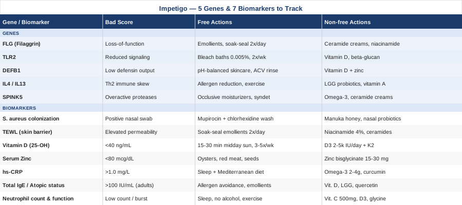
7 Biomarkers to Track if Impetigo Keeps Coming Back
Most people dealing with recurrent impetigo never look beyond the infection itself. But the infection is frequently a downstream signal rather than the root problem. Tracking specific biomarkers gives you a concrete, measurable picture of the factors driving recurrence — and actionable options for each one that go far beyond "be more careful with hygiene."
Biomarker 1: S. aureus Nasal and Skin Colonization
Why it matters: S. aureus colonizes the nasal passages in approximately 30% of the general population, and those carriers have substantially higher rates of recurrent skin infections because they continuously re-inoculate their own skin — particularly via hand-to-nose-to-skin contact. Household transmission is equally common: one colonized family member can seed an entire household without anyone realizing it. The colonization is typically asymptomatic, meaning you or your child can carry the bacteria indefinitely with no symptoms at all.
How to measure it: A nasal swab culture performed by a clinician, with sensitivity testing. Skin swabs from previously affected areas can also be cultured. A standard culture with sensitivity costs $30 to $100 out of pocket. MRSA-specific PCR nasal screening is faster and costs $50 to $150. If impetigo recurs every few months, testing the whole household simultaneously is worth requesting from a physician — isolated individual treatment while a colonized household contact goes untested is a common reason decolonization fails.
If colonization is confirmed: the plan without supplements
The most evidence-backed decolonization approach uses mupirocin nasal ointment (2%, applied twice daily for 5 days) combined with chlorhexidine body washes (4%, once daily for 5 to 7 days). This combination is clinically validated for reducing both nasal and skin S. aureus carriage. Household contacts should be screened and treated simultaneously, since re-inoculation from an untreated family member negates the individual effort. PubMed: mupirocin decolonization trials. Frequency: one course every 3 to 6 months under physician guidance if recurrence continues; not for continuous use due to mupirocin resistance risk.
If colonization is confirmed: the plan with supplements or additional tools
Manuka honey (UMF 10+ or MGO 263+) has demonstrated antimicrobial activity against S. aureus including MRSA in in vitro studies and wound care applications. Applied topically to active lesions or used as a purpose-formulated nasal gel, it is a low-risk adjunct. Nasal probiotic sprays containing Lactobacillus strains that competitively displace S. aureus from the nasal niche are an emerging strategy — early human studies show promise but evidence is not yet definitive. Side effects: minimal for both. Cycling: manuka honey can be used on active lesions continuously; nasal probiotics during high-risk periods or after antibiotic courses.
Biomarker 2: Skin Barrier Integrity — Transepidermal Water Loss (TEWL)
Why it matters: The outermost skin layer normally functions as a tight physical barrier that excludes bacteria. Transepidermal water loss (TEWL) measures how much water evaporates through the skin — a direct proxy for how well that barrier is sealed. Elevated TEWL means a more permeable, leakier skin surface that S. aureus finds far easier to colonize and invade. This is especially important in people with eczema, chronic dry skin, or filaggrin gene mutations (addressed later). Research has established that elevated TEWL in infancy predicts subsequent atopic disease and infection susceptibility in childhood and beyond. PubMed: TEWL and skin infection susceptibility
How to measure it: A tewameter or similar non-invasive device used in dermatology clinics. Not a standard primary care test — a dermatology referral is usually needed. Cost: often included within a consultation. Consumer-grade skin barrier/moisture devices exist at $100 to $300 but have variable accuracy compared to clinical instruments. Clinicians also assess barrier function indirectly through clinical examination of dryness severity and eczema scoring.
If skin barrier integrity is impaired: the plan without supplements
Emollient therapy is the cornerstone approach. The soak-and-seal method — applying a thick, fragrance-free emollient (petrolatum-based ointment preferred) within three minutes of bathing while skin is still hydrated — maximizes penetration and sealing efficacy. Apply twice daily. Avoid soap on affected areas; use a pH-balanced, non-foaming cleanser instead. Reduce hot water and prolonged shower exposure. In clinical trials in children with compromised skin barriers, consistent emollient use significantly reduced eczema severity and secondary infection rates compared to control groups.
If skin barrier integrity is impaired: the plan with supplements or equipment
Topical niacinamide (vitamin B3, 2–5% concentration) stimulates ceramide synthesis in keratinocytes, directly strengthening the barrier from within. Ceramide-dominant moisturizers — formulations with physiologic lipid ratios of ceramide, cholesterol, and free fatty acids — go beyond simple moisturization to restore structural components the damaged barrier cannot produce adequately. Systemically, evening primrose oil (500–1000 mg daily, rich in gamma-linolenic acid) shows modest barrier-supporting effects in atopic skin conditions in controlled studies. Side effects: niacinamide topically is very well tolerated; evening primrose oil may cause GI discomfort at higher doses. Cycling: topicals continuously; evening primrose oil as a 3-month trial, then reassess.
Biomarker 3: Vitamin D — 25-Hydroxyvitamin D
Why it matters: Vitamin D is not a bone health supplement with incidental immune effects — it plays a direct, upstream role in skin innate immunity by triggering keratinocytes to produce cathelicidin (LL-37), an antimicrobial peptide with potent killing activity against both S. aureus and S. pyogenes. When vitamin D is low, cathelicidin output drops — and so does a critical chemical layer of the skin's antibacterial defense. Multiple studies link low 25(OH)D to increased susceptibility to bacterial skin infections, impaired wound healing, and more severe atopic dermatitis severity. This is among the most actionable biomarkers in this list because deficiency is common and correction is straightforward. PubMed: vitamin D, cathelicidin, and skin immunity
How to measure it: Serum 25-hydroxyvitamin D (25(OH)D) — a standard blood test, widely available, typically covered by insurance when ordered for clinical indication. Out-of-pocket cost: $40 to $80. The standard "sufficiency" cutoff of 20 ng/mL is based on bone health data. For immune function optimization, most evidence supports targeting 40 to 60 ng/mL (100 to 150 nmol/L) — the range where cathelicidin production appears to be most robustly supported.
If vitamin D is low: the plan without supplements
Midday sun exposure of 15 to 30 minutes on arms and legs (without sunscreen) three to five days per week generates meaningful vitamin D cutaneously in summer months at mid-latitudes. Darker-skinned individuals require longer exposure for equivalent synthesis. At northern latitudes during autumn and winter, sunlight exposure alone is typically insufficient to maintain target levels. Retest 25(OH)D after three months of any approach.
If vitamin D is low: the plan with supplements
Vitamin D3 (cholecalciferol): 2,000 to 4,000 IU daily for maintenance in confirmed deficiency; 4,000 to 5,000 IU daily for repletion when baseline is below 20 ng/mL. Always paired with vitamin K2 (MK-7 form, 100 to 200 mcg daily) to direct calcium to bone rather than soft tissue. Retest 25(OH)D after three months to confirm levels have reached the target range, then adjust dose accordingly. Side effects: toxicity is rare but possible at sustained doses above 10,000 IU daily — monitor serum calcium if using higher doses long-term. Cycling: ongoing supplementation is appropriate for most people, especially those with limited sun exposure or dark skin at northern latitudes.
Biomarker 4: Serum Zinc
Why it matters: Zinc is essential for wound healing, skin barrier maintenance, neutrophil and T-cell activation, and — critically — the expression of antimicrobial peptides including beta-defensins. Zinc deficiency impairs virtually every arm of the immune response relevant to bacterial skin infection containment. It is more prevalent than most clinicians account for, particularly in children with limited dietary variety, individuals following plant-dominant diets, and people with gastrointestinal conditions affecting absorption. Even marginal deficiency that falls within broad laboratory normal ranges can measurably impair immune function.
How to measure it: Fasting serum zinc (or plasma zinc, which is slightly more precise). Cost: $40 to $80 out of pocket. Normal range: 70 to 120 mcg/dL; integrative practitioners typically target 80 to 100 mcg/dL. Alkaline phosphatase (ALP) on a standard metabolic panel is a useful indirect proxy — low-normal ALP alongside low-normal zinc suggests functional deficiency even when both values technically fall within reference ranges.
If zinc is low: the plan without supplements
Increase dietary zinc through oysters (the highest food source by far), red meat, beef liver, pumpkin seeds, and hemp seeds. For plant-based diets, soaking, sprouting, or fermenting legumes and grains reduces phytate content substantially and improves zinc bioavailability. In children, two to three weekly servings of zinc-dense animal foods can correct borderline deficiency without supplementation. Retest after four to six weeks of sustained dietary change.
If zinc is low: the plan with supplements
Zinc bisglycinate or zinc picolinate are the most bioavailable forms: 15 to 30 mg elemental zinc daily, taken with food to reduce nausea. The daily sustained upper limit is 40 mg; above that, copper absorption becomes significantly impaired. If supplementing beyond 30 mg for more than a few weeks, add 1 to 2 mg copper daily to prevent depletion. Topical zinc oxide on active lesions directly reduces bacterial load and accelerates re-epithelialization — this is backed by wound care evidence and is a useful adjunct during healing. Side effects: nausea and metallic taste at higher oral doses; copper depletion with chronic high-dose use without copper co-supplementation. Cycling: 3-month trial with retesting; adjust dose based on levels.
Biomarker 5: High-Sensitivity CRP
Why it matters: C-reactive protein is a systemic inflammation marker. Chronically elevated hs-CRP — even at sub-clinical levels — signals a state of ongoing background inflammation that can paradoxically blunt acute immune responses and slow wound resolution. More usefully for impetigo, elevated CRP points toward upstream factors that consistently increase infection vulnerability: poor diet, unmanaged eczema, sleep deprivation, metabolic imbalance, or chronic psychological stress. Addressing these underlying drivers, rather than just treating each infection as it occurs, is what actually breaks the recurrence pattern.
How to measure it: High-sensitivity CRP (hs-CRP) blood test, widely available, $20 to $60 out of pocket. Target: below 1.0 mg/L for optimal immune health. Above 3 mg/L warrants investigation into underlying inflammatory triggers. Above 10 mg/L typically indicates acute infection or significant pathology and needs clinical evaluation.
If hs-CRP is elevated: the plan without supplements
Address the most impactful lifestyle drivers first: reduce ultra-processed foods and refined sugars (the single most powerful dietary lever for CRP reduction), prioritize 7 to 9 hours of quality sleep consistently (even one week of sleep restriction elevates CRP measurably), and establish routine moderate aerobic exercise — 150 minutes per week of brisk walking consistently reduces hs-CRP by 20 to 35% in randomized controlled trials. A Mediterranean-pattern diet (olive oil, fatty fish, vegetables, legumes, minimal refined foods) reduces hs-CRP significantly within 8 to 12 weeks of sustained adherence. PubMed: Mediterranean diet and CRP in randomized trials
If hs-CRP is elevated: the plan with supplements
Omega-3 fatty acids (EPA and DHA combined, 2 to 4 grams daily from high-quality fish oil or algal oil) are among the most evidence-backed anti-inflammatory supplements — well documented across multiple meta-analyses in reducing CRP. Curcumin with piperine (500 to 1000 mg turmeric extract standardized to curcuminoids, combined with black pepper extract for bioavailability) consistently reduces CRP in human trials. Magnesium glycinate (300 to 400 mg before sleep) supports sleep quality and reduces inflammatory signaling through multiple pathways. Side effects: omega-3 at higher doses may mildly extend clotting time; curcumin may interact with anticoagulant medications. Cycling: 3-month trial with retesting of hs-CRP; omega-3 and magnesium can be used continuously.
Biomarker 6: Total IgE and Atopic Status
Why it matters: People with atopic conditions — eczema, allergic rhinitis, asthma — are at significantly elevated risk for recurrent impetigo because the atopic immune phenotype (Th2-dominant, elevated IgE, reduced antimicrobial peptide production) creates a skin environment that S. aureus actively exploits. The bacteria produce toxins that suppress cathelicidin and defensin expression while promoting barrier breakdown, establishing a vicious cycle between atopic inflammation and bacterial infection. Total IgE provides a single number that reflects the degree of atopic immune skewing, and knowing it guides targeted interventions rather than generic advice.
How to measure it: Total serum IgE, with specific IgE panels for common allergens when indicated. Cost: $50 to $150 depending on panel scope. An allergist or clinical immunologist can interpret results in context. General reference: total IgE above 100 IU/mL in an adult suggests atopic background; in children, normal ranges are age-specific. Very high IgE (above 1000 IU/mL) warrants specialist assessment to rule out more significant immune conditions including hyper-IgE syndrome, where recurrent skin infections are a cardinal feature. PubMed: IgE, atopic dermatitis, and S. aureus
If IgE is elevated: the plan without supplements
Allergen identification and load reduction is the starting point: skin prick or specific IgE blood testing to identify triggers (house dust mites are most common, followed by pet dander, mold, and specific foods). Reducing allergen exposure — dust mite mattress covers, air filtration, pet dander management — reduces the Th2 immune drive that worsens barrier function and antimicrobial defense. Consistent emollient therapy and wet wrap therapy during eczema flares significantly reduce IgE-driven skin barrier breakdown. Referral to an allergist is warranted when total IgE is above 500 IU/mL.
If IgE is elevated: the plan with supplements
Vitamin D at optimal levels (40 to 60 ng/mL) modulates Th1/Th2 balance, directly reducing the atopic immune skewing associated with high IgE. This is one of the strongest mechanistic reasons to maintain optimal D levels specifically in atopic individuals. Lactobacillus rhamnosus GG and Bifidobacterium lactis have the best evidence base among probiotics for reducing atopic dermatitis severity and secondary skin infections in children; most supporting studies use 5 to 10 billion CFU daily for a minimum of 3 months. Quercetin (500 mg twice daily) acts as a natural mast cell stabilizer reducing IgE-mediated degranulation — mechanistically sound with early clinical support. Side effects: probiotics are very well tolerated; quercetin may interact with certain medications. Duration: minimum 3 months before assessing response.
Biomarker 7: Neutrophil Count and Function
Why it matters: Neutrophils are the primary innate immune cells responsible for rapid killing of invading bacteria at the skin surface. Their speed of migration, phagocytic capacity, and oxidative burst intensity determine whether a S. aureus seeding event is contained within hours or progresses to a visible clinical lesion. Quantitative deficiencies (neutropenia) show up on routine blood work. More subtle qualitative impairments — reduced oxidative burst, impaired migration — can exist with a perfectly normal neutrophil count and go undetected for years. In children with frequent skin infections and no obvious predisposing condition, a neutrophil function assessment is a conversation worth having with an immunologist.
How to measure it: Complete blood count (CBC) with differential is the starting point — widely available, costs $20 to $50. Normal absolute neutrophil count (ANC): 1,500 to 8,000/mcL. If the ANC is consistently within normal range but recurrent bacterial skin infections continue without explanation, referral to a clinical immunologist for neutrophil function testing (oxidative burst assay, phagocytosis assay) is appropriate; specialized testing costs $200 to $500 and is often covered when recurrent infections are the documented clinical indication.
If neutrophil count or function is suboptimal: the plan without supplements
Prioritize sleep — even moderate restriction (6 hours versus 8 hours per night) measurably impairs neutrophil chemotaxis and intracellular killing capacity within days. Reduce alcohol intake: alcohol is a direct neutrophil function suppressant that affects migration and oxidative burst. Maintain moderate, consistent exercise while avoiding overtraining, which creates a period of functional immunosuppression. If ANC is consistently below 1,500/mcL, hematology referral is essential before any self-directed intervention.
If neutrophil count or function is suboptimal: the plan with supplements
Vitamin C (500 to 1000 mg daily) is among the most evidence-supported supplements for neutrophil function — neutrophils concentrate vitamin C to levels 50 to 100 times higher than plasma concentrations, using it to support oxidative killing and post-phagocytosis repair. Vitamin D at optimal levels also directly enhances neutrophil function through multiple pathways. Glycine (3 to 5 grams daily) supports glutathione production, which is central to neutrophil oxidative burst activity. Side effects: vitamin C above 2 grams daily may cause loose stools — reduce dose if this occurs. Cycling: all three can be used continuously at the doses described. PubMed: vitamin C and neutrophil immune function
The biomarker picture is most useful when viewed as a whole rather than in isolation — an individual with low vitamin D, elevated IgE, and confirmed nasal S. aureus carriage has a convergence of modifiable risk factors, not simply bad luck. Genetics, explored next, helps explain why some people land in that situation in the first place.
The Genetic Side of Impetigo: 5 Variants Worth Knowing
Genetics does not determine fate with impetigo. But if you have done a genetic panel — 23andMe, AncestryDNA with health add-ons, or a clinical genomic panel — understanding which variants you carry can substantially sharpen your prevention strategy. The five genes below are the most clinically relevant for understanding skin barrier integrity and innate immune recognition, the two biological systems that most determine whether S. aureus or S. pyogenes can establish a foothold.
FLG: The Filaggrin Gene
Filaggrin is the most structurally critical protein in the outermost skin layer. It aggregates keratin filaments during epidermal cell maturation, creating the dense, tightly sealed, water-retaining surface that physically excludes bacteria. Loss-of-function mutations in the FLG gene — present in approximately 10% of European-ancestry populations and lower frequencies in other groups — produce a structurally deficient skin barrier that allows S. aureus to adhere and penetrate far more easily than in unaffected individuals.
This is among the most replicated findings in dermatology genetics. People with FLG loss-of-function variants have elevated S. aureus skin colonization rates, higher rates of atopic dermatitis, and more frequent secondary bacterial skin infections. The landmark paper establishing this connection — Palmer et al., 2006, Nature Genetics — identified FLG mutations as the primary genetic cause of atopic dermatitis; subsequent work confirmed the direct link to S. aureus colonization and infection susceptibility.
If your FLG variant is a loss-of-function: the plan without supplements
Aggressive barrier protection is non-negotiable and lifelong. Apply petrolatum-based ointment (plain petroleum jelly is adequate and cost-effective) twice daily, using the soak-and-seal technique within three minutes of every bath or shower. Avoid all fragranced products and surfactant-heavy body washes. Wear 100% cotton directly against the skin. Maintain indoor humidity at 40 to 50% during dry seasons. These interventions functionally compensate for what the filaggrin-deficient barrier cannot do on its own. Frequency: twice daily, year-round, indefinitely.
If your FLG score is bad: the plan with supplements or equipment
Ceramide-dominant moisturizers — formulations with the physiologic 3:1:1 ceramide/cholesterol/fatty acid molar ratio — provide the lipid components that a filaggrin-deficient skin cannot adequately organize. Topical niacinamide (4%) stimulates ceramide synthesis in keratinocytes directly. Wet wrap therapy during flares (wet dressings over emollient-soaked skin left on for 2 to 8 hours) provides intensive barrier support when skin is most vulnerable. Humidifying the sleeping environment during dry months addresses the passive TEWL increase that FLG mutations worsen at night. Side effects: all approaches are very well tolerated; wet wraps may cause skin maceration if overused — limit to active flare management.
TLR2: Toll-Like Receptor 2
TLR2 encodes a pattern recognition receptor on innate immune cells that specifically detects cell wall components of gram-positive bacteria — exactly the category that includes S. aureus and S. pyogenes. Variants in TLR2 that reduce receptor expression or downstream signaling efficiency mean the body's alarm system fires more slowly in response to these bacterial invaders, giving them more time to establish and proliferate before the immune response mobilizes.
If your TLR2 variant affects receptor function: the plan without supplements
The practical implication of impaired TLR2 signaling is that reducing bacterial burden before it exceeds the delayed detection threshold becomes more important. Dilute bleach baths (0.005% sodium hypochlorite — approximately half a teaspoon of standard household bleach per gallon of water, twice weekly) reduce S. aureus skin burden substantially before colonization reaches the density that overwhelms a slower-responding innate immune system. This protocol is clinically validated in atopic dermatitis management and broadly safe at the correct dilution. PubMed: bleach baths and S. aureus burden reduction
If your TLR2 score is bad: the plan with supplements or equipment
Vitamin D at optimal levels (40 to 60 ng/mL) upregulates TLR2 expression and enhances downstream innate immune signaling through NF-κB pathways — making this one of the most directly relevant supplement interventions for anyone with TLR2 variants. Beta-glucan (from Saccharomyces cerevisiae or oats, 250 to 500 mg daily) is an innate immune modulator that activates Dectin-1 receptors with overlapping downstream signaling with TLR pathways; human trials show increased phagocytic activity and reduced infection frequency with consistent use. Side effects: minimal. Cycling: 3-month trial initially, then reassess.
DEFB1: Defensin Beta 1
Beta-defensin 1 is an antimicrobial peptide constitutively expressed by skin keratinocytes — meaning it is always present as a baseline chemical defense layer, unlike inducible peptides that require an immune trigger to be produced. Certain DEFB1 single nucleotide polymorphisms reduce this constitutive expression, leaving the skin with less persistent antimicrobial activity at rest. This is especially relevant as a first-responder defense against gram-positive bacteria like S. aureus and S. pyogenes.
If your DEFB1 variant is present: the plan without supplements
Maintaining skin acidity — the natural skin pH range of 4.5 to 5.5 — supports defensin function and inhibits S. aureus adherence, which prefers a more neutral pH environment. Use pH-balanced skincare products and avoid alkaline soaps. A dilute apple cider vinegar rinse (1 part vinegar to 10 parts water, applied with a cloth and left briefly before rinsing) can restore skin acidity without disrupting commensal bacteria significantly. Frequency: daily to every other day on non-lesional skin as part of the hygiene routine.
If your DEFB1 score is bad: the plan with supplements or equipment
Vitamin D directly upregulates both cathelicidin and beta-defensin expression in keratinocytes through the vitamin D response element in their promoter regions — this is among the most direct evidence-based rationales for maintaining 25(OH)D at 40 to 60 ng/mL in people with DEFB1 variants. Zinc supports defensin expression through zinc finger transcription factor pathways. If you carry DEFB1 variants, these two interventions rise to the top of the priority list. Doses and cycling as outlined in the biomarker sections above; neither requires cycling when monitored by serum levels.
IL4 and IL13 Pathway Variants
Variants in IL4 and IL13 encode cytokines that drive Th2-skewed immune responses — the immune phenotype underlying atopic dermatitis. This Th2 bias suppresses the Th1-mediated immune responses that are most effective against bacterial infections, while simultaneously promoting IgE elevation, reduced antimicrobial peptide production, and the skin barrier dysfunction described in the IgE biomarker section. People with these variants sit at the intersection of allergic immune dysregulation and recurrent bacterial skin infection susceptibility.
If IL4/IL13 variants are present: the plan without supplements
Allergen load reduction is the most powerful non-supplement approach — reducing environmental Th2 triggers (dust mites, pet dander, food allergens if relevant) gradually shifts immune balance away from Th2 excess. Regular moderate aerobic exercise (30 to 45 minutes, 4 to 5 days per week) consistently modulates Th1/Th2 balance in a favorable direction in human studies. For children, current pediatric allergy guidance supports early introduction of allergenic foods to prevent entrenchment of Th2 immune programming.
If IL4/IL13 score is bad: the plan with supplements or equipment
Lactobacillus rhamnosus GG and Bifidobacterium lactis are the best-studied probiotics for shifting immunity toward Th1 in atopic conditions — with the most evidence in pediatric populations. Dose: 5 to 10 billion CFU daily for a minimum of 3 months of continuous use before assessing response. Dietary vitamin A — retinol from liver, eggs, and dairy, or beta-carotene from orange and leafy vegetables — supports epithelial immunity and moderates Th2 skewing. Supplemental preformed vitamin A should remain below 5,000 IU daily as sustained high doses are hepatotoxic. Side effects: probiotics are very well tolerated; vitamin A toxicity is possible at high doses, particularly in children.
SPINK5: Serine Protease Inhibitor Kazal Type 5
SPINK5 encodes LEKTI, a protease inhibitor that regulates enzymes responsible for controlled shedding of the outer skin layer. Variants in SPINK5 allow skin proteases to become overactive, accelerating stratum corneum breakdown and weakening the barrier — most severely in Netherton syndrome, but milder variants contribute to atopic skin fragility more broadly in the general population. This overactive protease activity compounds the barrier defects seen with FLG mutations if both are present.
If your SPINK5 variant is present: the plan without supplements
Reduce anything that further activates already-overactive skin proteases: limit prolonged water immersion, avoid enzyme-containing laundry detergents in direct contact with skin, and use thick occlusive moisturizers that slow stratum corneum degradation. Detergent-free cleansers (syndets) are preferable to soap. Frequency: twice-daily emollient application with particular attention to the immediate post-bathing window.
If your SPINK5 score is bad: the plan with supplements or equipment
Ceramide/cholesterol/fatty acid barrier repair creams at physiologic lipid ratios are the most targeted topical strategy for SPINK5-related barrier fragility. Omega-3 fatty acids (EPA plus DHA, 1 to 3 grams daily) support the lipid composition of the skin barrier, providing the raw materials needed to maintain intercellular lipid lamellae even when protease activity is high. Green tea extract applied topically has demonstrated protease-inhibiting activity in early studies, offering a potential adjunct for reducing overactive protease damage at the skin surface. Side effects: minimal for all approaches. Omega-3 can be used continuously at these doses.
With the biomarker and genetic landscape mapped, it's worth zooming out to look at the broader immune and lifestyle factors that research suggests drive susceptibility — and that standard medical advice rarely addresses in enough depth.
10 Science-Backed Principles From Huberman Lab Research on Immune Function That Apply Directly to Impetigo
The Huberman Lab podcast has synthesized an enormous body of peer-reviewed research on immune function, skin biology, and lifestyle-based disease prevention across many episodes. What follows draws on the key findings most relevant to bacterial skin infection susceptibility — principles that challenge the standard infection-management narrative and point toward durable prevention rather than repeated treatment.
1. Vitamin D Functions as an Immune Hormone, Not a Supplement
The dominant clinical approach to vitamin D — test, identify deficiency, prescribe a supplement, move on — misses the point. Vitamin D functions as a steroid hormone, directly activating gene expression in immune cells and keratinocytes. Its role in inducing cathelicidin and beta-defensin production means it is embedded in the first-line response to S. aureus. Maintaining 25(OH)D at 40 to 60 ng/mL is not about supplementation strategy — it is about keeping a critical immune signal adequately loaded.
2. Sleep Is the Most Powerful Immune Intervention Available
No supplement, diet, or biohacking protocol compensates for chronically poor sleep when it comes to immune function. Sleep deprivation — even at moderate levels — measurably suppresses neutrophil killing capacity, reduces cathelicidin expression, elevates CRP, and increases skin TEWL within days. For anyone dealing with recurrent impetigo, optimizing sleep architecture (consistent schedule, dark environment, temperature 65 to 68°F, no alcohol within 3 hours of sleep) is among the highest-leverage changes available.
3. Nasal Breathing Has Direct Microbiological Consequences
Mouth breathing bypasses nasal filtration, mucociliary clearance, and the nitric oxide production that occurs in the nasal passages — all of which reduce bacterial and viral load before it reaches the respiratory tract. Nasal breathing also maintains a healthier nasal microbiome, which competes with S. aureus colonization. Habitual mouth breathers — particularly children with nasal obstruction — may warrant ENT assessment, as restoring nasal airway patency can reduce nasal S. aureus burden.
4. Cold Exposure Activates Innate Immunity in Measurable Ways
Brief deliberate cold exposure — cold showers, cold water immersion — activates the sympathetic nervous system and triggers release of norepinephrine, which has direct effects on innate immune cell mobilization. Regular cold exposure (2 to 4 minutes at 50 to 60°F, several times weekly) has been associated with increased natural killer cell activity and reduced rates of upper respiratory and skin infections in small human studies. This is not a first-line intervention, but it is a low-cost, low-risk adjunct for individuals with recurrent infections and no contraindications.
5. The Gut-Immune Axis Directly Shapes Skin Defense
The gut microbiome regulates systemic immune tone — including the Th1/Th2 balance that determines skin antimicrobial peptide production and atopic risk. Gut dysbiosis drives low-grade intestinal permeability and inflammatory signaling that reaches the skin via the gut-skin axis. Dietary strategies that support gut microbiome diversity (fiber from varied plant sources, fermented foods, minimizing unnecessary antibiotics) produce measurable downstream improvements in skin immune function and atopic disease severity.
6. Sunlight Has Immune Effects Beyond Vitamin D Synthesis
Direct UV exposure on skin induces not only vitamin D production but also triggers local production of nitric oxide (which has antimicrobial properties), modulates skin dendritic cell activity, and influences the skin microbiome composition. Regular moderate sun exposure — in amounts that tan lightly without burning — appears to have immune effects on the skin that exceed what vitamin D supplementation alone replicates. This distinction matters practically: supplements are important when sun exposure is limited, but they are not functionally equivalent to sunlight.
7. Cortisol Suppresses Antimicrobial Peptide Expression Directly
Chronic psychological stress drives sustained cortisol elevation, which has been shown to directly suppress cathelicidin and beta-defensin expression in keratinocytes — reducing the skin's chemical antibacterial defense. Chronic stress also worsens skin barrier function through cortisol-induced changes in skin lipid production. This creates a specific, measurable biological pathway from psychological stress to increased S. aureus susceptibility — not a vague correlation, but a mechanistically documented route. Stress management is not optional in a genuine prevention protocol.
8. Exercise Dose and Timing Matter More Than Most People Realize
Moderate consistent exercise (150 minutes per week of moderately intense activity) enhances innate immune surveillance, increases natural killer cell activity, and reduces systemic inflammation. Intense or prolonged exercise (marathon-level exertion, overtraining) creates a post-exercise immunosuppressive window of 24 to 72 hours during which infection risk increases. For anyone with recurrent impetigo, this distinction matters: exercise is protective at moderate doses and immunosuppressive at excessive ones.
9. Light Exposure Timing Sets Immune Circadian Rhythms
The immune system operates on a circadian clock. Neutrophil activity, cytokine release, and antimicrobial peptide production all follow time-of-day patterns. Circadian disruption — from shift work, irregular sleep/wake timing, or excessive artificial light at night — measurably dysregulates these immune rhythms. Getting bright outdoor light in the morning (10 to 30 minutes within the first hour of waking) and minimizing bright light in the 2 hours before sleep helps calibrate immune circadian timing, which most people overlook entirely in infection prevention.
10. Zinc and Magnesium Depletion Are More Common and Consequential Than Usually Treated
Both zinc and magnesium are frequently consumed in inadequate amounts relative to the amounts lost through sweat, stress, and modern agricultural soil depletion. Zinc's role in immune function is covered in the biomarker section. Magnesium deficiency impairs sleep quality (reducing the restorative immune benefits of sleep), elevates cortisol, and impairs the NF-κB inflammatory signaling that coordinates the initial response to bacterial skin invasion. Both should be assessed, not assumed to be adequate based on typical dietary intake.
These principles represent a systems view of infection susceptibility — one that conventional dermatology visits rarely have time to address but that substantively changes the trajectory for people stuck in recurrence cycles.
Complementary Approaches With Clinical Backing
The approaches below are not replacements for appropriate medical treatment of active impetigo, which requires antibiotic therapy when indicated. They are adjuncts and prevention strategies, selected specifically for their relevance to bacterial skin infection and supported by human clinical data rather than theoretical plausibility alone.
Microbiome-Directed Therapies
Microbiome-directed therapy for impetigo encompasses strategies that shift both the skin and gut microbial environments away from conditions favorable to S. aureus dominance. The core scientific rationale is that a healthy commensal microbial community — particularly Staphylococcus epidermidis, Cutibacterium species, and certain Corynebacterium strains — actively competes with S. aureus through competitive exclusion and direct antimicrobial compound production. This is not a theoretical concept: S. epidermidis strains produce serine protease Esp and bacteriocin-like compounds that specifically inhibit S. aureus biofilm formation and survival.
A 2021 study published in Nature Medicine demonstrated that transfer of selected S. epidermidis strains producing antimicrobial compounds to atopic skin reduced S. aureus burden significantly in a human pilot trial. Separately, oral probiotic interventions with Lactobacillus rhamnosus GG in children have demonstrated reduced rates of atopic disease flares and associated secondary skin infections across multiple randomized trials. PubMed: LGG probiotics and skin infection outcomes
Practically: take a well-characterized oral probiotic (LGG or B. lactis at 5 to 10 billion CFU daily) continuously, especially during and after antibiotic courses when microbiome disruption is at its peak. Integrate fermented foods daily. Switch from antibacterial body washes to pH-balanced microbiome-preserving cleansers. After any antibiotic course, this active restoration window — particularly the first 4 to 8 weeks — represents the highest-leverage period for re-establishing protective microbial diversity.
Saline Nasal Irrigation
Saline nasal irrigation mechanically reduces S. aureus nasal colonization by flushing bacteria, biofilms, and inflammatory debris from the nasal passages. Since nasal carriage is a primary route of S. aureus self-inoculation to the skin — particularly through hand-to-nose-to-skin contact, which occurs dozens of times per day — reducing nasal bacterial load is a mechanistically rational preventive measure for recurrent impetigo, especially when mupirocin-based decolonization has not produced durable results.
Clinical data on saline irrigation primarily derives from rhinosinusitis management, but its anti-staphylococcal effect has been studied more directly in atopic populations. Studies have shown reductions in nasal S. aureus burden in atopic patients following regular saline irrigation, with directionally positive effects on skin infection frequency as a secondary outcome. The safety profile is excellent when sterile water is used. PubMed: saline irrigation and nasal S. aureus colonization
Practical protocol: use isotonic or mildly hypertonic saline (0.9% or 2% NaCl) in distilled or pre-boiled and cooled water via a neti pot or squeeze bottle, once to twice daily. Never use unfiltered tap water for nasal irrigation — tap water carries rare but serious Naegleria fowleri risk in nasal irrigation applications. Initial setup costs $10 to $30; ongoing cost is minimal. For families dealing with repeated S. aureus-related skin infections, this is a simple, inexpensive daily habit worth establishing.
Light Therapy — Blue Light
Blue light (wavelengths 400 to 470 nm) has direct antibacterial effects through activation of endogenous bacterial porphyrins, causing photodynamic oxidative damage to bacterial cell walls and DNA. Unlike antibiotics, this mechanism does not induce bacterial resistance — making it an increasingly studied approach for S. aureus including MRSA. Blue light also has anti-inflammatory effects on keratinocytes and has been shown to reduce skin S. aureus burden in atopic patients in controlled settings.
Clinical studies have shown that twice-weekly blue light therapy (20 to 30 minutes per session) reduces S. aureus colonization on atopic skin and improves eczema severity scores over 4-week treatment courses. A pilot randomized controlled trial published in 2019 specifically showed significant reduction in S. aureus burden on arm skin in atopic dermatitis patients, with corresponding improvement in skin infection-related outcomes. Evidence is early-stage but mechanistically compelling and building. PubMed: blue light therapy and S. aureus on skin
Consumer blue light devices ($50 to $200) are available for home use. Recommended protocol based on published studies: 20 to 30 minutes per session, two to three times weekly on affected or historically affected skin areas. Always protect eyes during use — blue light is safe for skin but potentially damaging with direct eye exposure. Blue light is not appropriate for actively spreading or systemic impetigo — it is best applied during the resolution and post-resolution phase, and as a preventive measure during high-risk periods.
Chinese Herbal Medicine — Berberine
Several herbs in the traditional Chinese pharmacopoeia have demonstrated antimicrobial activity against S. aureus and S. pyogenes in vitro and in clinical contexts. The most studied bioactive compound for this purpose is berberine — an isoquinoline alkaloid found in Huang Lian (Coptis chinensis) and Huang Bai (Phellodendron bark) — which has demonstrated broad-spectrum antimicrobial activity, antibiofilm effects against S. aureus, and significant immune-modulating properties in multiple human trials. It also activates AMPK pathways that improve metabolic markers associated with elevated CRP and immune dysfunction.
A 2020 systematic review in Phytomedicine identified berberine's direct inhibitory activity against S. aureus — including MRSA — through multiple mechanisms including disruption of bacterial membrane integrity and suppression of virulence factor expression. Traditional formulas containing honeysuckle (Lonicera japonica, Jin Yin Hua) have also shown antimicrobial and anti-inflammatory efficacy in pediatric skin infection studies in Chinese clinical settings. PubMed: berberine and S. aureus antimicrobial activity
Practical considerations: berberine at 500 mg twice daily with meals (the dose used in most human metabolic and antimicrobial trials) is the most accessible and evidence-supported extract from this pharmacopoeia and commercially available without a prescription. Side effects include GI discomfort, particularly at initiation — start at 250 mg twice daily and increase after one week. Cycling: 8 to 12 weeks on, 4 weeks off; not for indefinite unmonitored use, as berberine interacts with several medications including metformin, cyclosporine, and anticoagulants. Do not use during pregnancy.
Low-Level Laser Therapy (Photobiomodulation)
Low-level laser therapy (LLLT) applies specific wavelengths of light (typically 630 to 1000 nm) at low irradiance to tissue, stimulating mitochondrial function, reducing local inflammation, and accelerating wound healing and re-epithelialization. For impetigo specifically, two mechanisms are clinically relevant: faster healing of existing lesions, reducing the open wound window during which re-infection can occur; and at shorter wavelengths, direct antibacterial photodynamic effects against S. aureus biofilms on skin.
A systematic review and multiple subsequent randomized controlled trials have confirmed LLLT's benefit for wound healing, including infected skin wounds — with consistent improvement in healing time and reduced infection rates compared to standard wound care alone. Studies specifically using wavelengths in the 630 to 660 nm range show the strongest anti-inflammatory and pro-healing effects; studies at 405 to 470 nm overlap with the blue light antibacterial range described above. PubMed: LLLT and wound healing outcomes
Practical application: clinical LLLT devices are available through physiotherapy clinics and some dermatology practices. Consumer-grade near-infrared devices ($150 to $500) vary in quality — look for devices with published wavelength and irradiance specifications. For impetigo, LLLT is most relevant as a wound-healing adjunct after the acute infection phase — applied to resolving lesions to speed re-epithelialization and reduce the scarring and pigmentary changes that sometimes follow. Not for application to actively infected open wounds. Sessions of 5 to 10 minutes over a small area, three to five times weekly, align with protocols used in published wound healing trials.
Conclusion
Recurrent impetigo is rarely explained by hygiene failures alone. The biology underneath — skin barrier integrity, nutritional status, nasal colonization, immune gene variants, microbiome composition, and systemic inflammation — determines who gets repeated infections and who doesn't. The seven biomarkers in this article give you a concrete starting point for identifying which of those factors are at play in your specific situation. The genetic context adds another layer of precision if you have that information available.
A practical next step does not require addressing everything at once. Starting with the most accessible tests — a nasal swab culture if recurrence is the issue, a vitamin D level, a basic zinc and hs-CRP panel, a CBC — and working from there is entirely reasonable. A dermatologist or allergist who understands the eczema-infection connection is the right specialist to partner with, particularly if IgE is elevated or atopic disease is part of the picture.
Impetigo responds well to a coordinated approach: reduce bacterial load, strengthen the barrier, and optimize the immune environment. The map is here. Where to start depends on what your tests reveal.