This article contains AI generated content.
Infrapatellar Contracture Syndrome: 6 Genes and 6 Biomarkers to Track
Introduction
If you are reading this after a knee that will not straighten, will not bend past a certain point, or simply feels "tight" months after an ACL reconstruction or a fracture repair, you already know that the standard advice does not quite fit your case. "Keep doing your physical therapy" and "give it time" are true in general, but they say nothing about why your knee scarred down harder than your surgeon's other patients, or why the stiffness kept building instead of fading on the timeline everyone expected.
Infrapatellar contracture syndrome, sometimes called patella infera or a severe form of arthrofibrosis, is not a single uniform process. It is the visible result of an internal fibrotic reaction, one shaped by inflammation levels, collagen turnover, immune recognition of surgical tissue, and in some people, genetic variants that tilt the whole system toward excess scarring. Generic rehab advice treats every stiff knee the same way. It rarely asks what is actually driving your particular tissue to over-produce scar matrix.
This article takes a more specific approach. Instead of stopping at "do your exercises," it looks at the blood markers and genetic variants that have been directly studied in postsurgical knee stiffness and arthrofibrosis, and lays out what can realistically be done about each one, with and without supplements or equipment, including realistic frequencies and honest side-effect notes.
None of this replaces your surgeon or physical therapist, and none of it promises a full reversal of established contracture. But better information changes decisions. Knowing which biomarkers reflect your current fibrotic activity gives you something concrete to track and discuss at your next appointment. Knowing which genetic variants have been linked to arthrofibrosis risk gives context, not a verdict. Together with a look at what current stretching science actually supports, and where complementary therapies have real supporting evidence, the goal here is to replace vague reassurance with a plan you can actually act on.
Summary
This article is built around one practical question: what, specifically, is making some knees scar down into contracture while others recover on schedule? The answer is not one thing. It is a combination of measurable biology, some of it in your bloodwork, some of it written into genes involved in inflammation, collagen turnover, and immune recognition.
You will find six biomarkers worth asking your doctor about, from a common one you may already have on a recent lab panel to a specialized fibrosis marker most clinics never mention. Each comes with how it is measured, roughly what it costs, and two levels of response: what you can change through habits alone, and what supplements or equipment add on top, including how often to use them and what to watch for.
You will also find six genetic variants that recent orthopedic research has tied to arthrofibrosis risk after ACL reconstruction, what each one plausibly does, and how its effect might be offset even though the gene itself cannot be changed. A closer look at stretching science, drawn from a widely discussed research-backed protocol, challenges some of the more aggressive manual therapy habits still common in stiff-knee rehab. Finally, a short review of complementary approaches, massage, photobiomodulation, biofeedback, and relaxation training, rounds out what has actual clinical evidence behind it for this specific problem, not just for general knee pain.
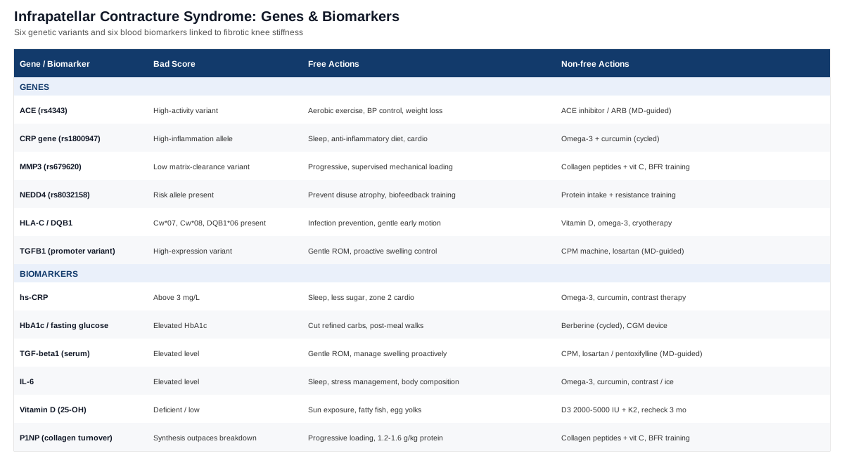
The Six Biomarkers Worth Tracking
Arthrofibrosis research has moved past treating knee stiffness as a purely mechanical problem. A 2022 systematic review of biochemical markers in postsurgical knee arthrofibrosis, published via PubMed, found that markers tied to TGF-beta signaling, collagen turnover, and extracellular matrix remodeling consistently distinguish stiff knees from knees that healed on schedule. A separate study identifying novel biomarkers in both animal models and clinical patients, available on PubMed, pointed to TGFBR1 correlating directly with range-of-motion loss. None of these markers are exotic. Most can be ordered through a regular blood draw, and the reasoning below follows the same logic Peter Attia, Thomas Dayspring, and Allan Sniderman use for cardiometabolic risk: track what is measurable, act on what is modifiable, and do not treat a single number as a verdict.
High-sensitivity CRP (hs-CRP)
hs-CRP reflects your overall systemic inflammatory tone. It is not specific to the knee, but chronic low-grade inflammation feeds the same fibroblast activity that drives excess scar tissue around the patellar tendon and fat pad. It also happens to be one of the most studied markers in longevity medicine, which means there is a large evidence base for how to move it.
How to measure it
A standard venous blood draw, typically 15 to 40 US dollars out of pocket if not covered by insurance, and often already included in routine metabolic or cardiac risk panels. Results above roughly 3 mg/L suggest meaningful systemic inflammation worth investigating further.If the score is bad, the plan without supplements
Prioritize sleep consistency (7 to 9 hours, stable wake time), reduce ultra-processed food and added sugar, add two to three weekly sessions of zone 2 cardio, address gum disease if present, and stop smoking or vaping if applicable. These changes typically show measurable hs-CRP movement within 8 to 12 weeks.If the score is bad, the plan with supplements or equipment
Omega-3 (EPA/DHA combined, 2 to 4 grams daily with food) has the strongest evidence for lowering CRP. Curcumin with piperine (500 to 1000 mg daily) is a reasonable add-on, cycled 8 to 12 weeks on, 2 to 4 weeks off, to avoid GI irritation. Contrast therapy (sauna followed by cold exposure, 3 to 4 times weekly) has emerging but weaker evidence. Side effects: fish oil can increase bleeding risk at high doses, especially alongside anticoagulants; curcumin can cause GI upset or interact with gallstone conditions; check with your physician before combining either with prescribed blood thinners.HbA1c and fasting glucose
Elevated blood glucose accelerates the formation of advanced glycation end-products, which stiffen collagen cross-links directly. A study screening dysglycemia in patients undergoing total knee arthroplasty, on PubMed, found a surprisingly high prevalence of undiagnosed elevated HbA1c in this surgical population, a population that overlaps heavily with people at risk of arthrofibrosis.
How to measure it
A fasting blood draw for HbA1c costs roughly 10 to 30 dollars. A continuous glucose monitor, useful for seeing real-time post-meal spikes, runs 50 to 100 dollars per month and is increasingly available without a prescription in many regions.If the score is bad, the plan without supplements
Cut refined carbohydrates and liquid sugar, take a 10 to 15 minute walk after meals, add two weekly resistance training sessions, and protect sleep, since even one night of poor sleep measurably worsens insulin sensitivity.If the score is bad, the plan with supplements or equipment
Berberine (500 mg, two to three times daily with meals) has glucose-lowering evidence comparable to some first-line medications in some trials; cycle it 8 to 12 weeks with a 2 to 4 week break to reduce GI adaptation issues. A CGM as ongoing equipment helps identify which specific foods spike your glucose. Side effects: berberine can cause GI upset and should not be combined with diabetes medication without medical supervision due to hypoglycemia risk.TGF-beta1 (serum)
TGF-beta1 is the closest thing arthrofibrosis research has to a root-cause marker. Immunostaining studies have repeatedly shown elevated TGF-beta and PDGF in arthrofibrotic tissue itself, documented on PubMed, and animal studies confirm that direct gene delivery of TGF-beta1 is sufficient to induce arthrofibrosis and chondrometaplasia, as shown in research indexed on PubMed. This is not a marker most primary care panels include, but it is worth requesting if you have a history of aggressive scarring.
How to measure it
A specialty ELISA blood test, typically 80 to 150 dollars, ordered through a functional medicine practice, sports medicine specialist, or research-affiliated lab. Not yet standardized for routine orthopedic use, so interpret trends rather than a single absolute number.If the score is bad, the plan without supplements
The counterintuitive finding in this area is that aggressive stretching or forceful manual therapy can actually spike TGF-beta1 through microtrauma, worsening the fibrotic cycle. Favor early, gentle, controlled range-of-motion work over forceful passive stretching, manage swelling proactively with elevation and compression, and avoid prolonged immobilization, which independently raises fibrotic signaling.If the score is bad, the plan with supplements or equipment
Continuous passive motion (CPM) machines, used for a few hours daily in the early postoperative weeks, have long been used to counter immobilization-driven stiffness. Prescription options like losartan (an angiotensin receptor blocker with independent anti-fibrotic data in other tissue types) or pentoxifylline are sometimes used off-label for fibrosis and should only be considered in direct discussion with your surgeon, never self-initiated. Side effects: losartan can cause low blood pressure and dizziness; pentoxifylline carries GI upset and bleeding-risk considerations.IL-6
IL-6 is an inflammatory cytokine that has shown direct predictive value for outcomes after ACL reconstruction, including pain and gait mechanics, according to a systematic review on PubMed. It sits upstream of much of the inflammatory-fibrotic feedback loop that keeps a knee swollen and stiff longer than expected.
How to measure it
Serum ELISA, roughly 50 to 100 dollars, usually ordered as part of a broader inflammatory panel rather than in isolation.If the score is bad, the plan without supplements
The same levers that reduce hs-CRP tend to reduce IL-6: consistent sleep, stress management, avoiding overtraining, and controlling body composition, since visceral fat is a meaningful IL-6 source.If the score is bad, the plan with supplements or equipment
Omega-3s and curcumin again apply here, at the same doses and cycling noted above. Contrast therapy and short-term ice application in the early postoperative window can blunt acute IL-6 spikes, though there is genuine debate over whether aggressively suppressing early inflammation with NSAIDs or excessive icing slows the healing signals a knee also needs; use short-term icing for comfort, but avoid routine high-dose NSAID use beyond what your surgeon specifically recommends.Vitamin D (25-OH)
Vitamin D directly influences fibroblast behavior. Research on dermal wound healing shows a synergistic relationship between vitamin D and low-concentration TGF-beta1 that shifts fibroblasts toward a more balanced, less scar-prone repair pattern, described on PubMed. Deficiency is common and easy to correct, making this one of the highest-value, lowest-cost markers on this list.
How to measure it
A standard 25-OH vitamin D blood test, 20 to 50 dollars, widely available and often bundled into annual physicals.If the score is bad, the plan without supplements
15 to 20 minutes of midday sun exposure on exposed skin several times weekly, and regular intake of fatty fish or egg yolks, though diet alone rarely fully corrects a real deficiency.If the score is bad, the plan with supplements or equipment
Vitamin D3, 2000 to 5000 IU daily, paired with vitamin K2 (about 100 mcg) to support proper calcium handling. Recheck levels every 3 months until stable, then every 6 to 12 months. Side effects: doses above 10,000 IU daily without monitoring can lead to hypercalcemia; this is a supplement that genuinely benefits from periodic blood testing rather than guessing.P1NP and collagen turnover markers
Procollagen type I N-terminal propeptide (P1NP) reflects the rate of new collagen synthesis. In a healthy remodeling knee, synthesis and breakdown stay roughly balanced. In arthrofibrosis, synthesis frequently outpaces breakdown, and matrix metalloproteinase activity (the enzymes responsible for that breakdown) becomes part of the picture, an interaction discussed in relation to CTGF and fibrotic remodeling in joint tissue on PubMed.
How to measure it
A specialty lab test, 60 to 120 dollars, most commonly used in osteoporosis monitoring but increasingly requested by sports medicine practices tracking soft tissue healing.If the score is bad, the plan without supplements
Progressive, controlled mechanical loading (not passive rest, and not aggressive overload) is the single strongest natural regulator of balanced collagen turnover. Adequate protein intake, roughly 1.2 to 1.6 grams per kilogram of bodyweight daily, supports proper matrix remodeling rather than excess scar deposition.If the score is bad, the plan with supplements or equipment
Collagen peptides (15 grams daily) combined with about 50 mg of vitamin C taken roughly 30 to 60 minutes before loading exercise has supporting evidence for connective tissue synthesis. Blood flow restriction (BFR) training equipment allows safe, low-load early mechanical stimulation without overloading a healing joint, typically 2 to 3 sessions weekly. Side effects: collagen peptides are generally well tolerated with occasional mild GI upset; BFR requires proper cuff pressure guidance from a trained clinician and is contraindicated with a history of blood clots or significant vascular disease.What Your Genes May Be Telling You
Genetic testing for orthopedic conditions is still an early field, and reputable voices in consumer genomics, including genomicist Ali Torkamani and health optimization educator Gary Brecka, both stress the same caution: a variant is a tendency, not a diagnosis, and most orthopedic gene-disease links come from single cohorts that need replication. With that caveat clearly stated, a notable 2024 study on genetics, sex, and platelet-rich plasma use in arthrofibrosis after ACL reconstruction, available on PMC, identified several specific polymorphisms tied to inflammation and extracellular matrix turnover that predicted who developed arthrofibrosis after surgery.
ACE (rs4343)
The ACE gene affects the renin-angiotensin system, and angiotensin II is known to promote fibroblast proliferation and amplify TGF-beta signaling in other fibrotic tissues. A less favorable variant here may mean a more fibrosis-prone baseline.
If the gene is bad, the plan without supplements
Regular aerobic conditioning and blood pressure management address the same renin-angiotensin pathway from the lifestyle side, and weight management reduces angiotensin II production from adipose tissue.If the gene is bad, the plan with supplements or equipment
This is one of the few areas where a prescription option, not a supplement, is the most direct compensation: ACE inhibitors or angiotensin receptor blockers like losartan have anti-fibrotic data in other organ systems and can be discussed with your physician if inflammation markers stay elevated. No supplement reliably substitutes for this pathway.CRP gene (rs1800947)
This variant is associated with a higher baseline inflammatory set point, tying directly back to the hs-CRP biomarker above. It does not guarantee high CRP, but it may mean your inflammatory system runs a notch hotter by default.
If the gene is bad, the plan without supplements
The same anti-inflammatory lifestyle levers described for hs-CRP apply here, and are worth applying more consistently if you know this variant is present.If the gene is bad, the plan with supplements or equipment
Omega-3 and cycled curcumin, as detailed in the hs-CRP section above, remain the most evidence-supported options, applied with the same 8 to 12 week cycling pattern.MMP3 (rs679620)
MMP3 encodes an enzyme responsible for breaking down excess extracellular matrix. A less favorable variant may mean slower clearance of collagen buildup, tilting the synthesis-versus-breakdown balance toward fibrosis.
If the gene is bad, the plan without supplements
Mechanical loading is the body's natural trigger for upregulating matrix remodeling enzymes. Progressive, supervised loading, rather than prolonged rest, is the most direct non-supplement lever here.If the gene is bad, the plan with supplements or equipment
The collagen peptide and vitamin C protocol described under P1NP above, combined with BFR training where appropriate, supports a more balanced turnover process.NEDD4 (rs8032158)
NEDD4 is an enzyme involved in tagging proteins for degradation, including receptors relevant to tissue remodeling and muscle maintenance. Its specific mechanistic role in arthrofibrosis is genuinely less well understood than the others on this list, and this finding should be treated as early and preliminary.
If the gene is bad, the plan without supplements
Because the mechanism is not fully mapped, the most defensible approach is generic and evidence-backed: preventing disuse muscle atrophy through consistent, guided activation work, including biofeedback-assisted training discussed later in this article.If the gene is bad, the plan with supplements or equipment
There is no targeted supplement protocol with credible evidence for this variant specifically. Standard muscle-preserving nutrition, adequate protein and resistance training, is more defensible than any specific product claim.HLA-Cw*07, Cw*08, and DQB1*06
These immune-recognition genes were flagged in an earlier study screening for genetic predisposition to arthrofibrosis after ACL reconstruction, on PubMed. The theory is that these variants shape how strongly the immune system reacts to surgical tissue debris or graft material, potentially amplifying the fibrotic scarring response. This remains an early, single-study finding.
If the gene is bad, the plan without supplements
Minimizing secondary inflammatory triggers matters most here: aggressive infection prevention, avoiding repeated surgical manipulations where clinically avoidable, and steady, gentle early mobilization rather than delayed, forceful mobilization later.If the gene is bad, the plan with supplements or equipment
No targeted supplement exists for this pathway. General immune-supportive measures, correcting vitamin D deficiency as detailed above, adequate sleep, and omega-3 intake, are the most defensible general supports, alongside cryotherapy and compression equipment to manage local inflammatory load after any procedure.TGFB1 regulatory variants
Promoter-region variants in the TGFB1 gene itself can influence baseline expression levels of the same TGF-beta1 protein discussed as a biomarker above. If you carry a higher-expression variant, the practical response mirrors the TGF-beta1 biomarker section in full, since the gene and the circulating protein reflect the same pathway.
If the gene is bad, the plan without supplements
Refer back to the TGF-beta1 biomarker plan: gentle controlled motion over forceful stretching, proactive swelling management, and avoidance of prolonged immobilization.If the gene is bad, the plan with supplements or equipment
CPM machines and, where appropriate and physician-supervised, the prescription options noted in the TGF-beta1 section remain the most relevant compensations.The Stretching Science That Changes the Rehab Conversation
A great deal of stiff-knee rehab advice still leans on an old assumption: stretch harder and more often, and the joint will loosen faster. Neuroscientist Andrew Huberman's widely discussed episode on research-supported stretching protocols, drawn from his lab's review of the flexibility literature and summarized on the Huberman Lab newsletter, challenges that assumption directly, and the underlying research is worth applying specifically to post-surgical knee stiffness.
1. Flexibility is trainable at any age, but the nervous system sets the limit, not just tissue length
Much of what feels like "tight tissue" is actually the nervous system limiting how far a joint is allowed to move, a protective reflex rather than a fixed structural wall. This matters directly for infrapatellar contracture, where fear of pain can add a neural restriction on top of an already mechanically stiff joint.
2. Short-term range-of-motion gains come mostly from increased stretch tolerance, not new tissue length
A single stretching session rarely changes tissue structure. It changes how much stretch your nervous system will tolerate before signaling to stop. Durable change requires repeated exposure over weeks, not a single aggressive session.
3. Static stretching outperforms ballistic and PNF stretching for long-term gains
A recent systematic review and meta-regression on the mechanisms behind range-of-motion improvement from stretching, indexed on PMC, confirms that static stretching produces reliable, moderate long-term gains, making it a more sensible default for a healing joint than aggressive dynamic or bounce-style stretching.
4. The minimum effective dose is smaller than most people assume
Roughly 5 minutes of accumulated static holds daily, 5 days a week, is enough to produce measurable range-of-motion improvement in the general flexibility literature. For a surgical knee, this reframes rehab as a low-dose, high-frequency habit rather than an occasional intense session.
5. Sets of 30-second holds outperform very brief stretches
Two to four sets of 30-second holds per stretch position is the pattern most consistently associated with meaningful gains, more effective than repeated very short holds of a few seconds each.
6. Intensity and duration can substitute for each other, to a point
Research comparing high-intensity, short-duration stretching against low-intensity, long-duration stretching found both approaches increase range of motion, with high-intensity short holds showing a modest edge. Neither requires the kind of forceful, pain-driven stretching still common in some manual therapy sessions.
7. Chronic stretching appears to remodel connective tissue stiffness, not just muscle
Sustained stretching programs measurably reduce passive muscle and connective tissue stiffness over weeks, which is directly relevant to a joint capsule and fat pad that have thickened and contracted after surgery.
8. Flexibility work may modulate pain perception and inflammation, not just mobility
Beyond joint mechanics, regular stretching has documented effects on pain modulation and may influence systemic inflammatory tone, connecting this strategy back to the hs-CRP and IL-6 biomarkers discussed earlier.
9. Aggressive, forceful stretching after surgery can backfire through microtrauma
This is the point that most directly challenges common stiff-knee rehab practice. Forceful stretching or manipulation can create microtears that reignite the same TGF-beta1-driven healing cascade responsible for the original contracture, potentially worsening fibrosis rather than resolving it. Gentle, frequent, tolerable-intensity stretching is better supported than occasional forceful sessions, particularly in a knee already prone to overscarring.
10. Combining stretch with active, controlled loading outperforms passive stretching alone
The best structural remodeling appears to come from combining flexibility work with active muscle engagement, not passive stretch alone, which lines up with the mechanical-loading logic behind the MMP3 and P1NP sections above: movement, not static rest or static stretch alone, is what tells tissue to remodel in a healthier direction.
Complementary Approaches Worth Considering
Several complementary modalities have direct clinical evidence in postsurgical knee stiffness specifically, rather than borrowed evidence from unrelated conditions. The four below meet that bar and can realistically be layered onto standard rehab.
Massage therapy
Massage directly addresses the mechanical and circulatory side of scar tissue formation around the patellar tendon and surrounding fascia, making it one of the more intuitive additions for infrapatellar contracture specifically, since the condition is defined by excess local scarring rather than a purely intra-articular problem.
A systematic review and meta-analysis of 11 randomized controlled trials covering 940 patients recovering from total knee arthroplasty, published on PMC, found that massage produced more significant pain relief and more pronounced range-of-motion improvement at postoperative days 7 and 14 compared to standard care alone.
In practice, this looks like scar and soft-tissue massage around the infrapatellar region, ideally started once the surgical site is fully closed and cleared by your surgeon, two to three times weekly for 15 to 20 minutes, performed by a licensed massage therapist or physical therapist familiar with postsurgical knees. Evidence is strongest in the first few postoperative weeks and becomes less clearly studied for long-established, chronic contracture.
Low-level laser therapy (photobiomodulation)
Photobiomodulation uses low-intensity light to influence cellular activity in healing tissue, and has specific postoperative knee data rather than only general pain-relief claims, which makes it relevant to active fibrotic remodeling rather than just symptom control.
A study on low-level laser therapy's effect on knee range of motion and functional recovery after total knee arthroplasty, available on PMC, used a protocol of roughly 6 J/cm² at 650 nm, applied for about 60 seconds per point, delivering a total dose near 48 J per session, across 12 sessions over six weeks, and found measurable improvements in swelling and function.
Realistically, this requires access to a clinic-grade device and a trained provider rather than an at-home unit, at least for the studied protocol, though consumer-grade photobiomodulation devices exist at lower doses. It is best used as an adjunct alongside active rehab, not a replacement for progressive loading and controlled motion work.
Electromyographic (EMG) biofeedback
One of the underappreciated contributors to knee stiffness after surgery is quadriceps shutdown, where the nervous system fails to properly reactivate the muscle, which in turn limits active range of motion and reinforces stiffness through disuse. EMG biofeedback directly targets this neuromuscular gap.
A randomized controlled trial examining EMG biofeedback targeting the vastus medialis after ACL reconstruction, on PubMed, found that adding biofeedback to standard rehabilitation in the first postoperative week, continued for six weeks, produced better knee extension outcomes than standard rehabilitation alone.
This typically requires a portable EMG biofeedback unit, either used in clinic with a physical therapist or, in some cases, a home unit under therapist guidance, applied during active quadriceps exercises several times weekly starting as early as tolerated after surgery. It is a genuinely evidence-supported way to address the muscle-activation piece of stiffness, distinct from the tissue-fibrosis piece the biomarker section addresses.
Relaxation training
Fear of pain and involuntary muscle guarding around a stiff, sensitive knee can mechanically limit range of motion independent of tissue-level fibrosis, which is where structured relaxation training earns its place on this list.
A randomized controlled trial on relaxation exercise therapy after total knee arthroplasty, indexed on PubMed, found improvements in pain, muscle strength, and kinesiophobia, the fear of movement that often keeps patients from engaging fully with their prescribed rehab exercises.
A practical version of this involves 10 to 15 minutes of guided progressive muscle relaxation or diaphragmatic breathing before rehab sessions, three to five times weekly, aimed specifically at reducing guarding behavior so that active range-of-motion work can proceed with less protective muscle tension. It is a low-risk addition with no meaningful side effects, though it works best as a complement to, not a substitute for, active mechanical rehab.
Conclusion
Infrapatellar contracture syndrome is not just a mechanical stiffness problem, and it does not respond well to one-size-fits-all rehab instructions. The biomarkers covered here, hs-CRP, HbA1c, TGF-beta1, IL-6, vitamin D, and collagen turnover markers like P1NP, give you a concrete, trackable picture of the inflammatory and fibrotic activity actually driving your stiffness, each with a realistic path to improve it, with or without supplements. The genetic variants tied to arthrofibrosis risk, from ACE and CRP polymorphisms to the HLA and TGFB1 pathways, add useful context about why your knee may be more scar-prone than average, even though the genes themselves cannot be edited. And the stretching science reviewed here makes one thing clear: gentle, frequent, consistent movement beats forceful, occasional stretching, especially in a joint already primed to overreact to tissue trauma.
None of this is a substitute for your surgeon's judgment on your specific knee. The useful next step is a practical one: ask for hs-CRP, HbA1c, and vitamin D on your next lab panel if you have not had them recently, bring this list to your physical therapist to discuss which complementary approaches fit your current healing stage, and track your range of motion consistently enough to notice whether small, steady changes are actually moving the needle. That is how better information turns into a better outcome.
Musculoskeletal: Tendon & Ligament Conditions
Autoimmune: Inflammatory Conditions