This article contains AI generated content.
· UpdatedIntercondylar Notch Stenosis — 5 Genes And 7 Biomarkers To Track
When the Problem Is Hiding in Plain Sight
Most people who deal with recurring knee instability, a ligament tear that happened without obvious cause, or joint stiffness arriving ahead of schedule never hear the words intercondylar notch stenosis in a clinical context. Not because the condition is rare — it is more common than its low profile suggests — but because the gap between anatomical risk assessment and standard clinical care remains wide. Unless an injury has already occurred and imaging is ordered for other reasons, this particular vulnerability often goes undetected.
The intercondylar notch is the groove at the distal femur through which both the anterior and posterior cruciate ligaments pass. When this groove is abnormally narrow — whether congenitally or due to progressive osteophyte formation driven by inflammation — the ACL is chronically compressed, frayed, and eventually impinged during normal knee movement. Research has consistently linked a lower notch width index to elevated ACL injury risk, yet few routine assessments include this measurement.
What makes this harder to manage is that generic advice — strengthen your legs, do your physical therapy, limit impact sports — is not wrong, but it is incomplete. A person whose notch is narrowing because of systemic inflammation and cartilage catabolism needs a different approach than someone whose risk is primarily structural and genetic. Treating these situations identically leaves real, addressable leverage on the table.
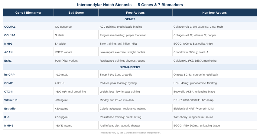
This article takes two precision angles. The first — and most practical for most readers — is biomarker tracking: seven laboratory markers that reflect the biochemical processes directly influencing notch geometry and the integrity of the ligaments within it. The second covers the genetic side: five variants with documented relevance to connective tissue quality, cartilage biology, and structural joint risk. Taken together, they offer a more actionable starting point than waiting for the next injury or the next scan. A third section draws on the work of researcher Keith Baar — one of the most useful and underappreciated voices in connective tissue science — and a final section covers complementary approaches with meaningful clinical evidence.
7 Biomarkers That Reveal What Is Actually Happening in Your Knee
The intercondylar notch may feel like a mechanical problem — and in some respects it is — but its progression is driven by biochemistry. Chronic inflammation thickens synovial tissue. Osteophytes grow from altered bone remodeling signals. Cartilage degrades when catabolic signaling outpaces repair. Hormonal changes alter tissue laxity and turnover. Tracking the right biomarkers puts numbers on these processes so that interventions can be targeted rather than generic.
1. High-Sensitivity C-Reactive Protein — Catching Inflammation Before It Narrows the Notch
Why it matters: Systemic low-grade inflammation is one of the primary biological drivers of joint space narrowing. In the knee, an inflamed synovial membrane can physically expand into the intercondylar notch, and persistent inflammatory signaling stimulates osteophyte growth at notch margins over time. hs-CRP is the most accessible and well-validated marker of this background inflammatory state — affordable, reproducible, and widely understood.
What it may reveal: Values below 0.5 mg/L reflect low systemic inflammation. Values between 0.5 and 1.0 mg/L occupy a gray zone. Values above 1.0 mg/L in a clinically stable, non-infected individual signal a persistent inflammatory state that may be actively driving joint tissue changes. Anything above 3.0 mg/L without an acute infectious cause warrants root cause investigation before other interventions are layered in.
How to Measure It
Standard blood draw, available at most labs and often included in routine preventive panels. Order specifically as hs-CRP rather than standard CRP for sensitivity at lower values. Cost: $10–$40. Collect fasting, ideally repeated on two separate occasions before drawing conclusions — CRP can spike temporarily with minor illness, poor sleep, or acute stress. Morning collection is standard.
If the Score Is Bad — The Plan Without Supplements
Dietary and behavioral changes move hs-CRP reliably. Removing ultra-processed foods, refined seed oils (high in linoleic acid), and high-glycemic staples is foundational. Sleep is underestimated here — even five consecutive nights of poor sleep measurably elevates CRP. Seven to nine hours with consistent timing is more impactful than most supplements. Zone 2 aerobic training (30–45 minutes at a conversational pace, 3–4 sessions per week) drives persistent anti-inflammatory adaptation through mitochondrial biogenesis. Visceral fat reduction is the highest-leverage long-term lever: adipose tissue continuously produces IL-6 and TNF-alpha, both upstream inducers of CRP.
If the Score Is Bad — The Plan With Supplements or Equipment
Omega-3 fatty acids (EPA + DHA, 2–4 g/day combined): Robust RCT evidence for reducing hs-CRP. Use triglyceride-form products for superior absorption. No mandatory cycling; use continuously. Monitor if on anticoagulants — some blood-thinning effect at higher doses. Curcumin phospholipid complex (500–1000 mg/day): Multiple controlled trials show meaningful CRP reductions; bioavailability is critical — avoid plain curcumin powder. Cycle 8–12 weeks on, 4 weeks off. Most common side effect is mild GI discomfort. Cold water immersion (10–15°C, 10–15 minutes, 3–4x/week): Activates norepinephrine and downstream anti-inflammatory pathways; increasingly validated in sports science and clinical literature. Requires only a bathtub and ice — no specialized equipment.
2. COMP (Cartilage Oligomeric Matrix Protein) — The Earliest Signal of Cartilage Stress
Why it matters: COMP is a glycoprotein released into circulation when the cartilage matrix is under mechanical stress or early degradation. It is one of the few serum biomarkers that rises before cartilage damage is visible on MRI, making it a genuine early warning tool. In notch stenosis, where the femoral cartilage walls of the intercondylar space are subject to abnormal contact forces from a compressed ACL, COMP can reveal whether mechanical stress is already translating into biological tissue damage — before the next imaging study shows anything conclusive.
What it may reveal: Elevated COMP (above approximately 12 U/L, though lab reference ranges vary) in someone with notch stenosis suggests active cartilage matrix loading or early breakdown. Serial tracking every 3–6 months can determine whether interventions are slowing or reversing the process — something imaging rarely captures at an actionable early stage.
How to Measure It
Serum COMP by ELISA; not included in standard panels and must be specifically ordered through a functional medicine or rheumatology-affiliated lab. Critical detail: collect the sample before any physical activity on the day of the test, ideally first thing in the morning — COMP rises transiently by 30–50% after exercise and will give a falsely elevated impression if collected post-activity. Cost: $60–$180.
If the Score Is Bad — The Plan Without Supplements
Reduce peak joint loading. Avoid sustained deep squats, heavy compressive loading through the knee, and repetitive stair descent under load. Substitute with cartilage-nutritive movement — cycling and swimming maintain interstitial fluid exchange in articular cartilage through gentle cyclic compression without excessive peak forces. Neuromuscular training targeting hip abductors and the vastus medialis oblique reduces valgus stress and redistributes load away from the medial compartment and notch area. Address foot pronation and gait abnormality through orthotics or gait retraining if indicated.
If the Score Is Bad — The Plan With Supplements or Equipment
Undenatured type II collagen (UC-II, 40 mg/day): Works through oral tolerance, modulating the immune response to cartilage collagen antigens. Clinical trials in OA populations have shown cartilage-protective effects and pain reduction over 24-week periods. Minimal side effects; well tolerated. Glucosamine sulfate (1500 mg/day as a single daily dose): Evidence is mixed, but the crystalline sulfate form has shown cartilage-preserving effects in multi-year controlled trials. Best assessed over at least 6 months. Collagen peptides (10–15 g/day with 50 mg vitamin C, taken 45–60 minutes before exercise): Based on Keith Baar's collagen timing research (discussed in depth below), this protocol doubles circulating collagen precursor availability during the exercise window — supporting anabolic collagen synthesis at the tissue level. Cycle: continuous. Side effects: negligible.
3. CTX-II (C-Terminal Crosslinking Telopeptide of Type II Collagen) — Tracking Active Cartilage Breakdown
Why it matters: Where COMP reflects mechanical stress on cartilage, CTX-II reflects actual enzymatic degradation of type II collagen — the primary structural protein of articular cartilage. Urinary CTX-II is among the most studied and validated biomarkers in OA research for quantifying cartilage catabolism at a molecular level. In notch stenosis, where ACL impingement creates repetitive micro-trauma to cartilage surfaces over time, tracking CTX-II can reveal whether that chronic mechanical stress is actively destroying cartilage tissue beyond the body's repair capacity.
What it may reveal: Elevated urinary CTX-II (typically above 300–500 ng/mmol creatinine, depending on the laboratory reference range) indicates that catabolic processes are winning over anabolic repair in the cartilage. Research has shown that high CTX-II predicts structural joint progression on imaging even in individuals without significant current pain, making it a meaningful tool for proactive surveillance rather than reactive response.
How to Measure It
Second morning urine void, assayed by ELISA and corrected for creatinine concentration. Must be specifically ordered; not part of routine panels. Available through specialty and functional medicine labs. Cost: $80–$200. Track at 3–6 month intervals during active intervention periods to assess direction of change over time.
If the Score Is Bad — The Plan Without Supplements
Body weight management has a direct and quantifiable impact — each kilogram of excess body mass adds approximately 3–4 kg of compressive force on the knee during walking. A 5–7% body weight reduction has been shown in controlled studies to measurably reduce cartilage degradation markers within 12 weeks. During periods of elevated CTX-II, reduce impact-loading activities and substitute with low-impact alternatives. Correct the biomechanical contributors: hip abductor weakness and poor deceleration mechanics are among the most modifiable drivers of cartilage overload in narrow-notch individuals — neuromuscular training programs specifically addressing landing mechanics are directly relevant.
If the Score Is Bad — The Plan With Supplements or Equipment
Boswellia serrata (AKBA form, 100–200 mg/day): One of the most robustly supported natural interventions for reducing cartilage degradation markers. Multiple RCTs have shown significant reductions in CTX-II alongside meaningful pain improvement in OA populations. Cycle: 8–12 weeks on, 4 weeks off. Side effects: occasional mild GI discomfort. Collagen peptides with vitamin C (timing as above): Support chondrocyte anabolic signaling during the exercise window to counterbalance catabolic activity. Functional unloading knee brace: For confirmed compartment loading issues, a valgus or varus unloading brace redistributes mechanical forces and can reduce the load-driven catabolic signal while the joint recovers. An orthopedic specialist can assess fit and indication.
4. Vitamin D (25-Hydroxyvitamin D) — The Bone Remodeling Regulator
Why it matters: Vitamin D is not merely a calcium and bone density story. Vitamin D receptors are expressed on chondrocytes, synoviocytes, and osteoblasts — all directly relevant to notch biology. Deficiency accelerates osteoclast activity, reduces chondroprotective signaling through IGF-1 pathways, and promotes synovial inflammation. Most directly relevant to progressive notch stenosis: osteophyte formation at notch margins — one of the primary mechanisms by which the notch narrows over time in adults — is facilitated by the inflammatory bone remodeling environment that vitamin D deficiency sustains.
What it may reveal: Serum 25-hydroxyvitamin D below 30 ng/mL is associated with greater knee joint inflammation markers, higher ACL re-injury rates following reconstructive surgery, and faster radiographic OA progression. Peter Attia and other longevity-focused clinicians target levels of 40–60 ng/mL as optimal for musculoskeletal health; values below 20 ng/mL represent a meaningful deficiency state for joint tissue biology.
How to Measure It
Serum 25-hydroxyvitamin D; widely available and frequently included in preventive annual panels. Cost: $30–$80. Measure twice per year — late winter and late summer — to capture the seasonal variation, which can swing 15–25 ng/mL between nadir and peak in individuals at northern latitudes.
If the Score Is Bad — The Plan Without Supplements
Direct skin exposure to midday UVB sunlight on large skin areas (arms, legs, and trunk) for 20–40 minutes, 4–5 days per week during spring, summer, and early autumn is effective for most people at latitudes below 40°. At higher latitudes or for individuals with darker skin tones or significant time indoors, sun exposure alone is insufficient in winter months and supplementation becomes necessary.
If the Score Is Bad — The Plan With Supplements or Equipment
Vitamin D3 with K2 (2000–5000 IU D3 combined with 100–200 mcg MK-7 K2, daily): K2 co-supplementation is not optional — it directs calcium into bone rather than soft tissue, reducing the risk of vascular calcification at higher vitamin D doses. Retest 25-OHD at 90 days to titrate dose to target. Side effects: hypercalcemia risk is realistic only at sustained doses above 10,000 IU/day without monitoring; K2 co-supplementation further reduces this risk. Narrowband UVB lamp (3–5 sessions/week, 5–10 minutes per session): Medical-grade home UVB devices effectively maintain vitamin D levels through winter for those who prefer a non-supplementation approach. Equipment cost: $150–$400. Practical and reliable for year-round maintenance when sun exposure is inadequate.
5. Estradiol — The Hormonal Architect of Notch and Ligament Biology
Why it matters: Estradiol's role in knee joint biology is far more direct than most clinical conversations acknowledge. Estrogen receptors are expressed in ACL fibroblasts, chondrocytes, synoviocytes, and osteoblasts — every tissue type relevant to notch health. Fluctuations in estradiol across the menstrual cycle are now recognized as a meaningful factor in ACL injury timing: injury rates cluster around the preovulatory estrogen peak when ligament laxity is highest. More relevant to progressive stenosis: declining estradiol during perimenopause is associated with accelerated chondrocyte loss, increased osteophyte proliferation, and joint space narrowing — changes that directly affect intercondylar notch geometry. In men, low estradiol accompanying low testosterone or impaired aromatase activity similarly compromises joint tissue biology.
What it may reveal: In premenopausal women, estradiol context can inform training load management. In perimenopausal and postmenopausal women, chronically suppressed estradiol may be an underappreciated driver of accelerating notch geometry changes. In men with low estradiol (typically below 20 pg/mL by sensitive assay), joint tissue quality is genuinely compromised regardless of other interventions.
How to Measure It
Serum estradiol (E2) by LC-MS/MS assay — not immunoassay, which is inaccurate below 50 pg/mL. In premenopausal women, collect on day 2–3 of the cycle for a reliable baseline. Cost: $40–$120. In postmenopausal women or men, timing is flexible.
If the Score Is Bad — The Plan Without Supplements
For women with low estradiol from lifestyle-related causes — undereating, relative energy deficiency in sport, chronic psychological stress — restoring caloric adequacy and moderating excessive training volume can meaningfully recover estrogen production within weeks. Resistance training in women supports estradiol through improved insulin sensitivity and reduced cortisol-mediated suppression of ovarian function. For men with low estradiol secondary to low testosterone: resistance training, sleep quality improvement, and visceral fat reduction are the highest-leverage non-pharmaceutical levers, as visceral fat impairs the hypothalamic-pituitary-gonadal axis.
If the Score Is Bad — The Plan With Supplements or Equipment
This is a medical decision, not a supplement decision. Bioidentical hormone therapy — transdermal estradiol (patch, gel, or spray) with micronized progesterone for women with an intact uterus — is the most direct and evidence-based intervention for perimenopausal and postmenopausal women with joint-related estrogen deficiency. Risks and benefits are individual and require physician assessment. For women who are not candidates for HRT, DIM (diindolylmethane, 100–200 mg/day) supports more favorable estrogen metabolism. Cycle: HRT typically continuous; DIM 3–4 months then reassess with labs. DIM may initially reduce estradiol slightly before metabolic improvement — retest at 6 weeks.
6. IL-6 (Interleukin-6) — The Inflammatory Amplifier in the Joint Space
Why it matters: IL-6 has a nuanced role in joint biology. Acutely, muscle-derived IL-6 during exercise has beneficial metabolic effects. But chronically elevated serum IL-6 — driven by visceral fat, poor sleep, sedentary behavior, or smoldering synovitis — promotes osteoclast activation, synovial tissue proliferation, and cartilage matrix degradation. In the context of intercondylar notch stenosis, IL-6-driven inflammatory bony remodeling is a primary mechanism through which the notch progressively narrows over years — often silently, without significant pain until late in the process.
What it may reveal: Baseline serum IL-6 above 2–3 pg/mL in a resting, non-infected individual reflects a background inflammatory milieu. Unlike CRP, which lags IL-6 by 6–12 hours, IL-6 can capture early or smoldering inflammatory activity before CRP has moved — making it more sensitive for early surveillance.
How to Measure It
Serum IL-6 by high-sensitivity ELISA; must be specifically requested. Sample handling is critical — serum must be spun and frozen promptly, as IL-6 degrades rapidly at room temperature. Available through functional medicine and hospital rheumatology lab panels, sometimes alongside TNF-alpha and IL-1beta in comprehensive inflammation profiles. Cost: $60–$150.
If the Score Is Bad — The Plan Without Supplements
Chronic resistance training is among the most potent behavioral interventions for reducing baseline IL-6 — multiple RCTs demonstrate significant reductions with 3x weekly programs over 8–12 weeks, through mechanisms including reduced visceral fat and improved mitochondrial anti-inflammatory signaling. Breaking up prolonged sitting independently matters: sedentary behavior elevates IL-6 even in people who exercise regularly, and simply interrupting sitting every 30–45 minutes with brief movement has measurable effects. Improving sleep quality — specifically targeting consistent sleep timing and keeping the sleep environment cool (18–19°C) — reduces the nighttime cytokine release that drives baseline IL-6 elevation.
If the Score Is Bad — The Plan With Supplements or Equipment
Tart cherry extract (anthocyanins, 480–960 mg/day) or tart cherry juice (240 mL twice daily): Multiple clinical studies demonstrate meaningful reductions in circulating IL-6 and related inflammatory markers. Note the high sugar content of juice form — extract is preferable for those monitoring glucose. Cycle: continuous or 6–8 week blocks. Magnesium glycinate (300–400 mg/day): Magnesium deficiency — prevalent in most adult populations — is independently associated with elevated IL-6. Glycinate form is best absorbed and best tolerated. Side effects: loose stools at high doses; glycinate form is the least problematic. Infrared sauna (50–60°C, 20 minutes, 3–4x/week): Consistent evidence from cardiovascular and chronic pain research for reducing systemic IL-6 with regular use. Home units range from $1,000–$4,000; commercial sauna access is a practical alternative.
7. MMP-3 (Matrix Metalloproteinase-3) — When the Joint Begins Degrading Its Own Scaffold
Why it matters: MMP-3 — stromelysin-1 — is an enzyme that degrades the extracellular matrix, including the collagen, proteoglycans, and fibronectin that give ligaments, tendons, and cartilage their structural integrity. In a knee with intercondylar notch stenosis, the ACL is under chronic mechanical stress at impingement points. Elevated MMP-3 in this context means that connective tissue is being enzymatically degraded precisely where it is most structurally vulnerable. MMP-3 is also a key driver of synovial tissue proliferation — the mechanism by which synovitis further narrows the notch physically.
What it may reveal: Serum MMP-3 above approximately 30 ng/mL in men or 43 ng/mL in women (ranges vary by lab and sex) reflects active joint tissue remodeling. Research in rheumatology populations has shown that elevated MMP-3 predicts structural joint erosion and progressive damage — making it a useful leading indicator for notch stenosis management rather than a trailing one.
How to Measure It
Serum MMP-3 by ELISA; available through functional medicine and rheumatology specialty labs. Some comprehensive joint inflammation panels include it alongside other MMPs. Cost: $70–$180. Track at 3–6 month intervals to assess whether interventions are moving the trend in the right direction.
If the Score Is Bad — The Plan Without Supplements
Address the root cause of MMP-3 elevation before increasing exercise load: active synovitis or chronic joint inflammation should be the first target. Aquatic therapy and joint-offloading bracing allow muscle maintenance without compounding MMP-3-driven matrix degradation. A period of elevated MMP-3 is a specific signal to reduce the volume and intensity of activities that load the knee heavily — not to push through. Anti-inflammatory dietary patterns (Mediterranean framework, reduced ultra-processed food intake) reduce the cytokine environment that upregulates MMP-3 gene expression.
If the Score Is Bad — The Plan With Supplements or Equipment
Green tea extract (EGCG, 400–600 mg/day): Epigallocatechin gallate is a well-studied natural MMP inhibitor with in vitro and human evidence for joint-protective effects. Use decaffeinated extract if caffeine-sensitive. Cycle: 8–12 weeks on, 4 weeks off. Rare hepatotoxicity risk at very high doses — stay within recommended ranges and avoid stacking with multiple other liver-metabolized compounds. Palmitoylethanolamide (PEA, 300–600 mg/day): Emerging evidence from chronic pain and joint inflammation research supports reduction of MMP activity and synovial inflammatory signaling with PEA. Well tolerated in clinical trials. Cycle: 8–12 week minimum before assessing benefit. Functional unloading knee brace: Mechanically redistributes forces away from the affected compartment, reducing the peak load stimulus that drives MMP-3 upregulation while the joint environment stabilizes. Orthopedic specialist assessment for proper fit is warranted.
The biomarker picture tells you what is happening now. The genetic picture tells you why your biological baseline may differ from someone with similar anatomy and lifestyle — and helps explain why two people with seemingly identical situations don't respond identically to the same interventions.
The Genetic Blueprint: 5 Variants That Shape Your Connective Tissue and Notch Risk
Genetics does not determine outcomes in intercondylar notch stenosis — but it does set the terrain. Understanding which variants you carry helps explain why certain individuals are more structurally vulnerable, more prone to cartilage breakdown, or less responsive to hormonal signals that would otherwise protect joint tissue. Consumer genetic platforms (such as 23andMe or AncestryDNA) provide raw data that can be analyzed through interpretation services like Athletigen; clinical-grade testing through a physician offers greater accuracy for specific single-nucleotide polymorphisms.
1. COL5A1 — The Structural Blueprint of Your Ligaments
What it affects: COL5A1 encodes the alpha-1 chain of type V collagen, which regulates collagen fibril diameter and organization in tendons and ligaments. A well-studied polymorphism in the 3' untranslated region of COL5A1 — the C/T variant — significantly affects ACL injury susceptibility. The TT genotype is associated with lower risk; the CC genotype produces collagen fibrils with reduced diameter, lower tensile stiffness, and greater susceptibility to mechanical failure. This association has been replicated across multiple independent cohorts studying ACL rupture populations. In the context of notch stenosis, where the ACL is chronically compressed, inherent ligamentous laxity from an unfavorable COL5A1 variant substantially amplifies rupture risk.
If the Gene Is Unfavorable — The Plan Without Supplements
Neuromuscular training is the primary evidence-based compensation. Programs specifically designed around ACL injury prevention — emphasizing landing mechanics, hip abductor activation, and hamstring co-contraction — directly address the consequence of ligamentous laxity. Plyometric training with a stiff landing emphasis teaches the musculature to absorb forces that the ligament alone cannot safely handle in a CC carrier. Prophylactic functional bracing during high-risk activities (cutting sports, alpine skiing) is a reasonable mechanical safeguard when COL5A1 risk is confirmed.
If the Gene Is Unfavorable — The Plan With Supplements or Equipment
Gelatin or hydrolyzed collagen (15 g) plus vitamin C (50 mg), taken 45–60 minutes before exercise: Keith Baar's published collagen timing research (see next section) demonstrates that this approach roughly doubles circulating collagen precursor amino acids during the subsequent exercise window, directly supporting ligament collagen synthesis. Cycle: daily before training sessions. Side effects: minimal. Zinc (15–25 mg/day) and copper (1–2 mg/day): Both are required cofactors for lysyl oxidase, the enzyme that crosslinks collagen chains into mechanically competent mature fibers. Deficiency in either mineral — common in athletes with restricted dietary variety — degrades collagen maturation regardless of substrate availability. Cycle: continuous; reassess serum levels at 3 months. Heavy slow resistance (HSR) training: Loading tendons and ligaments with slow-tempo, high-tension exercises (3–5 seconds eccentric, 3 seconds concentric) promotes collagen fiber remodeling and increases tensile stiffness of connective tissue over 12+ weeks. Well-supported for tendon adaptation; principles extend to ligament biology.
2. COL1A1 — The Foundation Collagen Gene
What it affects: COL1A1 encodes the alpha-1 chain of type I collagen — the most abundant structural collagen in tendons, ligaments, and bone. The Sp1 binding site polymorphism (rs1800012) alters the ratio of COL1A1 to COL1A2 chains, producing a collagen molecule with subtly different mechanical properties. The "S" allele (carrying the Sp1 site) is associated with reduced tensile strength in connective tissues and has been linked in multiple research populations to elevated risk of ACL rupture, Achilles tendon injury, and stress fractures. In a narrow-notch knee where the ACL is already under impingement stress, reduced baseline collagen quality from an unfavorable COL1A1 variant meaningfully elevates failure risk at lower force thresholds.
If the Gene Is Unfavorable — The Plan Without Supplements
Progressive heavy loading of the ACL through controlled resistance training is the primary structural intervention. Collagen remodeling within the ligament requires mechanical stimulus — loading without exceeding the tissue's current capacity. Training should specifically emphasize controlled deceleration mechanics and avoid unplanned directional changes. Address footwear: appropriate arch support and cushioning reduces the torsional and compressive forces transmitted through the knee during dynamic activities.
If the Gene Is Unfavorable — The Plan With Supplements or Equipment
The collagen timing protocol (15 g gelatin or hydrolyzed collagen with vitamin C, pre-exercise) applies directly and is arguably more important for COL1A1 S-allele carriers, who need greater support for collagen crosslinking during remodeling windows. Vitamin C (500 mg alongside collagen): Prolyl hydroxylase — the enzyme responsible for collagen crosslink formation — requires vitamin C as an obligate cofactor. Without adequate vitamin C, newly synthesized collagen has weaker crosslinks regardless of the substrate provided. Most people are above scurvy threshold but below optimal collagen quality threshold. Copper adequacy: Lysyl oxidase copper-dependence is a second crosslinking vulnerability often overlooked — assess dietary copper intake and supplement to ensure 1.5–2 mg/day if diet is restricted. Whole food sources: liver, shellfish, dark chocolate, legumes.
3. MMP3 Gene — The Matrix Remodeler Variant
What it affects: A functional polymorphism in the promoter region of the MMP3 gene — the 5A/6A variant (rs3025058) — alters baseline MMP-3 transcriptional activity. The 5A allele is associated with significantly higher MMP-3 expression. In practical terms, a 5A carrier has a biological baseline that more aggressively degrades extracellular matrix in connective tissue. In the context of notch stenosis, a 5A/5A or 5A/6A individual breaks down ACL collagen fibers at the impingement zone and cartilage proteoglycans at the notch walls more rapidly than a 6A/6A individual — even under equivalent mechanical conditions. Gary Brecka and other functional genetics practitioners have highlighted MMP gene variants as underappreciated drivers of accelerated connective tissue aging.
If the Gene Is Unfavorable — The Plan Without Supplements
MMP-3 activity is partly regulated by the mechanical loading environment. Excessive peak loading — rapid impact, high-velocity joint forces, poorly controlled deceleration — is a primary trigger for MMP-3 upregulation. Shifting training toward slow-tempo, controlled loading with adequate inter-session recovery gives connective tissue time to remodel without exceeding the catabolic threshold. An anti-inflammatory dietary pattern (Mediterranean framework, reduced refined carbohydrate, increased polyphenol intake) reduces the upstream cytokine environment that drives MMP-3 gene expression. This is particularly important for 5A carriers, for whom the gene is poised to amplify any inflammatory signal it receives.
If the Gene Is Unfavorable — The Plan With Supplements or Equipment
Green tea extract (EGCG, 400–600 mg/day): A direct natural MMP-3 enzyme inhibitor with evidence from joint disease research. Particularly relevant for 5A carriers who have chronically higher baseline MMP-3 activity. Cycle: 8–12 weeks on, 4 weeks off. Boswellic acids (AKBA, 100–200 mg/day): Inhibit both 5-lipoxygenase and MMP pathways simultaneously, addressing two branches of catabolic activity. The collagen timing protocol is especially justified for MMP3 5A carriers — stimulating anabolic collagen synthesis during exercise windows helps counterbalance an elevated catabolic background that cannot be fully corrected by behavioral means alone.
4. ACAN (Aggrecan) — The Cartilage Cushion Gene
What it affects: ACAN encodes aggrecan, the large proteoglycan that gives articular cartilage its compressive stiffness and resilience through water-binding capacity. Variants in the ACAN gene — particularly in the variable number tandem repeat (VNTR) region — influence cartilage proteoglycan content and quality. For intercondylar notch stenosis, this is directly relevant: the cartilage lining the notch walls is aggrecan-dependent for its compressive resistance. An individual with inherently lower aggrecan expression or turnover will develop thinner, less resilient cartilage — meaning the notch walls offer less of a buffer against ACL impingement forces and are more susceptible to the cartilage degradation that narrows the effective notch space over time.
If the Gene Is Unfavorable — The Plan Without Supplements
Protect cartilage from chronic overloading as the primary strategy. Favor low-impact activity and avoid extended periods of high compressive loading — heavy deep squats, running on hard surfaces for extended durations, and sustained kneeling. Systematic neuromuscular training to distribute knee loads more evenly across joint surfaces is the main modifiable tool. Body weight management is particularly important for ACAN variant carriers, since excess compressive loading is harder to compensate through cartilage of inherently lower quality — even modest excess weight creates disproportionate degradation risk.
If the Gene Is Unfavorable — The Plan With Supplements or Equipment
Chondroitin sulfate (800–1200 mg/day): As a building block for aggrecan synthesis, supplemental chondroitin sulfate provides substrate for proteoglycan production. Evidence quality is moderate; best results appear with 6+ months of continuous use. Side effects: minimal. Oral high-molecular-weight hyaluronic acid (80–200 mg/day): Emerging evidence supports oral HA for joint fluid viscosity and cartilage matrix support. For confirmed cartilage thinning on imaging, injectable viscosupplementation with HA is a more established clinical option — worth discussing with an orthopedic specialist when ACAN risk is combined with imaging evidence of thinning.
5. ESR1 (Estrogen Receptor Alpha) — The Hormonal Gatekeeper of Joint Tissue
What it affects: ESR1 encodes estrogen receptor alpha, which mediates estrogen's direct effects on bone remodeling, cartilage integrity, and ligament biology. Common ESR1 polymorphisms — including PvuII (rs2234693) and XbaI (rs9340799) — are associated with differences in bone mineral density, cartilage quality, and tissue responsiveness to estrogen. For women, an unfavorable ESR1 variant means that even physiologically normal estrogen levels may not adequately protect joint tissues — effectively amplifying the consequences of the perimenopausal estrogen decline on the knee joint. Functional genetics practitioners including Gary Brecka have highlighted ESR1 variants as underappreciated contributors to accelerated musculoskeletal aging in women, particularly in the context of notch geometry changes.
If the Gene Is Unfavorable — The Plan Without Supplements
Resistance training becomes more important, not less, when ESR1 receptor sensitivity is impaired. Mechanical loading of bone and joint tissue provides an estrogen-independent anabolic signal that partially compensates for reduced receptor responsiveness. Phytoestrogen-rich foods — particularly fermented soy products and ground flaxseed — provide weak estrogen receptor ligands that may partially activate impaired receptors; evidence for joint-specific benefit is limited but the dietary pattern is otherwise supportive. Minimizing cortisol-driven inflammatory signaling (through sleep, stress management, and zone 2 training) reduces the inflammatory burden that ESR1 variants cannot adequately counter through estrogen receptor pathways.
If the Gene Is Unfavorable — The Plan With Supplements or Equipment
For postmenopausal women with confirmed ESR1 variants, the clinical rationale for discussing hormone therapy with a knowledgeable physician is arguably strengthened — reduced receptor activity means that even the partial estradiol remaining post-menopause is less effective, accelerating the tissue consequence. Calcium (1000–1200 mg/day from food and supplement combined) with vitamin D3 and K2 becomes particularly important to protect bone and joint tissue from the downstream effects of impaired estrogen receptor signaling. Discuss bone density monitoring (DEXA) with a physician if ESR1 variant status is confirmed alongside other risk factors.
The genetic and biomarker frameworks together tell you the current status and baseline vulnerability of your knee. What the next section offers is something different: a research-based biological protocol that fundamentally challenges how most clinicians think about connective tissue repair — and gives you a practical playbook regardless of your genetic profile.
The Connective Tissue Research That Is Quietly Rewriting Physical Therapy
Among the most practically useful and genuinely counterintuitive voices in joint and connective tissue science is Dr. Keith Baar, muscle physiologist and connective tissue researcher at UC Davis. His work — discussed in depth across multiple appearances on Peter Attia's podcast The Drive and Rhonda Patrick's FoundMyFitness, and published in peer-reviewed journals — challenges longstanding assumptions about tendon and ligament biology that still govern most rehabilitation protocols. For someone managing intercondylar notch stenosis, these findings are directly relevant to ACL resilience, cartilage protection, and the limits of current return-to-play standards.
Collagen Synthesis Responds to Timing, Not Just Volume
Connective tissue does not benefit from prolonged, continuous loading. It responds optimally to short bouts of mechanical stimulation — as little as 6–10 minutes of appropriate exercise — followed by adequate rest. This is counterintuitive to most training frameworks, which assume that more sustained exercise means more tissue adaptation. For ligament biology, it simply does not work that way. The stimulus needed for collagen synthesis is brief; the subsequent rest period is when synthesis actually occurs.
Gelatin Plus Vitamin C Before Exercise Doubles Collagen Synthesis
Baar's research, published in the American Journal of Clinical Nutrition, demonstrated that consuming 15 grams of gelatin or hydrolyzed collagen alongside 50 mg of vitamin C approximately 45–60 minutes before exercise roughly doubled the circulating hydroxyproline and proline availability during the subsequent loading window — the precursors used to build new collagen fibers in loaded connective tissue. This is an inexpensive, practical, and specifically evidence-based tool that most rehabilitation programs do not incorporate. Shaw et al., American Journal of Clinical Nutrition, 2017
The ACL Can Be Trained — But Not Through Conventional Exercise Selection
The ACL is a ligament, not a muscle, but it still responds to mechanical loading through collagen remodeling. Isometric exercises at specific joint angles — around 20–30° of knee flexion for ACL emphasis — generate significant ligament tension without requiring the ACL to repeatedly traverse the narrowed notch during dynamic movement. For notch stenosis specifically, this is an important distinction: you can stimulate ACL remodeling and stiffening without the impingement risk of cutting and pivoting sports.
Estrogen Directly Suppresses Collagen Synthesis in Ligament Cells
Baar's laboratory has demonstrated that ACL fibroblasts cultured in high-estrogen conditions produce substantially less collagen than cells in low-estrogen or male hormone environments. This finding — counterintuitive to most clinical thinking — suggests that the preovulatory estrogen peak in the menstrual cycle may directly reduce ACL collagen production at the exact time when ligament laxity is also at its highest. The practical implication: high-risk dynamic activities may warrant modification during the preovulatory phase for women with notch stenosis.
Ligament Stiffness Is Trainable
Stiffness — not just strength — is the mechanical property that determines ACL failure resistance under impingement. Genetics (COL5A1, COL1A1), hormonal state, and training history all modulate ACL stiffness. Baar's work highlights that isometric loading and the timed collagen nutrition protocol are the primary tools available to increase stiffness in a structure most clinicians treat as fixed. This reframes the question from "can I protect my ACL?" to "what is the protocol for improving its mechanical properties?"
There Is a 6-Hour Collagen Synthesis Window After Loading
After a mechanical stimulus, collagen synthesis in connective tissue remains elevated for approximately 6 hours before returning to baseline — regardless of additional loading. This has a direct implication for rehabilitation programming: two short daily loading sessions separated by at least 6 hours may produce greater collagen adaptation than one longer session, because each stimulus opens a new synthesis window. Very few existing protocols are designed around this biology.
Vitamin C Is Non-Negotiable for Mature Collagen Structure
Prolyl hydroxylase — the enzyme that creates the hydroxylated prolines essential for strong collagen crosslinks — requires vitamin C as an obligate cofactor. Without adequate vitamin C, newly synthesized collagen lacks the crosslinks that give it tensile strength. Most people are above the scurvy threshold, but optimal collagen crosslinking may require higher tissue saturation than the standard recommended daily intake provides. Baar consistently recommends ensuring adequacy alongside any collagen supplementation protocol.
NSAIDs Actively Suppress Connective Tissue Adaptation
Regular non-steroidal anti-inflammatory drug use — reflexively prescribed and widely self-administered for joint pain — significantly reduces tendon and ligament collagen synthesis. The anti-inflammatory action that makes NSAIDs effective for pain also blunts the prostaglandin-mediated signaling that drives connective tissue adaptation to load. For someone with notch stenosis trying to improve ACL resilience through a systematic loading and nutrition protocol, chronic NSAID use directly undermines the goal. This is almost never mentioned in the clinical settings where NSAIDs are prescribed.
Open-Chain Leg Extensions Are Problematic at Specific Angles
Open-chain knee extension exercises (traditional leg extension machine) generate high ACL tensile strain between approximately 10° and 40° of knee flexion — exactly the range where a narrow-notch ACL is most vulnerable to impingement. Baar recommends avoiding open-chain extension in the vulnerable range for ACL-compromised individuals and preferring closed-chain alternatives (leg press, goblet squat, wall sit) that load surrounding structures while minimizing ACL impingement forces.
Return-to-Play Timelines Are Systematically Biologically Premature
Clinical ACL return-to-play timelines — typically 6–9 months post-reconstruction — are often based on functional testing thresholds rather than biological healing rates. Baar's research highlights that ligament collagen maturation — specifically the crosslinking and fiber organization that restore mechanical competence — takes considerably longer: potentially 12–18 months or more for full structural maturity. Athletes returned at 6–9 months may be functionally capable but biologically under-healed, with a collagen matrix still below the tensile properties needed to resist impingement-related failure in a narrow-notch anatomy. This finding is gaining traction in sports medicine, but has not yet uniformly changed clinical practice.
Cold Exposure and Load Timing Modulate Collagen Synthesis Rate
Temperature affects collagen gelation and tissue assembly. Baar has noted that cold applied immediately after loading may interfere with early collagen matrix organization in connective tissue — a biologically plausible mechanism behind findings that immediate post-exercise ice application may reduce adaptation in tendons, not just muscles. For notch stenosis management, this means that routine post-exercise ice applied to the knee — a ubiquitous practice — may have a more nuanced cost-benefit ratio than generally assumed, particularly during active rehabilitation phases focused on connective tissue remodeling.
Complementary Approaches With Meaningful Clinical Evidence
Low-Level Laser Therapy (Photobiomodulation)
Low-level laser therapy — also called photobiomodulation (PBM) — uses specific red and near-infrared wavelengths (typically 630–1000 nm) to penetrate tissue and stimulate mitochondrial activity, reduce inflammatory cytokine release, and support tissue repair processes. For intercondylar notch stenosis, PBM is relevant primarily as an anti-inflammatory tool targeting the synovitis and local inflammatory activity that drive both symptom burden and progressive notch narrowing through osteophyte formation. By supporting mitochondrial function in synoviocytes and chondrocytes and reducing IL-6 and TNF-alpha in the local joint environment, photobiomodulation addresses the inflammatory component of notch disease without the connective tissue adaptation costs of NSAIDs.
A systematic review and meta-analysis on LLLT in knee osteoarthritis found significant reductions in pain and meaningful functional improvements compared with sham treatments, with effect sizes comparable to pharmacological anti-inflammatory treatment — and without the collagen synthesis suppression that NSAIDs produce. Protocols in the evidence base typically use 830 nm wavelength diodes applied directly over the knee joint for 8–16 sessions over 4–8 weeks, with energy densities in the range of 4–8 J/cm². The research foundation for photobiomodulation in knee conditions continues to strengthen.
Home-grade PBM devices — red and near-infrared panels or targeted devices — are now widely available at $200–$700 and allow ongoing self-administration between clinical sessions. For notch stenosis, target the anterior and medial knee joint line for 10–20 minutes per session, 4–5 days per week. PBM will not mechanically reverse an established notch narrowing, but as a tool for managing the inflammatory process that drives progression, it is practical, safe, and increasingly evidence-supported with minimal side effect profile.
Tai Chi
Tai chi is a slow, deliberate, flowing movement practice that emphasizes controlled weight transfer, joint awareness, and graduated loading — qualities that make it directly relevant to intercondylar notch stenosis. It systematically trains the neuromuscular control of the knee through every degree of flexion and extension, progressively improving the hip abductor activation, quadriceps-hamstring co-contraction, and balance capacity that reduce dynamic valgus and landing mechanics associated with ACL impingement risk. Critically, it does so at a pace and force level that minimizes acute compressive loads on the joint while still providing the low-grade mechanical stimulus that maintains cartilage nutrition through cyclic fluid exchange.
The clinical evidence for tai chi specifically in knee osteoarthritis — the most closely related studied population — is among the strongest for any non-pharmacological intervention. A randomized trial comparing tai chi with standard physical therapy in knee OA patients demonstrated that 12 weeks of twice-weekly tai chi practice produced greater improvements in pain, function, and proprioception than the physical therapy comparator group. The proprioception improvement is particularly relevant for notch stenosis: impaired knee proprioception is an independent risk factor for ACL re-injury, and tai chi specifically targets mechanoreceptor sensitivity through its slow, attentive movement demands.
For practical implementation, 2–3 sessions per week of 30–45 minutes is sufficient to begin demonstrating neuromuscular benefit within 6–8 weeks. The simplified 24-form Yang style is widely taught in group classes and accessible through structured online instruction for beginners. Avoid deep horse-stance positions until sufficient quadriceps and hip stability is established, as deep sustained knee flexion under poor neuromuscular control increases rather than decreases impingement risk. Tai chi is appropriately low-impact for initiation even during periods of mild active knee irritation.
Yoga
Yoga is relevant to intercondylar notch stenosis through two distinct mechanisms: hip and surrounding structure mobility improvement — which directly reduces the aberrant knee loading patterns that increase impingement risk — and its modulation of the stress-cortisol-inflammation axis. Hip flexor tightness, reduced external rotator flexibility, and weak gluteal activation are among the primary biomechanical contributors to dynamic valgus and poor knee alignment during movement. A well-structured yoga practice targeting these patterns can meaningfully reduce the compressive and shear forces transmitted through the notch area during daily and athletic activities. Separately, its stress-regulating effects reduce the cortisol-mediated inflammatory signaling that accelerates cartilage catabolism.
Evidence for yoga in knee joint pathology shows meaningful improvements in pain, range of motion, and neuromuscular control following 8-week structured programs, with hip-opening and balance-focused practices showing particular relevance for knee mechanics. The important caveat for notch stenosis: deep knee flexion postures — full lotus, pigeon pose held deeply, hero pose, and similar — can increase intra-articular compressive forces and ACL impingement stress in a structurally narrowed notch, and should be approached carefully or avoided in the early stages of practice.
For practical application, 3–4 sessions per week of 30–45 minutes focusing on hip mobility, balance, and light body weight bearing is appropriate. Yin yoga — which emphasizes sustained, passive holds targeting connective tissue rather than muscle — is particularly relevant for its effects on fascia and ligamentous tissue health. Inform any general yoga instructor of the notch stenosis condition so that high-risk knee flexion poses can be modified. Progress deep flexion postures gradually and only as neuromuscular control and pain tolerance allow.
Massage Therapy
Massage therapy cannot change intercondylar notch geometry — but it addresses two meaningful contributors to functional limitation and symptom burden that are often underappreciated: soft tissue tension in the musculature surrounding the knee, and local vascular and lymphatic circulation to the joint. Chronic tightness in the quadriceps, iliotibial band, popliteus, and gastrocnemius alters biomechanical force transmission through the knee, increasing the compressive and shear loads directed at the notch area during movement. Targeted manual therapy to these structures can reduce this neuromechanical contribution to impingement without any joint-level intervention. Additionally, improved local circulation supports the synovial fluid dynamics that maintain cartilage nutrition in the notch area.
Controlled evidence for massage therapy in knee OA populations has shown consistent, modest benefits for pain and functional mobility. An 8-week Swedish massage protocol targeting knee OA patients produced significant improvements in pain and stiffness scores compared with waitlist control participants in a randomized trial. The mechanism appears to involve both direct mechanical tissue effects and modulation of local inflammatory mediators through improved circulation.
Practical application involves 45–60 minute sessions focusing primarily on the quadriceps, ITB, hip flexors, popliteal fossa, and gastrocnemius-soleus complex — rather than aggressive direct work on the knee joint capsule itself, which can aggravate active synovitis. Frequency of once or twice weekly during active management phases, reducing to monthly maintenance once symptoms stabilize. Self-massage using a foam roller, massage ball, or percussion device provides cost-effective daily maintenance between professional sessions and is particularly useful for managing quadriceps and ITB tension that contributes to altered knee mechanics.
Mindfulness Meditation and MBSR
Mindfulness-Based Stress Reduction (MBSR) is an 8-week structured program combining formal mindfulness meditation, body scan practice, and mindful movement, originally developed by Jon Kabat-Zinn at the University of Massachusetts Medical School. Its relevance to intercondylar notch stenosis is rooted in the well-documented relationship between chronic psychological stress, cortisol, and systemic inflammation. Chronically elevated cortisol suppresses chondrocyte anabolic activity, accelerates cartilage catabolism, and elevates both IL-6 and TNF-alpha — three mechanisms that directly drive the inflammatory progression of notch stenosis and impede the connective tissue remodeling that rehabilitation depends on. MBSR also addresses central sensitization, which frequently complicates the pain experience in chronic joint conditions regardless of the underlying structural pathology.
Meta-analyses of MBSR in chronic musculoskeletal pain conditions consistently show significant reductions in pain intensity, pain catastrophizing, and quality of life impairment. Importantly, several studies have measured inflammatory biomarkers directly — finding meaningful reductions in CRP and IL-6 in participants completing the 8-week protocol compared with controls, effects that persist at 3-month follow-up and reflect genuine biological change beyond subjective pain reporting.
MBSR programs are available through hospital integrative medicine departments, mindfulness centers, and validated online platforms (including the Palouse Mindfulness online program, which follows the original MBSR curriculum at no cost). The commitment structure is 8 weeks with one 2.5-hour weekly class session and daily home practice of 30–45 minutes. For intercondylar notch stenosis, MBSR is most appropriately integrated as a long-term background practice rather than an acute intervention — the stress-inflammatory pathway effects are cumulative and most meaningful over months of sustained engagement rather than as a short course.
A Smarter Starting Point
Intercondylar notch stenosis is not a condition that responds predictably to generic management. Its biological trajectory is shaped by a specific combination of inflammatory load, cartilage turnover rate, hormonal status, and connective tissue genetics — all of which vary meaningfully between individuals and all of which are, to varying degrees, measurable and addressable. The seven biomarkers covered here give you a concrete, trackable picture of what is happening in your knee's biological environment. The five gene variants give you a baseline understanding of why your tissues may respond differently from expectations. Keith Baar's connective tissue protocol offers a practical, inexpensive intervention framework that applies regardless of genetic profile. And the complementary modalities — particularly tai chi, photobiomodulation, and the MBSR protocol — provide evidence-based tools that can be layered alongside conventional care rather than substituted for it.
The next smart step is not to implement everything simultaneously. Start with the most accessible and informative biomarkers — hs-CRP and vitamin D are practical entry points for nearly everyone — and use the results to prioritize the next steps. Bring your findings to a clinician who is comfortable interpreting biomarkers in the context of musculoskeletal health, and discuss the genetic component if you have access to consumer genetic data. Progress with this condition is not guaranteed to be dramatic or fast, but it is measurable — and measurement is where meaningful management begins.
Musculoskeletal: Joint Conditions Tendon & Ligament Conditions Sports Injuries
Autoimmune: Inflammatory Conditions Connective Tissue Conditions