This article contains AI generated content.
· UpdatedIntra-Articular Loose Bodies — 6 Genes And 7 Biomarkers To Track
Introduction
Living with intra-articular loose bodies can feel like dealing with a joint that has turned against itself. The catching sensations, the unpredictable locking episodes, the swelling that appears without obvious provocation — these are symptoms that most people only partially understand, even after imaging confirms the diagnosis. And that partial understanding matters, because what you do next depends entirely on it.
Generic advice — rest, arthroscopic debridement, physical therapy — is not wrong, but it rarely addresses the underlying biology driving the problem. Two people can have the same MRI findings and experience radically different trajectories. One heals well after intervention; the other keeps forming new fragments. The difference often lies in what is happening at the cellular and molecular level: cartilage resilience, bone turnover rates, inflammatory burden, and genetic predispositions that no imaging study will reveal.
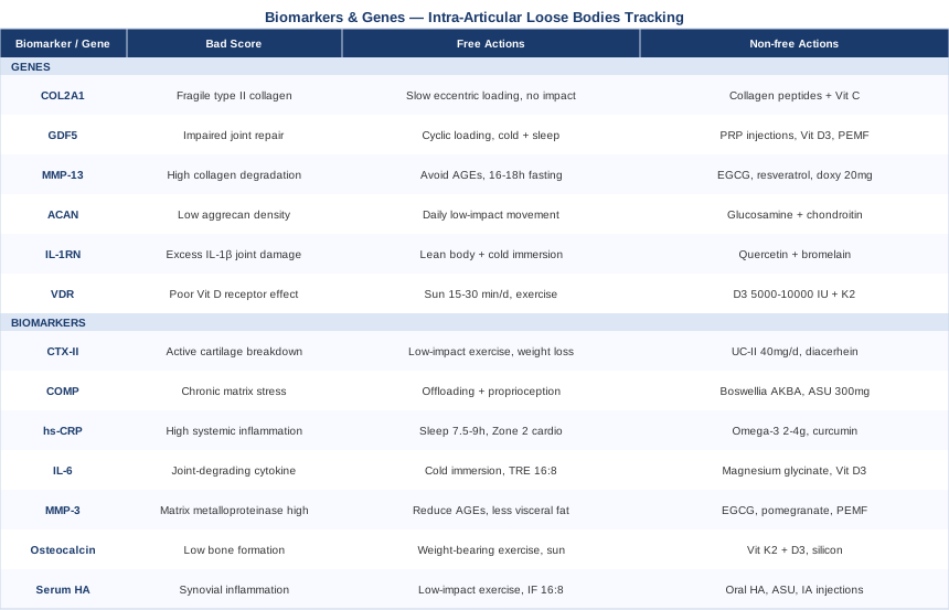
This article takes a more precise approach. The first section focuses on seven blood and urine biomarkers — specific, measurable molecules that reflect cartilage breakdown, bone remodeling, and synovial inflammation in real time. The second section covers six genetic variants that may explain why some individuals are constitutionally more prone to osteochondral fragility, poor repair signaling, or amplified inflammatory cascades. Both sections include concrete, actionable plans — with and without supplementation — for each finding.
Better information does not guarantee a perfect outcome, but it reliably leads to better decisions. When you know your CTX-II trajectory, your COMP levels, your GDF5 variant, and your VDR status, you stop guessing and start working with your own biology rather than against it.
7 Biomarkers That Reveal What Is Really Happening Inside Your Joint
Biomarkers are not abstract research constructs. They are specific, measurable molecules that tell a story your MRI cannot. For intra-articular loose bodies — whether arising from osteochondritis dissecans, synovial chondromatosis, or late-stage cartilage degeneration — a well-chosen panel reveals whether your joint environment is actively degrading, how much systemic inflammation is accelerating that process, and whether your body has the biological tools to repair what is breaking down.
The seven biomarkers below are selected for their clinical relevance to osteochondral pathology, their availability through specialty or standard laboratories, and the quality of evidence supporting their use in monitoring and intervention.
1. CTX-II — The Frontline Marker of Cartilage Collagen Breakdown
CTX-II (C-terminal crosslinked telopeptide of type II collagen) is the most validated biochemical marker of cartilage collagen degradation currently available. When chondrocytes die or cartilage matrix is enzymatically broken down, type II collagen is cleaved and its fragments — including CTX-II — are released into urine. Elevated CTX-II is consistently associated with cartilage loss, increased risk of osteoarthritis progression, and osteochondral defect formation. In the context of loose bodies, high CTX-II signals that the joint environment is actively destroying cartilage, which increases the likelihood that new osteochondral fragments will form even after existing ones are removed.
Multiple human studies confirm CTX-II as a strong predictor of radiographic joint space narrowing, with its urinary measurement correlating well with histological evidence of cartilage damage.
How to Measure It
CTX-II is measured in second-morning urine using an ELISA assay. It is not part of standard clinical panels and must be ordered through specialty laboratories such as Cyrex, Vibrant America, or research-affiliated clinical labs. Cost ranges from $100 to $250 USD. Results are expressed as nanograms per millimole of creatinine. Retest quarterly during active rehabilitation to track trajectory.
If the Score Is Bad: The Plan Without Supplements
The most impactful non-supplement intervention for elevated CTX-II is reducing axial joint loading without becoming sedentary. Switching from high-impact activity (running, jumping, heavy leg press) to low-impact alternatives like swimming, cycling, or water aerobics can meaningfully reduce cartilage collagen degradation. Body weight normalization, if applicable, is among the most effective levers: each excess kilogram adds approximately four kilograms of compressive force per step on the knee. Gait retraining with a physical therapist to reduce peak knee adduction moment can lower CTX-II over three to six months of consistent effort.
If the Score Is Bad: The Plan With Supplements or Equipment
The most evidence-backed supplement for reducing CTX-II is undenatured type II collagen (UC-II) at 40 mg per day. Unlike hydrolyzed collagen, UC-II works through oral tolerance mechanisms in the gut, dampening the immune response driving collagen breakdown. Several randomized controlled trials show meaningful CTX-II reduction and improved joint function with 90 days of use. Cycle: three months on, one month off. Side effects are minimal; mild GI discomfort has been reported rarely. Diacerhein at 50 mg twice daily has also shown CTX-II reduction in clinical trials, though GI side effects (loose stools) are common and limit use. For a device-based approach, continuous passive motion (CPM) equipment used post-arthroscopically for four to six hours per day has been shown to reduce cartilage breakdown markers and may be relevant for post-surgical management.
2. COMP — A Window Into Cartilage Matrix Stress
COMP (Cartilage Oligomeric Matrix Protein) is a structural glycoprotein that helps maintain cartilage architecture. When cartilage is subjected to excessive mechanical stress or enzymatic attack, COMP is released into the bloodstream. Serum COMP rises acutely after joint loading and chronically when cartilage is undergoing structural damage. In patients with osteochondritis dissecans or recurrent loose body formation, elevated serum COMP suggests ongoing osteochondral stress that makes healing difficult and recurrence more likely.
Research on COMP has demonstrated its usefulness as a disease activity marker in both early and late cartilage pathology, including in younger athletes at risk for OCD.
How to Measure It
COMP is measured via serum ELISA through specialty labs and some university-affiliated clinical diagnostics centers. Cost: $150 to $350 USD. Important: COMP rises transiently after exercise, so blood must be drawn after at least two hours of rest. Values are reported in ng/mL or U/L depending on the lab.
If the Score Is Bad: The Plan Without Supplements
Elevated chronic COMP calls for a structured mechanical offloading strategy — identifying and eliminating the specific loading patterns most damaging to the affected joint. For the knee: deep squats, impact landings, and heavy eccentric loading. For the elbow: overhead throwing mechanics and heavy pulling movements. Proprioceptive training — balance boards, single-leg stability work — has been shown to reduce abnormal joint forces and, over time, lower COMP in active individuals. Rest-to-load cycling (alternating loading and full-offloading days) is more effective than complete rest, which allows cartilage nutrition to deteriorate.
If the Score Is Bad: The Plan With Supplements or Equipment
Boswellia serrata extract (standardized to AKBA, 100–200 mg per day) has been shown in randomized trials to reduce serum COMP and improve cartilage volume on MRI after 90 days. Side effects are rare; occasional GI discomfort. Cycle three months on, one month off. Avocado-soy unsaponifiables (ASU) at 300 mg per day have demonstrated reductions in cartilage biomarkers including COMP in several European clinical trials. For equipment, offloading orthotic braces designed to shift weight away from the damaged compartment can reduce mechanical COMP release during daily activity. Reassess COMP every three months during any intervention period.
3. High-Sensitivity CRP — The Systemic Inflammation Signal
High-sensitivity C-reactive protein (hs-CRP) is produced by the liver in response to inflammatory cytokines, primarily IL-6. While it is not cartilage-specific, it is one of the most practical and widely available markers for tracking the systemic inflammatory burden that accelerates cartilage and bone degradation. Elevated hs-CRP is strongly associated with faster cartilage loss, reduced synovial health, and impaired healing after osteochondral injury. It is also easy to interpret, inexpensive, and frequently covered by insurance.
Peter Attia has repeatedly emphasized hs-CRP as a foundational longevity biomarker. A value below 0.5 mg/L is consistently associated with better joint and cardiovascular outcomes. Most clinical labs flag anything above 1.0 mg/L as elevated, but from a functional medicine standpoint, values between 0.5 and 1.0 mg/L are still worth addressing.
How to Measure It
Standard blood draw through any clinical or commercial lab (LabCorp, Quest, NHS). Cost: $10 to $50 USD; often included in comprehensive metabolic panels. Draw fasted, at rest. Repeat every three to six months during active treatment.
If the Score Is Bad: The Plan Without Supplements
The most powerful lifestyle lever for hs-CRP is sleep quality and duration. Chronically sleeping six or fewer hours per night elevates hs-CRP significantly; targeting 7.5–9 hours with consistent timing lowers it within four to six weeks. Regular Zone 2 aerobic exercise — three to four sessions per week at a pace where conversation is possible — is the second most effective intervention, lowering hs-CRP by up to 30% in controlled trials within 12 weeks. Eliminating ultra-processed foods and refined seed oils produces measurable reductions within three to four weeks for motivated individuals.
If the Score Is Bad: The Plan With Supplements or Equipment
Omega-3 fatty acids (EPA + DHA at 2–4 g per day) are among the most studied anti-inflammatory supplements and consistently lower hs-CRP in randomized trials. Choose a third-party-tested fish oil or algae-based omega-3; take continuously without cycling and retest every three months. Curcumin with piperine (500–1000 mg per day in a high-bioavailability form) also reduces hs-CRP and has cartilage-specific protective effects. Mild GI side effects possible at higher doses. For equipment, red and near-infrared light therapy panels used 10–20 minutes per day over the affected joint have shown reductions in systemic inflammatory markers and are well tolerated as an adjunct.
4. IL-6 — The Cytokine Driving Joint Degeneration
Interleukin-6 (IL-6) is a pleiotropic cytokine central to the inflammatory cascade driving cartilage destruction. In the joint, IL-6 amplifies matrix metalloproteinase (MMP) production, inhibits chondrocyte matrix synthesis, and promotes synovial tissue changes associated with progressive joint damage. In osteochondritis dissecans, elevated IL-6 is associated with poor fragment revascularization and healing. In synovial chondromatosis, IL-6 signaling may drive the metaplastic transformation of synovial tissue into cartilaginous loose bodies.
Research on IL-6 and joint pathology consistently identifies it as both a marker and a driver of worsening joint health.
How to Measure It
Serum IL-6 is available through most commercial labs but must be specifically requested, as it is not always included in standard panels. Cost: $50 to $150 USD. Draw fasted, in the morning. Values should ideally be below 2 pg/mL; anything above 5 pg/mL indicates significant systemic inflammatory activity.
If the Score Is Bad: The Plan Without Supplements
Cold exposure — cold water immersion at 12–15°C for 10–15 minutes, three to four times per week — has been shown to significantly reduce IL-6 over four to six weeks. Time-restricted eating with a 16:8 window reduces nocturnal IL-6 spikes, particularly when dinner is consumed earlier in the evening. Resistance training with moderate loads (60–70% 1RM) performed three times per week reduces circulating IL-6 within eight weeks; maximal-effort training acutely spikes it. Stress reduction through evidence-based practices (MBSR, consistent meditation) lowers chronic IL-6 by attenuating cortisol-driven inflammatory activation.
If the Score Is Bad: The Plan With Supplements or Equipment
Magnesium glycinate (300–400 mg at bedtime) reduces IL-6 by improving sleep quality and reducing cortisol-driven inflammation. No cycling required; long-term use is safe. Vitamin D3 + K2 (5000 IU D3 + 200 mcg MK-7 K2 daily) — numerous trials show IL-6 reduction with vitamin D repletion, especially when baseline 25-OH-D is below 40 ng/mL. Test vitamin D levels before starting. For equipment, near-infrared sauna sessions (45–60 minutes, three times per week) have demonstrated IL-6 reduction in several small randomized trials and are increasingly available through wellness centers.
5. MMP-3 — The Matrix Destroyer Worth Monitoring
Matrix Metalloproteinase-3 (MMP-3), also known as stromelysin-1, is one of the most destructive enzymes in joint pathology. It cleaves aggrecan, fibronectin, and other structural proteins essential to cartilage integrity, and activates other MMPs including the highly destructive MMP-13. Synovial fibroblasts and chondrocytes upregulate MMP-3 in response to IL-1β and TNF-α, creating a self-amplifying degradation loop. In the context of loose body formation, high serum MMP-3 indicates an environment where cartilage and subchondral bone are actively being dismantled — exactly the condition that generates new osteochondral fragments.
Studies on MMP-3 in joint disease show it to be a useful predictor of both disease severity and therapeutic response.
How to Measure It
Serum MMP-3 can be measured through specialty laboratories using ELISA assays, available through rheumatology-focused labs and some academic medical centers. Cost: $100 to $300 USD. Normal serum levels are typically below 59 ng/mL in women and 121 ng/mL in men (ranges vary by lab). Request it specifically as "serum MMP-3" or "stromelysin-1."
If the Score Is Bad: The Plan Without Supplements
Eliminating dietary triggers that drive MMP-3 upregulation is the first step. Advanced glycation end-products (AGEs) — concentrated in grilled, fried, and processed foods — directly stimulate MMP-3 in chondrocytes through RAGE receptor signaling. Replacing high-heat dry cooking with boiling, steaming, or slow cooking reduces AGE intake within days and lowers MMP-3 over four to eight weeks. Reducing visceral adiposity is equally critical: adipose tissue is a major source of the IL-1β and TNF-α that drive MMP-3 synthesis. Every 10% reduction in visceral fat is associated with meaningful MMP-3 decreases.
If the Score Is Bad: The Plan With Supplements or Equipment
Green tea extract (EGCG) at 400–800 mg per day is a natural MMP-3 inhibitor with supporting in vitro and early clinical data. Cycle eight weeks on, two weeks off. Pomegranate extract (250–500 mg per day, standardized to punicalagins) inhibits MMP-3 and has shown cartilage-protective effects in small clinical trials. Sub-antimicrobial doxycycline at 20 mg twice daily is an FDA-recognized MMP inhibitor used off-label for joint protection in some clinical settings — discuss with your physician; side effects include GI intolerance and photosensitivity. For equipment, a pulsed electromagnetic field (PEMF) device used 60 minutes per day has shown reductions in MMP-3 expression in chondrocyte studies and may slow osteochondral degradation over time.
6. Osteocalcin — The Bone Turnover Signal Most People Are Not Tracking
Osteocalcin is a protein produced by osteoblasts that plays a dual role: it is a marker of bone formation and a hormone-like molecule involved in energy metabolism, muscle function, and cartilage health. Contrary to intuition, lower osteocalcin is often more problematic than higher levels in the context of osteochondral pathology — it may reflect impaired osteoblast activity, poor subchondral bone quality, or insufficient mineralization at the osteochondral junction. In osteochondritis dissecans, subchondral bone insufficiency is a key mechanism of fragment separation, making osteocalcin a genuinely meaningful monitoring target.
How to Measure It
Osteocalcin (also called bone Gla-protein) is measured in serum and included in most bone metabolism panels. Available through LabCorp, Quest, and most clinical labs. Cost: $30 to $80 USD, often covered by insurance when ordered as part of a bone health evaluation. Mid-range values indicate healthy bone turnover; very low values may signal impaired bone formation.
If the Score Is Bad: The Plan Without Supplements
Weight-bearing exercise is the most powerful natural stimulus for osteocalcin production. Resistance training three to four times per week with progressive loading increases osteocalcin within 8–12 weeks. Optimizing natural sun exposure (for endogenous vitamin D synthesis) supports osteoblast signaling and should be addressed before reaching for supplements. Reducing alcohol intake and eliminating smoking — both of which suppress osteoblast activity — is a non-negotiable baseline.
If the Score Is Bad: The Plan With Supplements or Equipment
Vitamin K2 (MK-7 form, 150–200 mcg per day) activates osteocalcin protein through carboxylation, making it functionally active in bone mineralization. Studies show MK-7 significantly improves osteocalcin carboxylation and bone mineral density over 6–12 months. Always pair with Vitamin D3 (5000 IU per day) to support osteoblast activity and osteocalcin synthesis; D3 directs calcium toward bone when combined with K2. Silicon as orthosilicic acid (10 mg per day) enhances osteocalcin production and collagen synthesis in bone tissue, with well-established long-term safety. A whole-body vibration platform (15–30 Hz, 10–15 minutes per day) stimulates osteoblast activity and may raise osteocalcin in sedentary individuals.
7. Serum Hyaluronic Acid — The Joint Lubrication Marker
Serum hyaluronic acid (HA) reflects synovial health and the quality of joint lubrication. Hyaluronic acid is produced by synoviocytes lining the joint capsule and serves as the primary lubricant and shock absorber in synovial fluid. When the joint is chronically inflamed or mechanically stressed, HA is degraded faster than it can be replaced, reducing its molecular weight and lubricating capacity. Elevated serum HA indicates excessive HA release from stressed synovial tissue — a sign of active joint inflammation. Reduced synovial fluid HA concentration (measurable via joint aspiration) indicates compromised lubrication and increased susceptibility to osteochondral wear.
Research linking hyaluronic acid to joint health supports its role as both a diagnostic marker and a therapeutic target.
How to Measure It
Serum HA is available through specialty labs and is sometimes used in the diagnostic workup of inflammatory joint disease. Cost: $80 to $200 USD. Normal serum values are typically below 100 ng/mL; values above this threshold in the context of joint symptoms suggest active synovial inflammation. For a more direct measure, synovial fluid HA concentration and molecular weight can be assessed via joint aspiration — typically performed only when aspiration is already clinically indicated.
If the Score Is Bad: The Plan Without Supplements
Reducing mechanical joint irritation is the priority. Repeated microtrauma to the synovial lining — from impact, poor joint mechanics, or overuse — drives HA degradation. Switching to low-impact exercise, correcting joint alignment through orthotics or bracing, and addressing muscular imbalances causing abnormal joint tracking can meaningfully reduce synovial HA turnover over 8–12 weeks. Intermittent fasting (16:8 protocol) reduces systemic hyaluronidase activity and may help preserve HA integrity.
If the Score Is Bad: The Plan With Supplements or Equipment
Oral hyaluronic acid (80–200 mg per day, high-molecular-weight form) has been shown in randomized trials to improve synovial HA levels and reduce joint pain within four to eight weeks, with continued improvement at 12 weeks. Avocado-soy unsaponifiables (ASU) at 300 mg per day support synovial cell HA production. Intra-articular HA injections (viscosupplementation, three to five injections over three to five weeks) remain the most direct intervention, with variable but clinically meaningful benefit for many patients. For equipment, therapeutic ultrasound applied to the joint (1 MHz, pulsed mode, physiotherapy setting) has been shown to stimulate synoviocyte HA synthesis.
Understanding your current biomarker picture is the first layer of precision. Understanding why those numbers trend the way they do is the second — and that brings genetics into the picture.
What Your Genes May Be Telling You About Joint Resilience
Not everyone who experiences joint trauma develops loose bodies, and not everyone with early osteochondral damage progresses to full fragmentation. The difference often comes down to genetic variants that determine cartilage collagen quality, the inflammatory amplification threshold, repair enzyme efficiency, and osteochondral vascularization capacity. These are not destiny — they are predispositions that become relevant when you know they exist and adjust accordingly.
Most of these variants can be assessed through consumer genetic platforms (23andMe, AncestryDNA) combined with interpretation services like SelfDecode or Genetic Genie, or through comprehensive nutrigenomic panels offered through clinics applying frameworks developed by researchers like Ali Torkamani at Scripps Research and practitioners like Gary Brecka. The six genes below are among the most clinically relevant for osteochondral pathology.
COL2A1 — The Cartilage Collagen Blueprint
COL2A1 encodes the alpha-1 chain of type II collagen, the primary structural protein of articular cartilage. Pathogenic mutations cause skeletal disorders across a wide spectrum of severity, but common single nucleotide polymorphisms (SNPs) in and around COL2A1 are associated with reduced cartilage collagen cross-linking density and increased susceptibility to mechanical fragmentation. Athletes and active individuals with unfavorable COL2A1 variants may experience a lower threshold for osteochondral injury under normal training loads — making them more likely to develop defects that progress to loose body formation.
Research on COL2A1 variants and joint pathology shows consistent associations with cartilage quality and osteoarthritis risk across multiple populations.
If the Gene Is Bad: The Plan Without Supplements
The most effective non-supplement strategy is optimizing mechanical stimuli to collagen synthesis. Slow, controlled loading — eccentric exercises performed at three to four seconds per repetition — applies tensile strain that stimulates fibroblast and chondrocyte collagen production without peak impact forces that damage fragile collagen. A physiotherapy-supervised program three times per week, avoiding deep flexion under load until cartilage quality improves, is the appropriate starting point.
If the Gene Is Bad: The Plan With Supplements or Equipment
Hydrolyzed collagen peptides (10–15 g per day) combined with vitamin C (500 mg) taken 30–60 minutes before exercise have been shown in randomized trials to significantly increase collagen synthesis in joint tissue. The vitamin C is critical for hydroxylation of proline, a rate-limiting step in collagen assembly. Glycine (3–5 g per day) is the most abundant amino acid in collagen and consistently under-consumed in modern diets; supplementation supports COL2A1 protein assembly. No cycling required; long-term safety is well-established.
GDF5 — The Joint Repair Gene
GDF5 (Growth Differentiation Factor 5) is a bone morphogenetic protein critical to joint morphogenesis, cartilage repair, and osteochondral tissue regeneration. The rs143384 SNP in the GDF5 promoter region reduces gene expression by approximately 27% and has been associated with increased osteoarthritis risk in multiple genome-wide association studies across diverse ethnic groups. Reduced GDF5 signaling impairs the cartilage repair response after osteochondral injury, potentially contributing to the progression of osteochondral defects into loose bodies rather than natural healing.
If the Gene Is Bad: The Plan Without Supplements
GDF5 signaling can be partially upregulated through appropriate cyclic mechanical loading of joint tissue. Physiological compressive loading stimulates GDF5 expression in chondrocytes through mechanotransduction pathways — supporting structured rehabilitation over complete rest. Local cold application (10–15 minutes of ice at 4–8°C after exercise sessions) reduces post-exercise inflammatory suppression of GDF5 signaling. Prioritize sleep: GDF5 expression peaks during sleep, and growth hormone pulses during slow-wave sleep directly enhance GDF5-mediated repair cascades.
If the Gene Is Bad: The Plan With Supplements or Equipment
Platelet-rich plasma (PRP) injections deliver concentrated growth factors — including PDGF, TGF-β, and GDF5-pathway activators — directly into the joint. Multiple trials show PRP accelerates osteochondral healing, particularly in patients with osteochondritis dissecans. Best results are seen with two to three injections spaced four weeks apart. Vitamin D3 (5000 IU per day) upregulates GDF5 pathway components and is a foundational supplement when GDF5 variants are present. PEMF therapy (60 minutes per day) has been shown to stimulate GDF5 and BMP pathway activity in osteochondral tissue and is FDA-cleared for bone healing applications.
MMP-13 — The Cartilage Collagenase
MMP-13 (Matrix Metalloproteinase-13, or collagenase-3) is the primary enzyme responsible for degrading type II collagen in articular cartilage. It becomes chronically overactivated in inflammatory conditions, osteochondral injury, and in individuals with genetic variants that reduce MMP-13 regulatory efficiency. High MMP-13 activity at the osteochondral junction degrades the collagen network anchoring cartilage to subchondral bone — exactly the mechanism by which osteochondral fragments detach and become loose bodies. Multiple studies document MMP13 as a central driver of osteochondral breakdown.
If the Gene Is Bad: The Plan Without Supplements
Eliminating dietary AGEs is the most direct dietary intervention, as these compounds activate RAGE receptors and directly upregulate MMP-13 transcription in chondrocytes. Fasting (16–18 hour windows) reduces mTORC1 activity, which drives MMP-13 transcription in inflammatory conditions. Exercise intensity should be moderated during flare periods: intense anaerobic effort acutely spikes MMP-13 in joint tissue, while moderate aerobic exercise has a neutral to slightly inhibitory effect over time.
If the Gene Is Bad: The Plan With Supplements or Equipment
EGCG from green tea extract (400–800 mg per day) directly inhibits MMP-13 transcription with supporting in vitro and early clinical data. Cycle eight weeks on, two weeks off to prevent tolerance. Resveratrol (250–500 mg per day as trans-resveratrol) inhibits NF-κB-driven MMP-13 transcription. For a pharmaceutical option, sub-antimicrobial doxycycline at 20 mg twice daily remains the most validated MMP-13 inhibitor available — discuss with your physician. For equipment, photobiomodulation at 810–850 nm (20 minutes per session, three to four times per week) reduces MMP-13 expression in cartilage tissue and is among the best-supported device interventions for this specific molecular pathway.
ACAN — The Proteoglycan Foundation
ACAN encodes aggrecan, the large proteoglycan that gives articular cartilage its compressive stiffness by attracting water into the extracellular matrix. Variants in the ACAN gene have been associated with reduced aggrecan expression, premature disc degeneration, and increased susceptibility to cartilage mechanical failure. Cartilage with low aggrecan density loses compressive strength, making osteochondral fragmentation under normal loading more likely — even in the absence of acute trauma.
If the Gene Is Bad: The Plan Without Supplements
The most important intervention for ACAN insufficiency is joint hydration through movement. Articular cartilage has no blood supply; it is nourished by the compression-decompression cycle of movement, which pumps synovial fluid into the matrix. Daily low-impact joint cycling (stationary bike, swimming, gentle range-of-motion exercises) for 20–30 minutes maintains aggrecan hydration and mechanical function. Prolonged sitting accelerates aggrecan loss — moving hourly is a practical minimum for sedentary workers. Maintaining adequate dietary hydration further supports aggrecan hydration capacity.
If the Gene Is Bad: The Plan With Supplements or Equipment
Glucosamine sulfate (1500 mg per day) provides substrate for glycosaminoglycan synthesis, the building block of aggrecan. Long-term use (12+ months) shows moderate but consistent evidence for cartilage preservation in OA trials. Chondroitin sulfate (800–1200 mg per day) directly supports aggrecan structure by providing sulfated disaccharide chains; combining with glucosamine shows additive effects. Take continuously; cycling is not necessary. MSM (methylsulfonylmethane, 1000–3000 mg per day) provides sulfur for aggrecan sulfation and has anti-inflammatory supporting data from several small RCTs. Well-tolerated long-term.
IL-1RN — The Anti-Inflammatory Shield
IL-1RN encodes interleukin-1 receptor antagonist (IL-1Ra), the natural counterbalance to the highly destructive interleukin-1β. IL-1β drives MMP production, inhibits cartilage repair, and accelerates osteochondral degeneration. IL-1Ra blocks IL-1β from binding to its receptor, effectively neutralizing its effects. The Variable Number Tandem Repeat polymorphism in intron 2 of the IL1RN gene is associated with reduced IL-1Ra production and greater susceptibility to IL-1β-driven joint damage. Carriers of the A2 allele tend toward more severe inflammatory joint responses to injury and slower cartilage recovery.
Research on IL-1RN variants shows consistent associations with inflammatory joint pathology severity across multiple joint conditions.
If the Gene Is Bad: The Plan Without Supplements
The most powerful lifestyle lever is maintaining a lean body composition, particularly low visceral adiposity. Adipose tissue macrophages are the primary source of IL-1β in the absence of infection; every percentage point reduction in body fat measurably reduces IL-1β output. Cold water immersion (10–15 minutes at 12–15°C, three to five times per week) directly suppresses IL-1β production and increases the anti-inflammatory cytokine IL-10. Elimination of high-fructose corn syrup and refined sugars reduces NLRP3 inflammasome activation, the key molecular trigger for IL-1β release.
If the Gene Is Bad: The Plan With Supplements or Equipment
Quercetin with bromelain (500–1000 mg quercetin + 500 mg bromelain per day) reduces NLRP3 inflammasome-driven IL-1β production and has clinical trial support in inflammatory joint conditions. Cycle eight weeks on, two weeks off. Black seed oil (Nigella sativa, 2–3 g per day) inhibits IL-1β production through multiple pathways and has randomized trial evidence for inflammatory joint conditions. For pharmaceutical options, anakinra (recombinant IL-1Ra) is FDA-approved for rheumatoid arthritis and used off-label for joint protection in some clinical settings — requires physician management.
VDR — The Vitamin D Receptor and Osteochondral Healing
The VDR gene encodes the vitamin D receptor, which mediates the genomic effects of active vitamin D throughout the body. VDR is expressed in chondrocytes, osteoblasts, and synoviocytes, making it central to cartilage and bone metabolism. Key VDR polymorphisms — particularly FokI, BsmI, ApaI, and TaqI — affect receptor efficiency and have been associated with differential bone mineral density, cartilage quality, and osteoarthritis susceptibility. Multiple studies document consistent associations between VDR genotype and musculoskeletal outcomes. Practitioners applying genetic optimization frameworks — including Gary Brecka's work on nutrigenomics — frequently identify VDR variants as a critical modifier, explaining why some patients require significantly higher vitamin D supplementation to achieve equivalent biological effects.
If the Gene Is Bad: The Plan Without Supplements
Strategic sun exposure — 15–30 minutes of direct midday sun on arms and legs, without sunscreen, four to five times per week during spring and summer — is the most physiologically complete way to activate VDR, as it produces vitamin D3 alongside photoproducts that amplify VDR signaling. For those with receptor efficiency variants, the goal is to maximize serum 25-OH-D through natural means before testing. Weight-bearing exercise independently upregulates VDR expression in bone and cartilage tissue regardless of supplement status.
If the Gene Is Bad: The Plan With Supplements or Equipment
For individuals with unfavorable VDR variants, standard supplementation (1000–2000 IU) is frequently insufficient. Consider vitamin D3 at 5000–10,000 IU per day, monitored by serum 25-OH-D with a target of 60–80 ng/mL for osteochondral optimization. Always pair with vitamin K2 (MK-7, 200–300 mcg) to prevent soft tissue calcification, and magnesium glycinate (300–400 mg), which is essential for vitamin D activation and VDR function. Reassess 25-OH-D every three months until stable. A UVB full-spectrum light panel (30 minutes per day during winter months) can maintain endogenous vitamin D synthesis when natural sun is limited.
With both a biomarker panel and a genetic context in hand, the practical question becomes: what framework ties all of this together into daily decisions? That is where the following section offers something genuinely useful.
10 Insights From "Outlive" by Peter Attia That Apply Directly to Joint Health
Peter Attia's Outlive: The Science and Art of Longevity (2023) is not a joint health book, but its musculoskeletal chapter and underlying Medicine 3.0 framework are among the most practically useful things written about long-term joint preservation. Attia draws on hundreds of clinical studies to argue that medicine waits too long to intervene in musculoskeletal decline — by which point damage has compounded beyond easy reversal. For anyone dealing with loose bodies, this framing matters: the goal is not only to treat current fragments but to change the joint environment so new ones do not form.
1. The Centenarian Decathlon: Train for Your Last Decade
Attia's central longevity framework involves identifying the physical activities you want to sustain at age 80–90 and building backward from that goal, accumulating reserves of strength, stability, and mobility that account for natural attrition. For someone with loose bodies, the question is not just "how do I fix this now" but "what joint environment do I need to build to ensure function at 80?" This reframe shifts the entire management approach from reactive to proactive.
2. Zone 2 Training as a Systemic Anti-Inflammatory Tool
Three to four hours of Zone 2 aerobic exercise per week — distributed across four to five sessions at roughly 60–70% of maximum heart rate — consistently lowers hs-CRP, IL-6, and TNF-α in controlled research. For joint pathology driven by systemic inflammation, this is arguably the single most potent intervention with the fewest side effects. The low-impact nature of cycling or swimming makes it compatible with joint loading restrictions.
3. Muscle Mass as Joint Armor
Every pound of muscle surrounding a joint reduces the compressive load the cartilage must absorb per step. Attia references sarcopenia research showing that muscle mass in one's 40s and 50s is the strongest predictor of physical independence in the 80s. For loose bodies, the implication is direct: building and maintaining the muscles around the affected joint — quadriceps for the knee, rotator cuff for the shoulder, periscapular muscles for the elbow — is a structural protective strategy, not an optional add-on.
4. Grip Strength as a Musculoskeletal Proxy
Grip strength consistently emerges in Attia's analysis as one of the best predictors of all-cause mortality and musculoskeletal health. As a home-based metric, it reflects overall connective tissue integrity, neural drive, and systemic muscle protein turnover. A handheld dynamometer ($30–50) measured monthly gives a simple proxy for whether the musculoskeletal system is trending in the right direction during recovery.
5. Sleep and Growth Hormone — The Repair Window Most People Waste
Attia dedicates substantial attention to sleep's role in tissue repair, particularly growth hormone pulsing during slow-wave sleep. Growth hormone directly stimulates cartilage matrix synthesis, promotes osteoblast activity, and upregulates repair growth factors. Chronic sleep deprivation below seven hours consistently elevates inflammatory cytokines and reduces cartilage synthesis rates. Attia treats sleep optimization as upstream of virtually every other health intervention.
6. Protein Intake — Most People Are Under-Eating for Repair
Attia argues for protein intakes of 1.6–2.2 g per kg of body weight per day for active individuals — substantially above standard RDA recommendations. Collagen synthesis, cartilage repair, and muscle preservation around joints all depend on adequate amino acid availability. Most people with joint conditions eat well below this threshold, inadvertently limiting their own repair capacity.
7. Continuous Glucose Monitoring and Inflammatory Spikes
Postprandial glucose spikes above 140 mg/dL activate NF-κB in joint tissue, driving IL-1β and TNF-α production. Attia has popularized using continuous glucose monitors (CGM) for two to four weeks to map personal glucose responses to different foods — identifying hidden inflammatory triggers that standard dietary advice misses entirely. For individuals with recurrent joint flares, this level of personalization often reveals surprising culprits.
8. Stability and Proprioception — The Most Neglected Pillar
In Attia's framework, stability — the ability to control joint position through range of motion — is a separate training pillar from strength and aerobic capacity, and often the most neglected one. Poor joint proprioception after osteochondral injury creates compensatory movement patterns that accelerate cartilage damage. He advocates for structured stability training as a core component of any musculoskeletal health program, not a brief warm-up add-on.
9. The Omega-3 Index as a Functional Inflammation Metric
Attia recommends testing the omega-3 index — red blood cell EPA+DHA percentage — and targeting 8–12%, which requires active supplementation for most Western-diet individuals. This level is consistently associated with reduced systemic inflammatory drive and lower cartilage breakdown marker trajectories. It also provides a more meaningful measure than simply asking "do you take fish oil?"
10. Medicine 3.0 — Intervening Before the Threshold
The most important conceptual shift in the book is proactive, individualized early intervention rather than waiting for disease to become symptomatic or severe. Applied to intra-articular loose bodies: the time to address biomarkers, inflammation, and structural loading is before fragments require surgical removal, not after. Tracking CTX-II, COMP, and hs-CRP in the early stages of joint symptomatology creates an intervention window that does not exist in the standard wait-and-scan approach.
Complementary Approaches With Meaningful Clinical Support
Beyond biomarkers, genetics, and framework, several evidence-supported modalities can complement any protocol for managing joint pathology. The three below are selected for the quality of their human clinical evidence and their practical relevance to osteochondral conditions.
Low-Level Laser Therapy (Photobiomodulation)
Photobiomodulation (PBM) uses red and near-infrared light (typically 630–1000 nm) to modulate cellular energy metabolism, reduce inflammation, and promote tissue repair. In the context of intra-articular loose bodies, PBM is relevant for multiple mechanisms: it reduces synovial inflammation by lowering IL-1β, TNF-α, and MMP-3 expression; it promotes chondrocyte survival and matrix synthesis; and it enhances subchondral bone healing — all of which contribute to a more stable osteochondral environment with lower risk of new fragment formation. Several in vitro and preclinical studies have shown that PBM at 810–830 nm directly stimulates cartilage matrix production and reduces chondrocyte apoptosis.
Systematic reviews and meta-analyses of randomized controlled trials covering knee OA, rheumatoid arthritis, and other inflammatory joint conditions confirm that low-level laser therapy significantly reduces joint pain compared to sham. Clinical benefit is most consistent at wavelengths of 780–860 nm, with 4–12 joules per point and 8–12 sessions over two to four weeks. Effect sizes are moderate but clinically meaningful for pain and function.
For practical application, home-use PBM devices (Joovv, RedLight Rising, and similar brands) are available at $200–600 USD. Apply to the affected joint for 10–20 minutes per session, four to five times per week, with the device 0–2 cm from the skin. Allow two to three months of consistent use before assessing response. PBM is safe with minimal reported side effects. Avoid application over areas of known active malignancy; consult your physician before use near implanted electronic devices.
Tai Chi
Tai chi is a Chinese martial art practiced as slow, flowing movement combined with controlled breathing and mental focus. Its relevance to intra-articular loose bodies lies in its well-documented effects on proprioception, balance, neuromuscular joint control, and systemic inflammation — all of which reduce joint stress and the mechanical conditions that contribute to osteochondral fragmentation. Unlike most exercise interventions, tai chi is low-impact by design, making it suitable for individuals who cannot tolerate high-load exercise on the affected joint.
A 2009 randomized controlled trial (Wang et al., published in Arthritis & Rheumatism) demonstrated that 12 weeks of Yang-style tai chi significantly improved pain, physical function, and self-efficacy compared to controls in patients with knee osteoarthritis. Multiple subsequent trials have replicated these findings, including a 2016 trial in the Annals of Internal Medicine comparing tai chi to physical therapy and showing equivalent benefits. Meta-analyses consistently show small-to-moderate effects on joint pain and function across lower-limb conditions.
For practical application, beginners should start with a structured 8–12 week Yang-style or Sun-style tai chi class — in person or via quality video instruction — focusing on the 24-form sequence. Practice three to five times per week for 30–45 minutes per session. Because tai chi trains joint position awareness at low mechanical loads, it is particularly valuable during post-arthroscopic recovery, when proprioception has been disrupted. Side effects are essentially absent in patients without severe balance disorders.
Massage Therapy
Therapeutic massage targeting soft tissues surrounding the affected joint addresses several physiological mechanisms: it reduces periarticular muscle tension that increases joint compressive forces, improves local circulation and lymphatic drainage to reduce synovial inflammation, and decreases pain-related muscle guarding that distorts joint mechanics. While massage cannot directly treat loose body fragments, secondary improvements in joint mechanics, inflammation, and pain can meaningfully change the environment in which those fragments cause symptoms.
A 2015 randomized controlled trial found that Swedish massage combined with physical therapy produced greater improvements in knee pain and function than physical therapy alone in patients with knee OA. Several systematic reviews have concluded that massage therapy provides short-term pain relief and functional improvement for knee and other joint conditions, though evidence is limited by study quality and variable follow-up duration. Effects appear strongest for pain reduction and least consistent for long-term structural outcomes.
For practical application, focus on deep tissue massage targeting muscles directly surrounding the affected joint — quadriceps, hamstrings, and IT band for the knee; rotator cuff and pectorals for the shoulder. Sessions of 45–60 minutes, once per week initially and every two weeks for maintenance. Ask your therapist to incorporate myofascial release techniques around the joint capsule. Self-massage with a foam roller or massage gun for 15–20 minutes per day on periarticular muscles is a cost-effective adjunct between professional sessions. Avoid deep pressure directly over an acutely inflamed joint.
Conclusion
Intra-articular loose bodies are not purely a mechanical problem requiring a mechanical fix. They are a signal that the joint environment has reached a point of fragility — and without understanding and addressing the underlying biology, that environment will continue to generate problems regardless of how many fragments are removed. Tracking biomarkers like CTX-II, COMP, hs-CRP, IL-6, MMP-3, osteocalcin, and serum hyaluronic acid gives you real-time data on whether your joint is healing or still degrading. Understanding genetic variants in COL2A1, GDF5, MMP-13, ACAN, IL-1RN, and VDR helps clarify why certain approaches work for your biology and others fall short.
The most useful next step is not to implement everything at once, but to start with what is accessible: hs-CRP and osteocalcin through a standard blood panel, then CTX-II and COMP through specialty labs if available. Use those numbers to prioritize two or three targeted interventions. If you are working with a physician, orthopedic surgeon, or functional medicine practitioner, bring your biomarker data to the conversation. Specific numbers lead to specific plans — and that is when management stops being generic and starts matching the actual person being treated.
Musculoskeletal: Bone Conditions Joint Conditions Sports Injuries
Autoimmune: Inflammatory Conditions Connective Tissue Conditions