This article contains AI generated content.
· UpdatedIntraligamentous Ganglion Cyst — 5 Genes And 6 Biomarkers To Track
Introduction
An intraligamentous ganglion cyst occupies a very specific anatomical address — not next to a ligament, not adjacent to a joint capsule, but actually embedded within the ligament tissue itself. This location makes it harder to detect clinically, often invisible on plain films, and it produces symptoms — nerve compression, localized pain, restricted movement — that can seem disconnected from any obvious structural problem. Many people who reach this point have already had the diagnosis confirmed by MRI, already had the conversation about watchful waiting or surgical excision, and still feel like they're working with incomplete information.
What the standard pathway almost never addresses is the tissue biology that sets the stage for mucinous degeneration — the process by which normal, dense collagen-rich ligament tissue breaks down and accumulates fluid rich in hyaluronic acid. Two people can have identical mechanical loading histories and comparable imaging findings, yet only one develops a cyst. The difference increasingly points to individual variations in connective tissue metabolism, inflammatory signaling, and genetic architecture.
Generic advice — eat better, reduce stress, consider surgery — is not wrong, but it lacks resolution. It tells you nothing about whether your inflammatory markers are actively fueling matrix breakdown, whether your vitamin D level is suppressing the cytokine activity that drives fibrotic remodeling, or whether a genetic variant in your MMP3 promoter means you break down extracellular matrix faster than average under equivalent conditions. That kind of specificity is what makes interventions meaningful rather than merely hopeful.
This article takes a more granular approach. The first section covers six biomarkers that give a direct window into the connective tissue environment most relevant to intraligamentous cyst formation — each with measurement guidance, cost ranges, and specific protocols for improving a bad result. The second section explores five genetic variants that predispose toward the tissue changes underlying these cysts, along with targeted plans for compensating for each unfavorable profile. Additional sections examine nutritional science that challenges current mainstream guidance and complementary modalities with meaningful clinical evidence. Better information, consistently applied, really does lead to better outcomes.
6 Biomarkers That Reveal What Is Happening in Your Connective Tissue
Intraligamentous ganglion cysts don't emerge from a single cause. They arise from a tissue environment where the breakdown of extracellular matrix outpaces repair, where mucinous material accumulates because synthesis and degradation of hyaluronic acid have fallen out of balance, and where inflammatory signaling accelerates both processes. Six blood-based markers give you the most actionable window into that environment. They range from inexpensive and widely available to more specialized but increasingly accessible through direct-to-consumer and integrative labs.
1. High-Sensitivity C-Reactive Protein (hs-CRP)
Systemic low-grade inflammation is one of the most consistent upstream drivers of extracellular matrix degradation. hs-CRP doesn't measure connective tissue specifically, but chronic elevation above even nominally "normal" thresholds upregulates the matrix metalloproteinase enzymes that digest ligament collagen and proteoglycans. Peter Attia, whose clinical longevity framework has meaningfully shifted how many practitioners think about early disease prevention, recommends targeting hs-CRP at below 0.5 mg/L — well below the 1.0 mg/L that most standard labs flag as a concern. For context, values between 1.0 and 3.0 mg/L, while within the reference range, represent a sustained pro-degradation environment that compounds over time. Research linking hs-CRP to matrix metalloproteinase activity is available on PubMed.
How to Measure It
Routine blood test, available through any primary care provider or direct-to-consumer lab. Cost is typically $20–50. No fasting required. Request hs-CRP specifically, not standard CRP — the high-sensitivity version detects lower-level chronic inflammation more accurately. Test quarterly when making active changes; annually once stabilized.
If the Score Is High: The Plan Without Supplements
Target: below 0.5 mg/L
Sleep is the most impactful lever: chronic short sleep (under 7 hours) reliably elevates hs-CRP regardless of diet or exercise. Fix sleep architecture first. Next, remove seed oils and ultra-processed foods, which independently raise inflammatory markers, and replace them with extra-virgin olive oil, oily fish, and polyphenol-rich vegetables. Regular moderate aerobic exercise — 150 minutes per week at a pace that permits conversation — has consistent and well-documented anti-inflammatory effects without the inflammatory spike of high-intensity training. If visceral adiposity is present, fat loss tends to reduce hs-CRP substantially, as visceral fat is an active producer of pro-inflammatory cytokines.
If the Score Is High: The Plan With Supplements or Equipment
Omega-3 fatty acids (EPA + DHA): 2–4g of combined EPA/DHA daily from fish oil or algal oil. Take with a fat-containing meal. No cycling required; safe for continuous use. Monitor for additive blood-thinning effects if on anticoagulants. Occasional mild GI discomfort — take with food to minimize.
Curcumin with piperine: 500–1000 mg curcumin standardized to 95% curcuminoids, taken with 5–10 mg piperine to improve bioavailability, 2–3 times daily with meals. Cycle 8 weeks on, 2–4 weeks off. Well-tolerated; rare GI upset. Use caution with blood-thinning medications.
Red light therapy panel (660 nm / 850 nm): Daily sessions of 10–20 minutes over the trunk and affected area. Entry-level panels cost $150–400. Multiple studies show reduction in systemic inflammatory markers with consistent photobiomodulation use. See the complementary approaches section for additional detail.
2. Matrix Metalloproteinase-3 (MMP-3)
Where hs-CRP measures general inflammatory load, MMP-3 measures something more proximal to the ligament itself. Matrix metalloproteinase-3 — also called stromelysin-1 — directly degrades the proteoglycans and non-collagenous matrix proteins of connective tissue and activates other downstream MMPs, making it a kind of cascade initiator for extracellular matrix breakdown. Elevated circulating MMP-3 is documented in rheumatoid arthritis, ACL injury, chronic tendinopathy, and connective tissue disorders broadly. The mechanistic link to intraligamentous cyst formation is strong: mucinous degeneration begins with the enzymatic disruption of normal ligament matrix, and MMP-3 is one of the most active participants in that disruption. PubMed research on MMP-3 and connective tissue degeneration.
How to Measure It
A serum MMP-3 test is available through specialty labs and some academic medical centers, as well as a growing number of direct-to-consumer testing platforms. Cost is typically $80–200. Healthy adult reference ranges are generally 3–18 ng/mL, though lab-specific reference intervals apply. Values persistently above range warrant direct attention. Test alongside hs-CRP for a more complete picture of both systemic and tissue-level inflammatory activity.
If the Score Is High: The Plan Without Supplements
Mechanical overload is the primary non-inflammatory driver of MMP-3 upregulation in ligament tissue. Audit any repetitive loading patterns associated with the affected joint — occupational postures, sport-specific movement patterns, or habitual activities. Transitioning to graded isometric and low-load eccentric loading protocols reduces the repetitive dynamic stress that chronically activates MMP expression, while still providing the tensile stimulus that ligament fibroblasts need for structured remodeling. Chronic cortisol elevation from psychological stress independently upregulates MMPs — sleep and stress management here function as direct connective tissue interventions, not general wellness recommendations.
If the Score Is High: The Plan With Supplements or Equipment
Green tea extract (EGCG): 400–600 mg daily has demonstrated direct MMP-3 inhibitory activity in human tissue studies. Cycle 8 weeks on, 3–4 weeks off. Contains caffeine — account for total daily load. Take with food to reduce nausea risk. Not recommended on empty stomach.
Astaxanthin: 6–12 mg daily with fat. A marine carotenoid with documented antioxidant and anti-MMP activity in connective tissue contexts. Continuous use is generally safe; mild skin pigmentation change at very high doses. No strict cycling required at 6–12 mg.
Low-dose doxycycline (prescription only): At sub-antimicrobial doses of 20 mg twice daily, doxycycline is the most clinically validated pharmacological MMP inhibitor. Already used in periodontal disease for this exact mechanism. Requires physician involvement; primary concern with longer-term use is gut microbiome disruption — concurrent probiotic support is advisable.
3. Serum Hyaluronic Acid
Ganglion cyst fluid is not a non-specific effusion. It is primarily hyaluronic acid-rich mucoid material — and the aberrant accumulation of high-molecular-weight hyaluronan within ligament tissue is the defining event of mucinous degeneration. Serum hyaluronic acid provides a systemic proxy for how well your body is balancing HA synthesis (via hyaluronan synthase enzymes) and degradation (via hyaluronidases). An elevated serum HA in someone with an intraligamentous cyst raises the question of whether the imbalance is localized to the affected ligament or reflects a broader shift in connective tissue metabolism. It also directly informs the relevance of HAS2 genetic testing discussed in the next section.
How to Measure It
Serum hyaluronic acid is measured by ELISA-based assay, available at specialty labs and integrative medicine practices. Cost ranges from $100–300 depending on the provider. Healthy adult values are generally below 50 ng/mL; values above 100 ng/mL are clinically notable. Always interpret alongside a basic hepatic panel — elevated HA can reflect liver functional changes, and distinguishing connective tissue turnover from hepatic origin matters for interpretation.
If the Score Is High: The Plan Without Supplements
The balance between HA synthesis and degradation is heavily influenced by inflammation and mechanical environment. A sustained anti-inflammatory diet — minimizing processed sugars (which fuel the substrate precursors for HA synthesis) and seed oils — shifts the synthesis-degradation balance. Adequate zinc intake is important: zinc is a co-factor for hyaluronidase activity, and deficiency impairs HA breakdown. Moderate aerobic exercise stimulates healthy HA turnover in joint tissues and counteracts the accumulation pattern favored by prolonged immobility.
If the Score Is High: The Plan With Supplements or Equipment
Bromelain: 250–500 mg, 2–3 times daily, taken away from meals. Exhibits hyaluronidase-like proteolytic activity and has been studied for connective tissue management in soft tissue conditions. Cycle 6 weeks on, 2 weeks off. Avoid with pineapple allergy or if taking anticoagulants without monitoring. Generally well-tolerated.
Zinc bisglycinate: 15–25 mg daily with food. Direct support for hyaluronidase co-factor activity. Do not exceed 40 mg/day for sustained periods without monitoring serum copper, as high zinc competes with copper absorption. Cycling not strictly required at 15–25 mg.
Serrapeptase: 10–30 mg (10,000–60,000 IU) daily on an empty stomach. Systemic proteolytic enzyme with evidence for connective tissue applications. Cycle 4–6 weeks on, 2 weeks off. Use caution with anticoagulants; limited but promising human evidence specifically for connective tissue applications.
4. COMP (Cartilage Oligomeric Matrix Protein)
COMP is a structural glycoprotein present throughout tendons, ligaments, and cartilage, where it plays a critical role in organizing collagen fibrils and maintaining extracellular matrix architecture. When connective tissue is under stress or actively degrading, COMP is released into circulation — making serum COMP a sensitive and specific marker of connective tissue turnover. The research base is deepest in osteoarthritis and rheumatoid arthritis, but elevated serum COMP is relevant to any condition involving ligament matrix remodeling. In the context of an intraligamentous ganglion cyst, COMP functions as a direct "wear indicator" for the ligament matrix most relevant to cyst formation and recurrence. COMP as a connective tissue biomarker — PubMed research.
How to Measure It
Serum COMP is offered through expanded connective tissue or autoimmune panels at specialty labs and some rheumatology practices. Cost is approximately $150–350. Reference values depend on the assay but are generally below 10–12 U/L in healthy adults. Important: COMP is transiently elevated after physical activity — ideally draw blood after a 24-hour rest period from significant exercise to avoid spurious elevation.
If the Score Is High: The Plan Without Supplements
Elevated COMP above reference range signals that the connective tissue around the cyst site is under more mechanical stress than it can manage regeneratively. The primary intervention is load management — not complete rest, but structured graded loading. Evidence from tendinopathy research (heavy slow resistance training protocols) consistently shows that progressive tensile loading at sub-threshold intensity, over 8–12 weeks, promotes structural collagen remodeling without sustaining the inflammatory MMP activation that maintains elevated COMP. Working with a physical therapist experienced in tendinopathy or ligament rehabilitation is highly advisable here — the difference between a loading dose that stimulates repair versus one that perpetuates damage is not intuitive without guidance.
If the Score Is High: The Plan With Supplements or Equipment
Collagen peptides with vitamin C: 10–15g of hydrolyzed collagen peptides taken with 100–200 mg of vitamin C, 30–60 minutes before connective tissue loading exercise. Research from Keith Baar's laboratory at UC Davis supports this timing as the most effective window for enhancing collagen synthesis in response to mechanical stimulus. Daily use is appropriate; no cycling required. Well-tolerated; minimal side effects.
Manganese: 2–5 mg daily supports collagen cross-linking through manganese-dependent enzyme activity. Avoid doses above 10 mg/day long-term — excess manganese accumulation carries neurological risk. No strict cycling at the 2–5 mg range.
Bioavailable silicon (ch-OSA form): 10–25 mg daily. Orthosilicic acid stabilized with choline supports type I collagen synthesis in fibroblasts. Safe for continuous use at recommended amounts; evidence base includes human clinical trials in connective tissue-related outcomes.
5. TGF-β1 (Transforming Growth Factor Beta-1)
TGF-β1 is one of the most paradoxical cytokines in connective tissue biology. At acute, short-duration elevations, it promotes healing and matrix deposition — a pro-repair signal. When chronically elevated, it drives pathological fibrosis and aberrant tissue remodeling, including the mucinous and myxoid changes seen in degenerating ligaments and tendons. Several lines of evidence point to dysregulated TGF-β signaling as a contributor to the mucinous degeneration that precedes ganglion cyst formation. Measuring your serum TGF-β1 baseline contextualizes whether the fibrotic signaling environment in your connective tissue is amplified — and whether the TGFB1 genetic variants discussed in the next section are actively expressing themselves in measurable biology.
How to Measure It
Serum TGF-β1 is available via ELISA at specialty labs and some standard commercial laboratories through cytokine or autoimmune panels. Cost is approximately $100–250. Reference ranges vary by lab; healthy adult levels are generally below 15–20 ng/mL. Note that serum TGF-β1 reflects the systemic pool, not local tissue concentration — it is a directional signal, not a precise tissue measurement.
If the Score Is High: The Plan Without Supplements
Chronic psychological stress, short sleep, and visceral adiposity are the three most consistent non-supplement drivers of sustained TGF-β1 elevation. Sleep architecture comes first: 7–9 hours of adequate sleep per night meaningfully reduces pro-fibrotic cytokine production. Regular moderate aerobic exercise (not intense to the point of generating a major inflammatory response) has documented TGF-β1-lowering effects over 8–12 weeks. Diet: minimize advanced glycation end products (AGEs), which form primarily from high-heat cooking of processed meat and refined carbohydrates, and which potentiate TGF-β-driven fibrotic damage. Reduce alcohol intake, which independently elevates fibrogenic cytokines.
If the Score Is High: The Plan With Supplements or Equipment
Vitamin D3 + K2: 2000–5000 IU vitamin D3 with 100–200 mcg of MK-7 K2 daily. Vitamin D is one of the most extensively studied modulators of TGF-β signaling — maintaining 25-OH vitamin D above 40 ng/mL is a non-negotiable priority when serum TGF-β1 is elevated. Take with a fat-containing meal. Test and adjust annually.
Magnesium glycinate: 300–400 mg before bed. Magnesium deficiency — extremely common in modern populations — amplifies cytokine dysregulation including TGF-β. Glycinate form minimizes GI effects. Safe for continuous use; loose stools at higher doses are the primary side effect.
Berberine: 500 mg 2–3 times daily with meals has demonstrated TGF-β1 inhibitory effects on fibrotic signaling in connective tissue and hepatic studies. Cycle 8–12 weeks on, 4 weeks off. Begin at lower doses to assess GI tolerance. Has mild glucose-lowering effects — relevant for anyone with metabolic considerations.
6. 25-OH Vitamin D
Listed last, but arguably the most actionable marker on this list because deficiency is extraordinarily common and multidimensionally correctable. Vitamin D receptors are expressed in ligament fibroblasts, tenocytes, and synoviocytes — the cell types most directly involved in maintaining ligament matrix integrity. Vitamin D regulates collagen synthesis, modulates MMP expression, supports anti-inflammatory cytokine balance, and maintains the signaling environment at bone-ligament attachment sites. Low 25-OH vitamin D is consistently associated with higher rates of soft tissue injury, slower connective tissue healing, and elevated inflammatory markers — all directly relevant to the tissue environment in which an intraligamentous ganglion cyst forms and potentially recurs. Vitamin D and connective tissue healing research on PubMed.
How to Measure It
Standard blood test, routinely available and typically costing $30–60. The optimal functional range per Peter Attia and most integrative practitioners is 40–60 ng/mL (100–150 nmol/L in European units). Values below 30 ng/mL represent clinical deficiency; values between 30–40 ng/mL warrant supplementation in most contexts. Test at least annually; ideally twice yearly (end of winter and end of summer) to track the seasonal nadir.
If the Score Is Low: The Plan Without Supplements
Increase direct sun exposure — not through glass — for 15–25 minutes daily on large skin surface areas (back, arms, legs) between 10 am and 3 pm, accounting for latitude, season, and skin tone. Increase dietary sources: fatty fish (salmon, mackerel, sardines), egg yolks, and liver. Understand that food and sun combined rarely bring clinically deficient levels into the optimal range for people at higher latitudes, especially in winter — supplementation is usually necessary for anyone starting below 30 ng/mL.
If the Score Is Low: The Plan With Supplements or Equipment
Vitamin D3 + K2: For baseline levels below 30 ng/mL, start with 5000 IU D3 daily and retest after 3 months to adjust. Maintenance once in range is typically 2000–3000 IU daily. Always pair with 100–200 mcg MK-7 K2 to direct calcium appropriately. Take with a fat-containing meal for best absorption. Toxicity occurs above 150 ng/mL — annual testing keeps this in check.
UVB lamp: A targeted UVB phototherapy lamp provides an indoor alternative for those with limited sun access. Use per device manufacturer protocols, typically 5–15 minutes of exposure 3–4 times weekly. Avoid sunburn — start with shorter sessions.
The Genetic Variants Behind Connective Tissue Vulnerability
Biomarkers tell you what is happening right now. Genetic variants explain part of the why — the inherited tendencies that make one person's ligament biology more susceptible to mucinous degeneration under equivalent conditions. None of these variants are deterministic; they shift probability, not destiny. But knowing them allows you to target the specific biological pathways where your personal risk is concentrated, rather than applying a generic approach to a specific problem.
Genetic testing through platforms like 23andMe, AncestryDNA, or clinical genomics services provides raw data for most of these variants. Third-party interpretation tools (Genetic Lifehacks, Promethease) allow you to query your raw file for specific SNPs without a prescription.
MMP3 — The Extracellular Matrix Breakdown Accelerator
The MMP3 gene encodes stromelysin-1, and its best-characterized polymorphism involves a repeat sequence of either 5 or 6 adenosines in the gene's promoter region — commonly called the 5A/6A polymorphism. The 5A allele drives significantly higher MMP-3 expression than the 6A allele. Homozygous 5A/5A individuals break down extracellular matrix faster under equivalent mechanical and inflammatory conditions. This variant has been studied in rheumatoid arthritis, Achilles tendon rupture, ACL tears, and other connective tissue conditions characterized by excessive matrix degradation. For someone prone to intraligamentous cyst formation, an MMP3 5A/5A profile represents important biological context for why mucinous degeneration of the ligament may occur more readily — and why serum MMP-3 monitoring (biomarker 2, above) is particularly important for this genotype. MMP3 5A/6A polymorphism and connective tissue research on PubMed.
If the Gene Is Unfavorable: The Plan Without Supplements
The goal is to reduce the conditions that activate MMP3 expression: chronic mechanical overload, inflammatory cytokine signaling, oxidative stress, and cortisol. Load management is the most mechanical lever — transition to graded progressive loading protocols below the threshold of inflammatory activation, and avoid abrupt increases in activity volume or intensity. An anti-inflammatory whole-food diet reduces the substrate for MMP-activating cytokines: eliminate ultra-processed foods and high-omega-6 seed oils, prioritize polyphenol-rich vegetables, oily fish, and extra-virgin olive oil. Sleep and cortisol management directly regulate MMP expression — treating these as metabolic interventions, not lifestyle luxuries, is appropriate for this genotype.
If the Gene Is Unfavorable: The Plan With Supplements or Equipment
EGCG (green tea extract): 400–600 mg daily. Well-documented MMP-3 inhibitory activity. Cycle 8 weeks on, 3–4 weeks off. Take with food; contains caffeine. Frequency: daily during active cycles.
Astaxanthin: 6–12 mg daily with fat. Reduces the oxidative stress that triggers MMP cascade activation. No strict cycling needed at 6–12 mg; safe for continuous use. Side effects: mild skin pigmentation change at very high doses only.
Low-dose doxycycline (prescription required): 20 mg twice daily is the most direct pharmaceutical MMP inhibitor for someone with confirmed MMP3 5A/5A status and elevated serum MMP-3. Discuss with a physician. Cycling: periodic use (8–12 weeks) is preferable to indefinite use. Main side effect: gut microbiome disruption — support with probiotics.
Photobiomodulation (660/850 nm panel): 10–15 minutes daily over the affected joint area. Multiple peer-reviewed studies support MMP modulation through photobiomodulation — covered in detail in the complementary approaches section.
COL5A1 — The Ligament Structural Blueprint Gene
COL5A1 encodes the alpha-1 chain of type V collagen, which acts as a nucleation template regulating the diameter and organization of type I collagen fibrils — the structural backbone of ligaments and tendons. The rs12722 polymorphism in the 3' untranslated region of COL5A1 affects mRNA stability and therefore the amount of type V collagen produced. The CC genotype has been associated with increased soft tissue injury risk across multiple cohort and case-control studies, most notably from the research group at the University of Cape Town, who found this association across Achilles tendon rupture, ACL injury, and chronic tendinopathy. The direct mechanistic link to intraligamentous cyst formation is inferential: a structurally weaker ligament matrix — one with larger, less-organized collagen fibrils due to inadequate type V collagen template — is more susceptible to the micro-damage that initiates mucinous degeneration. COL5A1 rs12722 and soft tissue injury risk on PubMed.
If the Gene Is Unfavorable: The Plan Without Supplements
COL5A1 CC genotype is primarily managed through loading strategy and recovery planning. How you load connective tissue matters more than how much. Eccentric and isometric loading protocols — applied with adequate recovery time (72+ hours between heavy sessions targeting the same connective tissue) — favor higher-quality collagen synthesis at a pace the tissue can sustain. Avoid abrupt increases in training volume; use progressive overload principles with longer adaptation windows than typical muscle training recommendations. Warm up thoroughly before any connective tissue stress — raising tissue temperature before loading significantly improves viscoelastic properties.
If the Gene Is Unfavorable: The Plan With Supplements or Equipment
Collagen peptides + vitamin C: 10–15g hydrolyzed collagen with 100–200 mg vitamin C, 30–60 minutes before connective tissue exercise. This protocol — supported by Keith Baar's research — is the single most evidence-backed nutritional intervention for improving collagen synthesis quality. Daily use; no cycling required. Well-tolerated.
Lysine and proline: 500–1000 mg of each daily. These amino acids are rate-limiting for collagen synthesis and are relatively sparse in muscle meat-dominated diets. Safe for continuous use.
Copper bisglycinate: 1–2 mg daily. Copper is the co-factor for lysyl oxidase, which cross-links collagen fibrils to give them tensile strength. Deficiency produces structurally weak collagen regardless of synthesis rate. Balance with zinc intake — excess zinc without adequate copper causes relative copper deficiency.
TGFB1 — The Fibrosis and Remodeling Tendency Gene
The TGFB1 gene harbors two commonly tested variants that affect TGF-β1 production: the Leu10Pro polymorphism and the C-509T promoter SNP (rs1800469). High-producing variants are associated with greater tendencies toward fibrotic remodeling — a property that accelerates acute wound healing but creates elevated long-term risk of pathological tissue change when the signaling is sustained. In the setting of repetitive ligament microtrauma, a high-TGF-β1 genotype may favor the mucinous remodeling process that precedes ganglion cyst formation. This variant is particularly relevant in combination with the serum TGF-β1 biomarker discussed earlier — the genotype explains the baseline tendency; the measured serum level tells you whether it is currently active.
If the Gene Is Unfavorable: The Plan Without Supplements
Vitamin D sufficiency is among the most documented lifestyle-level modulators of TGF-β signaling — for this genotype, maintaining 25-OH vitamin D above 40 ng/mL is a primary priority, not an afterthought. Regular moderate aerobic exercise (below the intensity that generates a major inflammatory response) has documented effects on downregulating fibrotic cytokine pathways with consistent practice over 8–12 weeks. Minimize dietary advanced glycation end products (AGEs): reduce high-heat cooking of processed meats and fried foods, which potentiate TGF-β-driven tissue damage. Reduce alcohol, which independently amplifies fibrogenic cytokine production.
If the Gene Is Unfavorable: The Plan With Supplements or Equipment
Vitamin D3 + K2: A primary intervention for this genotype — 2000–5000 IU D3 with 100–200 mcg MK-7 K2 daily. Test baseline and maintain in the 40–60 ng/mL range. Continuous use is appropriate; annual retesting guides dosing adjustment.
Quercetin: 500–1000 mg daily. TGF-β1 inhibitory effects documented in multiple connective tissue contexts. Cycle 6–8 weeks on, 2–3 weeks off. Generally well-tolerated; take with food.
N-acetylcysteine (NAC): 600–1200 mg daily. Reduces the oxidative stress that amplifies TGF-β-driven fibrosis. Cycle 8–12 weeks on, 4 weeks off. Take with food to minimize nausea, which is the primary reported side effect.
HAS2 — The Hyaluronan Accumulation Gene
HAS2 encodes hyaluronan synthase 2, one of the three enzymes responsible for synthesizing hyaluronic acid. This gene is of direct mechanistic relevance: the mucoid fluid inside ganglion cysts is primarily composed of high-molecular-weight hyaluronan, and its accumulation within ligament tissue defines mucinous degeneration at the molecular level. Variants that increase HAS2 expression or activity shift the HA synthesis-to-degradation balance toward net accumulation, creating the molecular substrate for cyst formation. Specific HAS2 SNPs are less extensively characterized in the orthopedic literature than MMP3 or COL5A1 variants, but the biological logic is direct — and HAS2 genotyping is increasingly available through comprehensive genomic panels. This gene connects genetics most tightly to the serum hyaluronic acid biomarker discussed in section 3.
If the Gene Is Unfavorable: The Plan Without Supplements
Support endogenous hyaluronidase (the HA-degrading enzyme) through adequate zinc intake, which is a direct co-factor for hyaluronidase activity. Minimize dietary sugar and refined carbohydrates: the synthesis of hyaluronic acid depends on UDP-glucuronic acid and UDP-N-acetylglucosamine, both of which are made more available in high-sugar metabolic environments. Moderate exercise promotes healthy HA turnover in joint tissues; prolonged immobility shifts the balance toward accumulation. Anti-inflammatory diet patterns reduce the HAS2-activating inflammatory signals.
If the Gene Is Unfavorable: The Plan With Supplements or Equipment
Bromelain: 250–500 mg 3× daily between meals. Documented hyaluronidase-like proteolytic activity. Cycle 6 weeks on, 2 weeks off. Caution with anticoagulants; avoid with pineapple allergy.
Serrapeptase: 10–30 mg daily on an empty stomach. Supports connective tissue matrix turnover through proteolytic activity. Cycle 4–6 weeks on, 2 weeks off. Evidence base is still developing but showing real-world clinical utility.
Zinc bisglycinate: 15–25 mg daily with food. Direct hyaluronidase co-factor support. Balance copper intake if supplementing continuously — the zinc:copper ratio matters.
VEGFA — The Vascular Remodeling Gene
VEGFA encodes vascular endothelial growth factor A — the primary driver of new blood vessel formation. Ligaments under normal conditions have limited vascularity, but connective tissue remodeling under stress involves localized angiogenesis, and aberrant neovascularization is a recognized feature of degenerating tendons and ligaments. The rs2010963 G>C polymorphism in VEGFA is associated with differential VEGF production; the GG genotype produces higher VEGF, potentially amplifying the vascular and tissue remodeling environment that contributes to mucinous change and cyst expansion. This is the most mechanistically indirect of the five genes covered here — direct evidence linking VEGFA variants specifically to ganglion cyst formation is limited — but the vascular remodeling biology is coherent and worth knowing for anyone interested in a complete genetic picture.
If the Gene Is Unfavorable: The Plan Without Supplements
Avoid loading the affected ligament to the point of provoking a substantial acute repair response, which is the primary physiological trigger for VEGF upregulation. Manage metabolic conditions — elevated blood glucose and excess visceral adiposity both independently upregulate VEGF expression. Anti-inflammatory dietary patterns moderate the co-inflammatory signals that amplify VEGF production in connective tissue.
If the Gene Is Unfavorable: The Plan With Supplements or Equipment
Resveratrol: 150–500 mg daily with fat. Well-documented VEGF-modulating effects across multiple tissue types. Cycle 8 weeks on, 4 weeks off. Low side effect profile at moderate doses; mild GI effects occasionally reported.
Quercetin: 500–1000 mg daily — dual use here, as it also modulates TGF-β1 (above). Cycle as noted.
Omega-3 fatty acids: 2–4g EPA/DHA daily — consistently shown to reduce pathological VEGF upregulation in connective tissue and inflammatory contexts. Safe for continuous use; monitor with anticoagulants.
Deep Nutrition: 10 Things That May Reframe Your Approach to Connective Tissue
Catherine Shanahan, MD, wrote Deep Nutrition: Why Your Genes Need Traditional Food as a clinically grounded argument for how ancestral dietary patterns directly regulate genetic expression and tissue quality. The book draws on molecular biology, anthropology, and clinical practice — and it challenges standard dietary guidance in ways that are particularly relevant for connective tissue conditions. What follows are the ten most impactful ideas from that book for someone managing a ligament-level disorder.
Traditional Fats Are Not the Enemy of Connective Tissue — They Feed It
Shanahan argues that butter, lard, and tallow contain fat-soluble vitamins A, D, and K2 in highly bioavailable forms — nutrients that directly regulate collagen gene expression in fibroblasts and matrix mineralization at bone-ligament attachment sites. These vitamins are largely absent from processed food-equivalent fats and most fortified vegetable oils.
Oxidized Seed Oils Are Directly Incorporated Into Connective Tissue
Oxidized polyunsaturated fatty acids (PUFAs) from heated vegetable oils — canola, soy, corn — don't simply pass through the body. They incorporate into cell membranes and connective tissue matrices, creating structural fragility that normal lipids would not. The body builds collagen with whatever building material is available, including oxidized lipids that compromise structural integrity.
Sugar Glycates Collagen — Making Ligaments Brittle Over Time
The non-enzymatic reaction between sugar molecules and collagen proteins (glycation) produces advanced glycation end products (AGEs) that cross-link collagen abnormally, making it stiff, brittle, and mechanically compromised. This process accelerates connective tissue degeneration and is diet-reversible over months to years of reduced sugar intake.
Bone Broth Provides What Muscle Meat Diets Cannot
Proline and hydroxyproline — the two most abundant amino acids in collagen — are sparse in muscle meat but abundant in bone broth, skin, and cartilage. These are rate-limiting substrates for collagen synthesis. A diet composed only of muscle meat may provide adequate total protein while being functionally deficient in the specific amino acids the body needs to build and repair connective tissue.
Liver Is the Missing Cofactor in Modern Diets
Liver contains copper, zinc, vitamin A (retinol, not beta-carotene), and riboflavin at concentrations that no supplement stack fully replicates in bioavailable form. These cofactors are required for lysyl oxidase (collagen cross-linking), collagen gene transcription, and matrix metalloproteinase regulation. A population that has eliminated organ meats from its diet has removed the most nutrient-dense food for connective tissue maintenance.
The Four Pillars Work Synergistically, Not Independently
Shanahan identifies four traditional food categories: fresh vegetables and fermented foods; meat on the bone; organ meats; and traditional fats. The synergy between these categories creates tissue-supporting effects greater than any single component — bone broth minerals work with fat-soluble vitamins from liver; fermented foods support the gut-immune axis that modulates MMP expression. Modern evidence is increasingly supporting these combinations.
Vitamin A From Animal Sources Controls Collagen Gene Transcription
Retinol (animal-source vitamin A) activates nuclear receptors that regulate collagen gene transcription in fibroblasts. Beta-carotene from plant sources requires conversion to retinol — a conversion that is highly variable across individuals and is impaired in those with common BCMO1 gene variants. Many people assuming adequate vitamin A status from vegetables may be functionally retinol-deficient.
Fermented Foods Link Gut Health to Connective Tissue Inflammation
The gut microbiome modulates systemic inflammatory cytokine activity — including the MMP and TGF-β signaling pathways most relevant to connective tissue disorders. Shanahan's case for fermented foods as a connective tissue intervention is built on this gut-immune-tissue axis. Kefir, naturally fermented sauerkraut, and kimchi consumed regularly shift the inflammatory environment in ways that standard probiotic supplements may not replicate.
Refined Carbohydrates Deplete the Cofactors Required for Collagen Strength
High refined carbohydrate intake depletes copper and vitamin C — both required for lysyl oxidase, the enzyme responsible for collagen fibril cross-linking. This means that even if total collagen synthesis is adequate, the resulting collagen may lack the cross-linking density that gives ligaments their tensile strength. The mechanism connects diet quality directly to mechanical tissue vulnerability.
The Connective Tissue You Have Today Reflects Months of Prior Input
Ligament collagen has a half-life measured in months to years, not days. The connective tissue environment you're managing right now reflects the cumulative dietary and inflammatory inputs over a long prior window. Shanahan's most clinically useful framing is that sustained dietary change — not a short intervention course — is what shifts connective tissue quality meaningfully. This is a genuinely different timescale than most people expect, and adjusting expectations to that timeline prevents premature abandonment of effective strategies.
Complementary Approaches Worth Considering
The strategies above address biology and diet. The following modalities have human clinical evidence that makes them relevant as adjuncts — not replacements for medical care, but genuinely useful tools for the specific challenges that come with an intraligamentous ganglion cyst: pain management, connective tissue healing, and functional recovery.
Low-Level Laser Therapy (Photobiomodulation)
Photobiomodulation (PBM) uses low-intensity light in the red (630–680 nm) and near-infrared (800–880 nm) wavelengths to stimulate cellular energy production in mitochondria and modulate inflammatory signaling in tissue. For connective tissue conditions, the mechanism is particularly relevant: PBM has been shown to reduce MMP expression in fibroblasts, upregulate collagen synthesis, and reduce inflammatory cytokine activity in tendons and ligaments — all directly applicable to the biological environment surrounding an intraligamentous cyst.
A systematic review and meta-analysis in The Lancet (Bjordal et al.) found that low-level laser therapy at the correct dose and wavelength produced significant pain relief in tendinopathies, with effects superior to placebo. The same mechanisms support its application to ligament conditions. The specific dose studied most frequently uses 3–8 J/cm² per session at 820–904 nm wavelengths, applied directly over the affected joint area. Low-level laser therapy and connective tissue outcomes on PubMed.
For realistic application: consumer-grade PBM panels in the 660/850 nm range are widely available at $150–400. Use for 10–15 minutes over the affected joint, 5 days per week, for an initial 8-week trial period. Keep device 5–10 cm from skin. Avoid eye exposure to direct beam. PBM is not a substitute for load management but is a genuine complement to rehabilitation — it works best alongside structured physical therapy, not instead of it.
Mindfulness-Based Stress Reduction (MBSR)
MBSR is a structured 8-week program developed by Jon Kabat-Zinn that combines mindfulness meditation, body scanning, and yoga into a systematic approach to pain and stress management. Its relevance for intraligamentous ganglion cysts is multifold: chronic pain sensitization is a real phenomenon that can amplify pain perception from even modest local stimuli; psychological stress chronically elevates cortisol, which upregulates MMP expression and impairs collagen synthesis; and the central nervous system's contribution to pain experience in connective tissue conditions is substantial.
A randomized trial published in JAMA Internal Medicine (Cherkin et al., 2016) found that mindfulness meditation practice produced clinically meaningful improvements in chronic low back pain that were comparable to cognitive behavioral therapy and significantly superior to standard care at 6 and 12 months. While that population differs from intraligamentous cyst patients, the neurological mechanism of pain modulation and stress-hormone normalization applies broadly. MBSR and chronic musculoskeletal pain — research on PubMed.
Practical entry points: the formal 8-week MBSR program is available through hospital systems, online platforms (Palouse Mindfulness offers a free version), and apps such as Waking Up or Insight Timer. A minimum of 20–30 minutes of daily formal practice is needed to generate the neurophysiological changes documented in trials. The primary barrier is consistency, not technique — starting with a 10-minute daily practice and extending it gradually is more sustainable than trying to begin at full protocol intensity.
Massage Therapy
Manual massage therapy applied to the soft tissues surrounding the affected joint addresses several processes relevant to intraligamentous cyst management: reducing the protective muscle guarding that can alter joint mechanics and load distribution; improving local circulation and lymphatic drainage in tissues adjacent to the cyst; and providing neurological input that modulates central pain processing. It does not address the cyst directly, but the mechanical and neurological environment around the cyst affects both symptoms and, over time, the local inflammatory milieu.
A 2018 systematic review in the Journal of Orthopaedic & Sports Physical Therapy found moderate evidence that soft tissue mobilization and manual therapy produced clinically meaningful improvements in pain and function for connective tissue and musculoskeletal conditions, with effects sustained at short- to medium-term follow-up. Evidence specifically for intraligamentous ganglion cysts is absent — the evidence base applies to the surrounding soft tissue context, not the cyst itself. Massage and soft tissue conditions research on PubMed.
Practically, this means working with a licensed massage therapist with experience in sports or orthopedic massage — not generic relaxation massage. A protocol of weekly or biweekly sessions focused on the muscles and connective tissue surrounding the affected joint, combined with the loading and nutritional strategies above, offers the most realistic synergy. Communicate the cyst location and diagnosis clearly so the therapist avoids direct pressure on the cyst site.
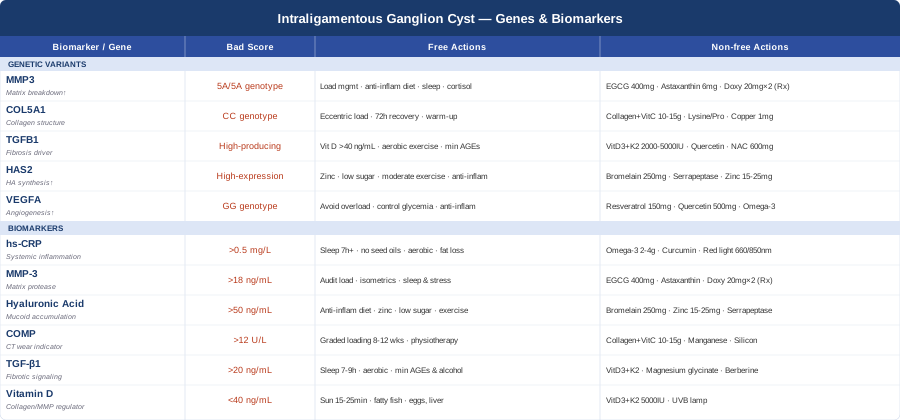
Conclusion
Intraligamentous ganglion cysts sit at the intersection of mechanical stress, connective tissue metabolism, and individual biological variation. Understanding where your own biology sits — through the six biomarkers and five genetic variants covered in this article — gives you a substantially more actionable picture than a generic "wait and see" approach. The interventions are not exotic: improving your hs-CRP, normalizing vitamin D, managing MMP-3, and supporting collagen synthesis quality through targeted nutrition and appropriately structured loading are all within reach without specialist gating. The genetic context adds precision to those efforts — it explains which pathways to prioritize and why some interventions will matter more for your profile than others.
The practical next step is to identify the most accessible starting point: order an hs-CRP and 25-OH vitamin D panel if you haven't already, consider adding MMP-3 if you have access to a specialty or integrative lab, and audit your diet against the connective tissue nutrition framework described above. If you have genetic testing data, query your raw file for the five variants covered here. Share this framework with your treating physician or physical therapist — the more information your care team has about your specific biology, the more precisely they can tailor their recommendations. Better information, applied consistently over the months that connective tissue biology actually requires, is a genuinely different strategy than watchful waiting.
Musculoskeletal: Joint Conditions Tendon & Ligament Conditions
Autoimmune: Inflammatory Conditions Connective Tissue Conditions