This article contains AI generated content.
Intramuscular Myxoma: 4 Genes and 6 Biomarkers to Track
Getting told you have an "intramuscular myxoma" often produces two conflicting feelings at once: relief that the word "benign" appeared somewhere in the report, and unease that nobody quite explained why this lump formed, whether it will come back, or what it has to do with the rest of your health. Generic soft-tissue-tumor advice tends to stop at "it's benign, we'll remove it if it bothers you," which is true but thin. It doesn't explain the molecular reason these tumors form, why a small subset of patients also have bone changes, or how pathologists actually rule out the more dangerous look-alikes.
The more useful answer lives one level deeper, in the specific gene mutation that drives most of these tumors and in the panel of pathology and imaging markers used to confirm the diagnosis. That detail matters practically: it changes how confident you can be in the diagnosis, what follow-up (if any) makes sense, and whether anyone should look at your bones as well as your muscle.
This article works through that detail in two connected layers. First, the genetics — what GNAS and a few related genes do, and why identifying (or ruling out) their mutations changes the clinical picture. Second, the biomarkers pathologists and radiologists actually use on a specimen or scan, what each one costs, and what a "concerning" result should prompt you to do next. A short detour into a book about diagnostic reasoning, and a look at supportive, evidence-based habits for recovery and bone health, round out the picture.
None of this replaces a conversation with your surgeon or pathologist. But understanding the mechanism behind your own report tends to turn a vague diagnosis into a manageable one.
Summary
Most intramuscular myxomas are driven by a single, acquired mutation in one gene, and that fact alone answers several questions patients usually ask: why did this happen, could I have inherited it, and will it happen again elsewhere. It also explains an unusual but well-documented pairing between this soft-tissue tumor and a specific bone condition, which is worth knowing about even if it applies to only a minority of cases. Beyond the driver gene, two other genes matter mainly because they help rule out mimics that look similar under the microscope but behave very differently.
On the biomarker side, a handful of stains, molecular tests, and imaging signs are what actually separate a straightforward myxoma from something that needs a different treatment plan altogether. Knowing what CD34, S100, MDM2/CDK4, beta-catenin, tumor cellularity, and a specific MRI signal pattern are checking for gives real context to a pathology report that otherwise reads like a foreign language. The sections below walk through the plan for each finding, what can realistically be done without any supplements or devices, and where monitoring tools, bone-health supplementation, or imaging equipment genuinely add value — plus a look at what a classic book on medical diagnostic reasoning gets right about avoiding misdiagnosis in cases exactly like this one.
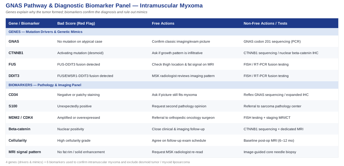
What Recent Genetics Research Reveals About Intramuscular Myxoma
Unlike inherited conditions where a "bad" gene variant sits in every cell of your body from birth, intramuscular myxoma is driven by a somatic mutation — one that arises only in the tumor's own cells, not in your blood, your other tissues, or your children's DNA. That distinction matters immediately: there is no blood test, no ancestry report, and no supplement regimen that changes this mutation, because it isn't a trait you carry. What genetic testing actually offers here is diagnostic clarity, not personal risk prediction. The following genes are the ones worth understanding if you or a family member has received this diagnosis.
GNAS: the mutation behind most intramuscular myxomas
The GNAS gene encodes the alpha subunit of a G-protein (Gsα) that normally switches on and off in response to hormonal signals, activating the cAMP/PKA signaling pathway inside cells. In the great majority of intramuscular myxomas, tumor cells carry an activating point mutation at codon 201 (typically Arg201His or Arg201Cys) that locks this switch in the "on" position. The result is a cell population that produces excess mucopolysaccharide-rich extracellular matrix — the soft, gelatinous, myxoid tissue that gives these tumors their name, their characteristic feel on exam, and their gelatinous appearance on imaging and pathology.
This isn't a fringe finding. Molecular studies using Sanger sequencing and next-generation sequencing have detected GNAS mutations in a large majority of confirmed intramuscular myxomas, and refining these detection methods has been an active area of pathology research over the past two decades. One study using highly sensitive molecular inversion probe sequencing found GNAS mutations in nearly all cases tested, including some that earlier, less sensitive methods had missed (see Bekers et al., 2019). An earlier study similarly concluded that GNAS1 mutations occur "more commonly than previously thought" once testing methods improved (see Delaney et al., 2009), and cytogenetic work has mapped the mutation directly to the GNAS locus in tumor karyotypes (see Panagopoulos et al., 2017).
The plan without medication or supplements
Because the mutation is confined to tumor tissue, there is no lifestyle change, diet, or supplement that reverses it or prevents it from having occurred — claims to the contrary would not be accurate. What genuinely helps at this stage is understanding the natural history: intramuscular myxomas are slow-growing, do not spread, and in appropriately selected small, asymptomatic cases can reasonably be watched with periodic clinical exams rather than removed immediately. If surgery is chosen, the practical, no-cost step is simply ensuring the specimen is sent for confirmatory testing (either GNAS sequencing or supportive immunohistochemistry, covered below) rather than relying on appearance alone, since a small number of more aggressive myxoid tumors can mimic this one visually.
The plan with monitoring tools, testing, or medical equipment
Where equipment genuinely adds value is in confirmation and surveillance. GNAS mutation testing (typically PCR-based sequencing performed on the biopsy or resection specimen) is the most specific way to confirm the diagnosis when the picture is ambiguous; expect this to be ordered by the pathologist rather than something you request directly, and it is usually a one-time test on tissue already collected, not a recurring lab draw. Follow-up MRI, if recommended, is usually spaced months to a couple of years apart depending on whether the tumor was removed or is being observed — there is no universal fixed interval, and your surgeon should set this based on tumor size and location. There are no supplement protocols, dosing cycles, or side-effect profiles to discuss for the GNAS mutation itself, because none exist that act on it; be cautious of any product marketed to "fix" a somatic tumor mutation, since this is not how the biology works.
GNAS again, in the context of Mazabraud syndrome
A minority of people with intramuscular myxoma — historically estimated at a small single-digit percentage, though more recent series suggest it may be underdiagnosed — also have fibrous dysplasia of bone, a separate condition where normal bone is replaced by fibrous, weaker tissue, also driven by activating GNAS mutations arising early in development. This combination is called Mazabraud syndrome, first described in the surgical literature decades ago (see Gianoutsos et al., 1990) and still actively studied for the full range of its skeletal and soft-tissue manifestations (see Hagelstein-Rotman et al., 2022).
This is the one place where the underlying gene has real, practical downstream consequences worth actively managing — not because you can fix the mutation, but because the fibrous dysplasia component carries genuine risks of bone pain, deformity, and fracture that respond to established treatment.
If bone involvement is suspected: the plan without supplements
The starting point is simple and free: if you've been diagnosed with intramuscular myxoma, mention it to your physician, and ask whether a skeletal survey or targeted imaging of nearby bone is warranted, particularly if you have unexplained bone pain, a history of fractures, or multiple myxomas. Weight-bearing activity within pain-free limits helps maintain bone strength generally, and avoiding high-impact sports in areas of known fibrous dysplasia reduces fracture risk without costing anything.
If bone involvement is confirmed: the plan with supplements and equipment
For confirmed fibrous dysplasia, calcium and vitamin D adequacy is a baseline recommendation, dosed to reach normal vitamin D blood levels (typically 800–2000 IU daily of vitamin D3, adjusted by a physician based on blood testing) rather than a fixed one-size figure; side effects at these doses are rare but include mild GI upset, and levels should be rechecked every 6–12 months rather than continued blindly. For more significant disease with pain or fracture risk, bisphosphonate therapy (such as intravenous pamidronate or zoledronic acid, prescribed and administered by a specialist, typically in cycles spaced months apart) has evidence for reducing bone pain and turnover in fibrous dysplasia, though it does not reverse the underlying lesion; known side effects include flu-like symptoms after infusion and, rarely with long-term use, jaw bone issues, so it is reserved for confirmed, symptomatic disease rather than used preemptively. DXA bone density scanning (a low-cost, low-radiation scan, typically $50–150 out of pocket where not covered) is reasonable to establish a baseline and monitor response every one to two years.
CTNNB1: the gene that rules out a more aggressive mimic
Desmoid-type fibromatosis (aggressive fibromatosis) is a locally infiltrative soft-tissue tumor that can resemble a myxoid mass clinically and, in some cases, on initial pathology review, but behaves very differently — it tends to grow into surrounding tissue and recur after incomplete removal. The great majority of sporadic desmoid tumors carry a specific activating mutation in CTNNB1, the gene encoding beta-catenin, most often at codon 41 or 45. A systematic review found CTNNB1 mutation testing has very high specificity for desmoid-type fibromatosis when the diagnosis is in question (see Sakai et al., 2020), and histopathology studies have linked specific mutation subtypes to nuclear beta-catenin staining patterns and clinical behavior (see Huss et al., 2013).
The plan without special testing
If your tumor's imaging and clinical exam are entirely typical for intramuscular myxoma — well-circumscribed, gelatinous on MRI, not infiltrating adjacent structures — additional CTNNB1 testing usually isn't necessary; standard histology and the biomarkers discussed later already distinguish the two conditions reliably in most cases. Asking your surgeon directly, "does this look and behave like a myxoma, or is there any infiltrative pattern that concerns you," is a free, useful question before requesting extra molecular work.
The plan with molecular testing
Where the specimen shows an infiltrative growth pattern, spindle-cell architecture, or recurrence after removal, CTNNB1 sequencing or nuclear beta-catenin immunohistochemistry (performed by the pathology lab on existing tissue, typically bundled into surgical pathology costs rather than billed separately, though standalone molecular panels can run several hundred dollars) clarifies the diagnosis. This changes management meaningfully: desmoid tumors are often managed with a more cautious, sometimes non-surgical watch-and-wait approach given high recurrence rates after surgery, which is the opposite of the more straightforward surgical approach typical for myxoma.
FUS and DDIT3: the genes that rule out myxoid liposarcoma
The other major mimic is myxoid liposarcoma, a malignant fatty tumor that can look deceptively similar to intramuscular myxoma on a quick glance at imaging or a small biopsy sample, but requires oncologic surgery, often radiation, and ongoing surveillance. Myxoid liposarcoma is defined molecularly by a fusion between the FUS gene (or occasionally EWSR1) and DDIT3, present in the vast majority of cases and detectable by FISH or RT-PCR testing (see Powers et al., 2010, and a more recent review of the DDIT3 fusion biology at Diaz-Perez and Kerr, 2024).
The plan without molecular testing
Location and imaging pattern do a lot of the differentiating work before any lab test is needed: myxoid liposarcoma overwhelmingly arises in the deep soft tissue of the thigh and typically shows some fat signal or enhancing solid nodules on MRI, whereas intramuscular myxoma is more uniformly gelatinous with a lower likelihood of solid enhancing components. A careful radiologist reading the MRI with this distinction in mind is, in many straightforward cases, enough to keep confidence high without further testing.
The plan with FISH or molecular equipment
When imaging is ambiguous, when the patient is younger, or when the tumor arises in a thigh location classically associated with myxoid liposarcoma, FISH testing for the FUS-DDIT3 (or EWSR1-DDIT3) fusion is the definitive step; this is a lab-based test performed on the biopsy tissue, generally costing in the low hundreds of dollars where billed separately, with results in about one to two weeks. There is no supplement or device equivalent here — this is purely a diagnostic decision point, and its value is in avoiding either an unnecessarily aggressive workup for a benign myxoma or, more dangerously, under-treating an actual sarcoma.
Taken together, these four genetic findings form a decision tree rather than four separate worries: GNAS confirms the myxoma itself, and CTNNB1 and FUS/DDIT3 testing exist specifically to make sure nothing more serious is hiding behind a similar appearance. That same logic carries directly into the biomarker panel pathologists use on the actual tissue sample.
Six Pathology and Imaging Biomarkers Worth Understanding
If genetics answers "why did this form," biomarkers answer "how do we know for certain what this is." These are the stains, molecular tests, and imaging signs that show up on an actual pathology or radiology report, and each one carries a specific piece of the confirmation puzzle.
CD34
CD34 is a cell-surface marker that stains diffusely and strongly positive in the great majority of intramuscular myxomas, a pattern strong enough that one study proposed it as a practical, sensitive screening marker alongside GNAS sequencing (see research on CD34 as a sensitive marker for intramuscular myxoma, 2019). It matters because it's cheap and fast relative to molecular sequencing — a standard immunohistochemistry stain, usually costing well under 100 dollars as part of a surgical pathology panel, with same-week turnaround.
How to measure it: performed automatically by the pathology lab on the resected or biopsied tissue; you don't order this yourself. If the stain is unexpectedly negative or patchy, the plan without extra cost is simply asking the pathologist whether the overall picture still fits myxoma; the plan with additional testing is reflex GNAS sequencing or an expanded immunohistochemistry panel to resolve the ambiguity.
S100 protein
S100 is typically negative in intramuscular myxoma, and that negative result is itself informative — it helps exclude nerve sheath tumors and some cartilage-forming lesions that can occasionally sit in a similar location and cause diagnostic confusion (see the broader clinicopathologic characterization of myxoid soft-tissue lesions in van Roggen et al., 2001).
How to measure it: again, a routine, low-cost immunohistochemistry stain done automatically as part of the diagnostic panel. If S100 comes back unexpectedly positive, the practical next step without added technology is a second pathology opinion on the same slides; with added resources, referral to a sarcoma-specialized pathology center for a full panel is the more thorough option, particularly for a first-time diagnosis in an atypical location.
MDM2 and CDK4
These two markers, checked together by immunohistochemistry or FISH, are the standard way to rule out atypical lipomatous tumor and well-differentiated liposarcoma, fatty tumors that can occasionally show myxoid change and confuse the picture on a small sample (see Clay et al., 2016 on the appropriate use of MDM2/CDK4 testing in problematic lipomatous tumors). Amplification of MDM2 (detected by FISH) or overexpression of both proteins (by immunohistochemistry) points away from a simple myxoma and toward a lesion needing wider surgical margins.
How to measure it: immunohistochemistry is inexpensive (typically bundled into surgical pathology fees); FISH testing for MDM2 amplification, when needed, is a distinct send-out test that can add a few hundred dollars and one to two weeks. If either marker is positive, the plan without further equipment is prompt referral to an orthopedic oncology surgeon rather than a general surgeon for re-excision planning; the plan with equipment is a staging MRI or CT to assess local extent before any second surgery.
Nuclear beta-catenin
As discussed under CTNNB1 above, nuclear (rather than only membranous or cytoplasmic) beta-catenin staining supports a desmoid tumor diagnosis rather than myxoma, though it is an imperfect marker on its own — Huss et al. (see 2013) found nuclear positivity correlates with mutation status but isn't present in every genetically confirmed case, so a negative stain alone doesn't fully exclude the diagnosis if other features are worrying.
How to measure it: a standard immunohistochemistry stain, low cost, run alongside the initial panel or added as a reflex test. If nuclear positivity is found unexpectedly, the plan without molecular testing is close clinical and imaging follow-up given desmoid tumors' unpredictable behavior; the plan with equipment is confirmatory CTNNB1 sequencing and a dedicated MRI protocol to map the tumor's relationship to surrounding muscle and neurovascular structures before deciding between surgery and observation.
Tumor cellularity
Cellularity — how densely packed the tumor cells are relative to the myxoid matrix — is a straightforward, no-extra-cost feature the pathologist grades directly during routine microscopic review. It carries real prognostic weight: in a study of intramuscular myxoma and Mazabraud syndrome, higher cellularity was statistically associated with a greater chance of local recurrence after removal (see Majoor et al., 2019).
How to measure it: read directly from the standard hematoxylin and eosin slides already prepared for diagnosis — no additional stain or fee involved. If cellularity is reported as high, the plan without extra equipment is simply agreeing on a follow-up exam or imaging schedule with your surgeon rather than assuming the case is closed after removal; the plan with equipment is a baseline post-operative MRI at a defined interval (commonly around 6–12 months, per your surgical team) to catch any early recurrence while it's still small and easy to manage.
MRI signal pattern (T2 hyperintensity and peripheral fat rim)
Before any tissue is even taken, MRI itself functions as a biomarker. Intramuscular myxoma classically shows marked T2 hyperintensity (reflecting its high water and mucin content) along with a thin peripheral rim of fat and surrounding muscle edema, a pattern detailed in a review of soft-tissue myxomatous lesions with pathologic correlation (see Petscavage-Thomas et al., 2014). This pattern is genuinely useful for pre-surgical planning and for reassurance when it's textbook-typical.
How to measure it: a standard MRI with and without contrast, typically ranging from a few hundred to over a thousand dollars depending on your location and insurance coverage, ordered by your physician once a mass is felt or found incidentally. If the imaging pattern is atypical — solid enhancing components, poorly defined margins, no fat rim — the plan without additional equipment is requesting review by a musculoskeletal radiologist rather than a general radiologist; the plan with more resources is proceeding to image-guided core needle biopsy for tissue diagnosis before any surgery is scheduled.
Between the genetic drivers and these six biomarkers, the diagnostic picture for intramuscular myxoma is unusually well mapped compared to many soft-tissue conditions — which is exactly why a second opinion or a request for specific testing is a reasonable thing to ask for, not an overreaction. That idea, that good diagnostic reasoning is a skill and not just a matter of ordering more tests, is the subject of a book worth knowing about.
What "How Doctors Think" Gets Right About Cases Like This One
How Doctors Think, by physician Jerome Groopman, isn't written about myxoid tumors specifically, but its central argument maps almost perfectly onto the diagnostic path described above: most missed or delayed diagnoses aren't caused by a lack of tests, but by cognitive shortcuts that stop a clinician from ordering the right one. For a condition where the correct answer depends on distinguishing a benign tumor from two genuinely dangerous mimics using specific molecular markers, this matters more than it might for a simpler diagnosis.
1. Representativeness bias can end the workup too early
If a mass "looks like" a typical myxoma on the first read, it's tempting to stop there. Groopman's point is that the moment something feels representative of a familiar pattern, doctors often stop asking "what else could this be" — precisely the habit that CD34, S100, and FUS/DDIT3 testing exist to override.
2. Availability bias favors the diagnosis seen most recently
A radiologist who just read three myxoma cases may anchor on that pattern; one who just missed a sarcoma may over-test the next ambiguous mass. Either way, recent experience — not just your actual scan — can shape the read.
3. "Satisfaction of search" stops the eye once one abnormality is found
Once a myxoid mass is identified, it's easy to stop scanning for a second, subtler feature (like a small solid enhancing nodule) that would change the diagnosis entirely. This is a direct argument for a second, deliberate look at imaging rather than reflexive re-reads of the report.
4. Confirmation bias makes doctors seek data that fits their first guess
Once "benign myxoma" is written down, subsequent findings tend to get interpreted to fit that label. Asking explicitly "what would change your mind about this diagnosis" forces a clinician to name the specific test that would falsify their first impression.
5. Patients under-describe atypical symptoms out of politeness
Groopman notes that patients often soften or omit details that don't fit the story they think the doctor wants to hear. For a myxoma diagnosis, this might mean not mentioning that the mass grew quickly, or that it's newly painful — details that specifically warrant re-imaging.
6. A good history question beats a reflexive extra test
Rather than ordering every possible molecular panel, Groopman argues the sharper move is often a better question: has this changed in size or texture, is there a family history of soft-tissue or bone conditions, and does anyone else in the family have unexplained bone pain (relevant, given Mazabraud syndrome).
7. Diagnostic momentum is hard to reverse once labeled
Once "myxoma" appears on a chart, later clinicians tend to inherit that label rather than re-examine it, even when new symptoms arise. This is a real argument for requesting that any new or changed symptom be evaluated fresh, not filtered through the old diagnosis.
8. Uncertainty should be stated, not hidden
Groopman is critical of clinicians who project more certainty than the evidence supports. A pathology report that says "favor benign myxoma" versus one confirmed by GNAS sequencing and a clean biomarker panel represent two very different confidence levels, and patients deserve to know which one they have.
9. A second opinion is a diagnostic tool, not an insult
The book frames requesting a second pathology read, especially on an unusual or borderline slide, as a normal part of good care rather than a sign of distrust — directly relevant given how visually similar myxoma, desmoid tumor, and myxoid liposarcoma can be on a rushed read.
10. The best question a patient can ask is "what else could this be?"
Groopman's single most quoted piece of advice is this literal sentence, asked directly of your physician. For intramuscular myxoma, the honest, complete answer to that question is exactly the two or three mimics discussed above — and hearing your doctor name them, unprompted, is a reasonable bar for confidence in the diagnosis.
Diagnostic clarity is one half of managing this condition well; the other half is what you do with your body during recovery and, for the subset of patients with Mazabraud-associated bone involvement, over the longer term. A few supportive, evidence-based approaches are worth knowing about here — with the honest caveat that none of them treat the tumor itself.
Supportive Approaches Worth Considering Alongside Medical Care
None of the following changes a GNAS mutation or shrinks a myxoma. Their value is in supporting recovery after surgery, managing the anxiety that comes with any tumor diagnosis while confirmatory testing is pending, and, for Mazabraud syndrome specifically, protecting bone health. Evidence here is drawn from surgical and musculoskeletal populations generally rather than intramuscular myxoma specifically, since condition-specific trials don't exist for a tumor this uncommon — that's stated plainly rather than implied.
Massage therapy and soft tissue mobilization
After surgical removal of a myxoma, scar tissue and localized muscle adhesion around the incision are common and can limit range of motion for weeks to months. Soft tissue mobilization techniques address exactly this kind of restriction, not the tumor biology itself.
A systematic review of soft tissue mobilization for adhesion-related pain and restricted function found consistent improvements in scar mobility and reported pain following structured treatment (see Wasserman et al., 2019), though the review focused on abdominal surgery rather than limb muscle surgery specifically.
Realistically, this means waiting until your surgeon confirms the incision has fully healed (usually two to four weeks post-op) before starting gentle scar massage, working with a physical therapist familiar with post-surgical soft tissue work two to three times weekly initially, and stopping if it increases pain rather than gradually easing it.
Mindfulness meditation and MBSR
The stretch of time between finding a mass, getting imaging, and receiving a final pathology report (sometimes weeks) is a well-documented source of anxiety, independent of the eventual diagnosis. Mindfulness-based approaches are among the better-studied non-drug tools for this kind of waiting-period anxiety.
A randomized trial of a mobile-app-delivered mindfulness intervention in patients awaiting colorectal cancer surgery found meaningful reductions in preoperative anxiety compared with usual care (see Rocamora González et al., 2022), a population facing a comparable diagnostic-uncertainty window.
A realistic starting point is a structured 8-week MBSR course or a reputable app-based program, practiced 10–20 minutes daily; there are no dosing concerns, though people with significant trauma history sometimes find unstructured mindfulness practice brings up difficult material and should work with a trained instructor rather than going it alone.
Tai chi
For the minority of readers with confirmed Mazabraud syndrome and fibrous dysplasia, fall prevention and balance carry real weight, since a fall onto weakened bone raises fracture risk in ways it wouldn't for someone with normal bone density.
A meta-analysis of tai chi for fall prevention in older adults found a consistent reduction in fall rates across pooled trials (see Huang et al., 2017), though this evidence comes from general older-adult and osteoporosis populations rather than fibrous dysplasia specifically.
Two to three sessions weekly of a beginner-level tai chi class, ideally one geared toward older adults or fall prevention rather than a martial-arts-focused class, is a reasonable and low-risk starting point; the main caution is avoiding any pose involving high impact or deep lunging directly over a known area of bone weakness without your physician's clearance first.
Progressive muscle relaxation
For the practical anxiety and muscle tension that surrounds surgery specifically (rather than the general diagnostic waiting period), progressive muscle relaxation has a more direct evidence base in surgical populations.
A randomized trial in patients undergoing colorectal cancer surgery found progressive relaxation exercise improved physiological parameters, pain, and anxiety levels compared with standard care (see Ozhanli and Akyuz, 2022).
This is easy to self-administer using a free recorded script, done in the days leading up to surgery and again during early recovery, 10–15 minutes once or twice daily; it carries essentially no side effects, though it shouldn't be done directly over a fresh surgical site in a way that strains the incision.
Conclusion
Intramuscular myxoma is, for most people, exactly what it sounds like on first hearing: a benign, slow-growing tumor driven by an acquired GNAS mutation, confirmed through a well-established combination of immunohistochemistry, occasional molecular testing, and characteristic imaging. The genetics don't change with lifestyle or supplements, but the biomarkers built around that genetics — CD34, S100, MDM2/CDK4, beta-catenin, cellularity, and MRI signal pattern — are exactly the tools that separate a straightforward diagnosis from one of its two more serious mimics, desmoid tumor and myxoid liposarcoma. For the smaller group with Mazabraud syndrome, the bone side of the equation deserves its own attention, with real, evidence-backed options for bone health rather than vague reassurance.
The single most useful next step is a direct conversation with your surgeon or pathologist, armed with specific questions: was GNAS mutation testing performed or considered, were CD34 and S100 both checked, does the tumor's location and MRI pattern fit typical myxoma without solid enhancing components, and — if you have any bone pain, prior fractures, or multiple myxomas — has fibrous dysplasia been ruled out. A precise question, asked once, tends to do more for your peace of mind than weeks of searching on your own.
Musculoskeletal: Bone Conditions Muscle Conditions