This article contains AI generated content.
· UpdatedIntraosseous Ganglion Cyst of the Knee — 6 Biomarkers and 5 Genes to Track
Introduction
If you have been told you have an intraosseous ganglion cyst in your knee, you have probably heard some version of the same advice: rest, monitor it, consider surgery if the pain becomes unbearable. That advice is not wrong, but it is incomplete. It treats the cyst as an isolated finding rather than as a signal from a deeper biological environment that made it possible in the first place.
Intraosseous ganglion cysts are benign, fluid-filled lesions that form within bone tissue, typically in the subchondral bone just beneath the cartilage of the knee. They arise when mechanical stress, tissue remodeling dysregulation, or synovial fluid intrusion creates a cavity that fills with gelatinous material. Generic advice rarely touches on why that remodeling went wrong, or what could be done to change the biological conditions that allowed it to happen.
That is where a more targeted approach becomes genuinely useful. By looking at specific biomarkers circulating in your blood and urine, you can measure, in real time, whether your cartilage is degrading, whether your bone is resorbing faster than it forms, and whether your inflammatory burden is feeding the cycle. By understanding relevant gene variants, you can identify whether your connective tissue, collagen, or tissue-remodeling enzymes are working against you at a structural level — and find evidence-based ways to compensate.
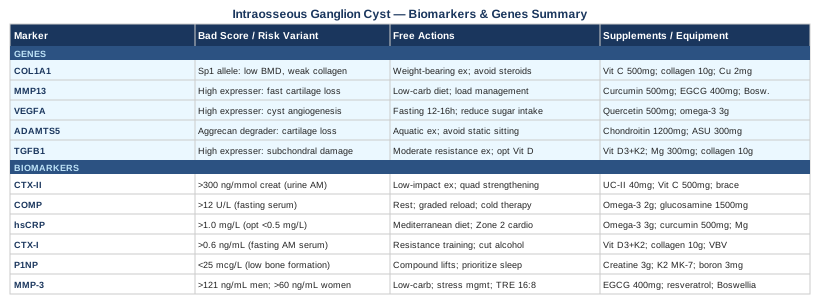
This article covers both angles. The first section focuses on six biomarkers that are directly relevant to the biology of intraosseous cyst formation and progression, with practical plans for what to do when any of them is out of range. The second section explores five genes with meaningful connections to subchondral bone and cartilage health, along with compensation strategies. Additional sections draw on insights from evidence-based longevity medicine and cover complementary methods with legitimate clinical backing. Better information does not guarantee better outcomes, but it gives you something that a vague prognosis never can: a concrete set of levers to pull.
6 Biomarkers Worth Tracking
Understanding the biological environment around an intraosseous ganglion cyst requires looking at cartilage degradation, bone remodeling balance, systemic inflammation, and tissue-remodeling enzyme activity. These six markers collectively paint a picture that no single test could.
1. CTX-II (Carboxy-Terminal Crosslinked Telopeptide of Type II Collagen)
Why it matters: CTX-II is the most specific circulating marker of type II collagen degradation — the structural collagen of articular cartilage. Intraosseous ganglion cysts frequently form in subchondral bone beneath areas of cartilage stress or thinning. When cartilage degrades, the mechanical load it used to absorb transfers to the subchondral bone, altering remodeling dynamics and increasing the risk of fluid intrusion and cyst formation. Persistently elevated CTX-II does not diagnose a cyst, but it signals that the tissue environment is deteriorating. Multiple studies in patients with early osteoarthritis have shown that elevated urinary CTX-II predicts radiographic progression years before imaging catches it.
How to measure it
CTX-II is measured in a second-morning void urine sample, corrected for creatinine. Blood serum versions also exist. It is available through specialty functional medicine labs and some rheumatology reference labs. Cost range: $50–$150. Standard labs do not routinely offer this test; you may need to request it explicitly. A commonly cited concern threshold is above 300–400 ng/mmol creatinine, though lab-specific reference ranges apply.
If the score is bad, the plan without supplements
The most effective non-supplement intervention is reducing mechanical overload on the joint while maintaining active muscle support. Switch from high-impact activities (running, jumping) to low-impact alternatives such as swimming or cycling. Physical therapy focused on quadriceps strengthening is critical — every kilogram of quadriceps force reduces medial knee joint load substantially. Addressing body weight if elevated is among the most impactful moves available: each pound of body weight removed reduces knee joint force by approximately four pounds during walking. Prioritize sleep of seven to nine hours, since cartilage matrix repair occurs largely during slow-wave sleep phases. Retest CTX-II every three to six months to track direction.
If the score is bad, the plan with supplements or equipment
Undenatured type II collagen (UC-II) at 40 mg/day has been tested in multiple randomized controlled trials for cartilage-related outcomes, with several showing reductions in pain and improvements in joint function. Vitamin C at 500–1000 mg/day is an essential cofactor for collagen hydroxylation — the step that determines collagen quality, not just quantity. Boron at 3–6 mg/day supports collagen cross-linking. For equipment, an offloading knee brace during physical activity reduces compressive forces on the affected compartment. Cycle collagen supplements at eight weeks on, two weeks off. High-dose vitamin C above 1000 mg/day can cause gastrointestinal distress and should be introduced gradually.
2. COMP (Cartilage Oligomeric Matrix Protein)
Why it matters: COMP is released from cartilage matrix when it is mechanically stressed or undergoing active degradation. Unlike CTX-II, which reflects irreversible collagen breakdown, COMP elevates rapidly with acute loading stress and can normalize with appropriate rest — making it useful as a dynamic marker of current joint demand. In patients with established joint disease, persistently elevated serum COMP has been associated with faster cartilage volume loss on MRI. For someone managing an intraosseous cyst, COMP can reveal whether daily loading patterns are placing the joint under levels of demand it cannot recover from.
How to measure it
COMP is measured from fasting morning serum. It is available through specialty rheumatology reference labs, including Quest Diagnostics Specialty Testing and similar. Cost range: $100–$300. A commonly referenced upper normal in adults without joint disease is approximately 12 U/L, though ranges vary between assay systems.
If the score is bad, the plan without supplements
Load management is the primary lever. Implement a structured rest period from high-demand activities, followed by graded reloading guided by a physiatrist or sports medicine physician. Cold therapy for fifteen to twenty minutes post-activity reduces local inflammatory mediators that prolong COMP elevation. Progressive strength training, when introduced correctly, actually reduces long-term COMP by improving joint mechanics and distributing load more evenly across the articular surface. Reassess every three to four months.
If the score is bad, the plan with supplements or equipment
Omega-3 fatty acids at 2–4 g/day (combined EPA and DHA) have anti-inflammatory effects that may reduce the inflammatory amplification of COMP release. Glucosamine sulfate at 1500 mg/day has shown in several European trials a modest slowing of COMP rise over time. Chondroitin sulfate at 1200 mg/day adds proteoglycan substrate and mild anti-inflammatory action. Run a twelve-week trial before evaluating effect. Glucosamine should be used cautiously in individuals with insulin resistance or diabetes, as it may subtly affect glucose metabolism. Chondroitin at higher doses may interact with anticoagulant medications.
3. hsCRP (High-Sensitivity C-Reactive Protein)
Why it matters: hsCRP is the most accessible marker of systemic low-grade inflammation available. While intraosseous ganglion cysts are not primarily inflammatory lesions, chronic inflammation drives the tissue-remodeling dysregulation — particularly MMP overactivation and osteoclast dominance — that creates and sustains the pathological environment around these lesions. Elevated hsCRP has been consistently associated with faster cartilage loss and worse joint outcomes across multiple longitudinal studies. Lowering systemic inflammation is one of the most modifiable and evidence-backed interventions available regardless of other treatment choices.
How to measure it
hsCRP is a standard blood test available at virtually every clinical laboratory. Cost range: $15–$50. It is often included in comprehensive metabolic or cardiovascular panels. Target: below 1.0 mg/L for low inflammatory burden; below 0.5 mg/L is optimal. Values above 3.0 mg/L indicate high-grade inflammation that warrants investigation for underlying drivers (infection, metabolic disease, autoimmune condition).
If the score is bad, the plan without supplements
The Mediterranean dietary pattern has the strongest evidence base for reducing hsCRP in clinical populations — multiple meta-analyses confirm significant reductions within eight to twelve weeks of adherence. Eliminating ultra-processed foods, refined sugars, and seed oils high in omega-6 fatty acids is the most impactful single dietary step. Zone 2 aerobic training — steady-state cardio at a conversational pace — for 150 minutes per week has documented anti-inflammatory effects via improved mitochondrial function. Consistent sleep of seven to nine hours and stress management (which directly regulates cortisol and NF-κB, the main driver of CRP production) complete the non-supplement approach. Retest every six to eight weeks to confirm direction.
If the score is bad, the plan with supplements or equipment
Omega-3 fatty acids at 3–4 g/day EPA and DHA — from high-quality triglyceride-form fish oil or algae-based EPA/DHA — reduce hsCRP by fifteen to thirty percent in intervention trials. Curcumin with piperine at 500–1000 mg/day has shown significant CRP reductions in multiple randomized trials, particularly the BCM-95 or MERIVA phospholipid-complexed forms that absorb more reliably. Magnesium glycinate at 300–400 mg/day addresses the magnesium deficiency that impairs the anti-inflammatory signaling pathway regulated by NF-κB. Curcumin at higher doses may interfere with CYP3A4 drug metabolism — check for interactions with any current medications. High-dose omega-3 may modestly thin blood; relevant if undergoing surgery.
4. CTX-I (C-Terminal Telopeptide of Type I Collagen)
Why it matters: CTX-I measures the rate of bone resorption — specifically, how quickly type I collagen in bone is being broken down by osteoclasts. Subchondral bone adjacent to an intraosseous ganglion cyst often shows abnormal remodeling, with resorption rates that outpace formation. High CTX-I suggests the bone matrix in the cyst vicinity is being actively degraded, which can expand the lesion and further weaken the structural integrity of the joint. This marker is also the standard tool for monitoring response to anti-resorptive medications (like bisphosphonates) if those are ever indicated.
How to measure it
CTX-I is measured from a fasting, morning blood sample — timing is critical because levels fluctuate by up to forty percent throughout the day. It is also measurable in urine. Cost range: $50–$150. Available through most reference labs. General reference ranges for active adults: 0.3–0.6 ng/mL in serum (varies significantly by age, sex, and menopausal status). Post-menopausal women may have physiologically higher values that still require clinical context.
If the score is bad, the plan without supplements
Progressive resistance training is the single most powerful intervention to shift bone remodeling toward formation and away from resorption. Impact loading appropriate to fitness level — even brisk walking — stimulates mechanosensitive osteoblast activity. Reducing alcohol intake is important: alcohol directly increases osteoclast activity and impairs osteoblast function even at moderate consumption levels. Adequate dietary protein at 1.6–2.0 g per kilogram of body weight provides the amino acid substrate for bone matrix formation. Sun exposure for ten to twenty minutes daily (when available and appropriate for skin type) optimizes vitamin D without supplementation.
If the score is bad, the plan with supplements or equipment
Vitamin D3 combined with K2 (MK-7 form) is the foundational intervention: 2000–5000 IU of D3 with 180–200 mcg of MK-7 daily. Vitamin D normalizes calcium-phosphate metabolism; K2 directs calcium into bone rather than soft tissue by activating osteocalcin. Collagen peptides at 10–15 g/day provide glycine and proline for bone matrix synthesis. In severe cases, a physician may evaluate prescription-level anti-resorptives. For equipment, whole-body vibration platforms used at 30–50 Hz for ten minutes three times per week have shown modest benefits for bone density in several clinical studies. Monitor serum 25-OH vitamin D every six months; target 50–70 ng/mL. Vitamin K2 should be discussed with any physician managing anticoagulation therapy.
5. P1NP (Procollagen Type I N-terminal Propeptide)
Why it matters: P1NP is the mirror image of CTX-I: it measures the rate of new bone formation. The CTX-I to P1NP ratio is what truly reveals the state of bone remodeling balance. When CTX-I dominates, bone is being lost. When P1NP leads, bone is being built. For an intraosseous ganglion cyst surrounded by compromised subchondral bone, you want P1NP elevated and CTX-I restrained. The World Health Organization has designated P1NP as the reference standard bone formation marker for clinical research, reflecting its reliability and sensitivity.
How to measure it
P1NP is measured from a standard blood draw, preferably fasting and morning (to pair correctly with CTX-I for ratio calculation). Cost range: $50–$200 through specialty labs. Not universally available through standard panels — request explicitly. In adults without metabolic bone disease, typical reference ranges are 25–100 mcg/L, though values must be interpreted alongside CTX-I and clinical context.
If the score is bad, the plan without supplements
Progressive resistance training — particularly compound movements like squats, deadlifts, and step-ups — is the most potent stimulator of P1NP production. The osteogenic response is load-dependent: higher mechanical strain within safe limits produces greater bone formation signaling. Prioritizing sleep is equally important: the majority of growth hormone secretion, which drives bone formation, occurs during slow-wave sleep stages. Dietary protein adequacy, particularly leucine-rich sources, amplifies the anabolic response to resistance exercise. Retest after twelve weeks of consistent training to observe P1NP response.
If the score is bad, the plan with supplements or equipment
Creatine monohydrate at 3–5 g/day has shown increases in bone formation markers in several randomized trials, particularly when combined with resistance exercise. Vitamin K2 (MK-7) at 180–200 mcg/day activates osteocalcin, a bone protein directly involved in P1NP-linked formation pathways. Boron at 3–10 mg/day increases levels of estrogen, testosterone, and vitamin D — all of which stimulate osteoblast activity and P1NP production. Magnesium glycinate at 300–400 mg/day acts as a cofactor for the enzyme systems underlying bone matrix synthesis. Creatine is generally well-tolerated but causes transient water retention during the first two weeks; kidney function should be monitored with long-term use above 5 g/day.
6. MMP-3 (Matrix Metalloproteinase-3)
Why it matters: MMP-3, also called stromelysin-1, is a tissue-remodeling enzyme that degrades multiple components of the extracellular matrix: type IV collagen, fibronectin, proteoglycans, and laminin. It also plays a critical role as an activator of other MMPs — including MMP-13, the most potent cartilage-degrading enzyme. In the context of intraosseous ganglion cysts, elevated MMP-3 reflects a tissue environment where degradation is outpacing repair at the matrix level. Serum MMP-3 is well-validated as a circulating biomarker in inflammatory joint diseases and correlates with both local tissue destruction and systemic inflammatory burden.
How to measure it
MMP-3 is measured from serum through specialty rheumatology reference labs. Cost range: $100–$400. Not available through standard metabolic panels — requires specific request through labs such as Labcorp Specialty Testing or equivalent. Reference ranges vary by sex: typically below 59.7 ng/mL for women and below 121 ng/mL for men, though assay-specific ranges apply.
If the score is bad, the plan without supplements
Dietary pattern revision has the most evidence behind it for reducing MMP-3: specifically, eliminating refined carbohydrates and seed oils high in omega-6 fatty acids (which drive arachidonic acid metabolism and downstream MMP activation). Chronic psychological stress sustains cortisol elevation, which drives NF-κB activation and consequent MMP upregulation — stress reduction is not optional here. Time-restricted eating within a sixteen-hour fast, eight-hour eating window has shown MMP-reducing effects in several metabolic studies, likely through improved insulin sensitivity and reduced inflammatory signaling. Heat and cold contrast therapy applied locally to the knee reduces pericellular MMP activity in superficial tissue layers. Retest every three to six months.
If the score is bad, the plan with supplements or equipment
EGCG (epigallocatechin gallate) from green tea extract at 400–800 mg/day is among the most studied MMP-3 inhibitors available without a prescription, with in vitro and human data supporting its effectiveness. Resveratrol at 250–500 mg/day inhibits the NF-κB transcription factor that drives MMP-3 gene expression. Boswellia serrata (AKBA-standardized) at 200–400 mg/day has well-documented anti-MMP effects and is one of the most clinically studied botanical anti-inflammatories. These should not all be combined simultaneously; start with one for eight to twelve weeks before evaluating and adding another. High-dose EGCG (above 800 mg/day) has documented hepatotoxicity risk and should not exceed this range. Resveratrol may interact with anticoagulant medications and estrogen-sensitive conditions.
The Genetic Angle: 5 Genes That May Influence Your Risk and Recovery
Biomarkers tell you what is happening right now. Genes explain part of why the tissue environment is predisposed to the patterns you see in those biomarkers. For intraosseous ganglion cysts specifically, direct genetic research is limited — this is a relatively rare condition and large genetic association studies do not yet exist for it. What does exist is substantial genetic research on subchondral bone remodeling, cartilage integrity, and tissue-remodeling enzyme activity — the same biological processes that determine who develops this type of lesion. The five genes below are drawn from that adjacent evidence.
1. COL1A1 — Collagen Type I Alpha 1
What it does: COL1A1 encodes the alpha-1 chain of type I collagen, the primary structural protein in bone, tendon, and connective tissue. A well-studied polymorphism in the Sp1 binding site of COL1A1 (referred to as the GTITG variant) has been associated in multiple studies with reduced bone mineral density and altered collagen structural quality. Bone with structurally compromised collagen scaffold is more susceptible to mechanical failure and fluid intrusion — the preconditions for subchondral cyst formation.
Without supplements: Weight-bearing and impact exercise (walking, low-level plyometrics appropriate to fitness level) stimulates bone formation signals regardless of COL1A1 variant. Avoid prolonged immobilization. Reduce corticosteroid use where clinically possible — corticosteroids suppress collagen synthesis directly through COL1A1 transcriptional repression.
With supplements or equipment: Vitamin C at 500–1000 mg/day is essential for the hydroxylation of proline and lysine residues during collagen assembly — a step that determines collagen fiber quality and tensile strength. Collagen peptides at 10–15 g/day with vitamin C taken thirty to sixty minutes before exercise have shown significant improvements in tendon and ligament collagen content in RCT data (Keith Baar's work at UC Davis is particularly relevant here). Copper at 2–3 mg/day activates lysyl oxidase, the enzyme responsible for cross-linking collagen fibers. Run an eight-week minimum trial. Do not exceed 3 mg/day copper for sustained periods — excess copper accumulation can be toxic. Monitor via periodic serum copper and ceruloplasmin.
2. MMP13 — Matrix Metalloproteinase 13 (Collagenase-3)
What it does: MMP13 encodes collagenase-3, the most potent collagen-degrading enzyme known, with high specificity for type II cartilage collagen. Promoter variants that increase MMP13 expression have been associated in genome-wide studies with faster cartilage loss in osteoarthritis. Because subchondral bone lesions — including ganglion cysts — frequently form beneath zones of cartilage thinning, an overactive MMP13 variant creates a fundamentally unfavorable tissue trajectory. This gene essentially determines the speed at which cartilage collagen is enzymatically dismantled.
Without supplements: Load management reduces the mechanical stimuli that activate MMP13 expression. High-glycemic diets — specifically elevated glucose and advanced glycation end-products — are potent MMP13 activators; reducing refined carbohydrate intake is a meaningful and underappreciated strategy. Evidence suggests that prolonged NSAID use, while reducing acute pain, may impair cartilage repair signaling over time and is not an appropriate long-term strategy for addressing MMP13 overactivation.
With supplements or equipment: Curcumin (BCM-95 or MERIVA form) at 500–1000 mg/day is among the most studied MMP13 inhibitors available — multiple in vitro studies in chondrocytes and clinical data from OA trials support its relevance. EGCG at 400–800 mg/day inhibits MMP13 at the transcriptional level in cartilage cell research. Boswellia serrata (AKBA form) at 200–400 mg/day rounds out the botanical MMP13 suppression toolkit. Run a single agent for twelve weeks before combining. Curcumin at doses above 1000 mg/day may interfere with drug metabolism via CYP3A4 — check for interactions with any current prescription medications.
3. VEGFA — Vascular Endothelial Growth Factor A
What it does: VEGFA controls angiogenesis — the formation and maintenance of new blood vessels. Intraosseous ganglion cysts are characterized by a fibrous, vascularized cyst wall, and VEGF signaling is implicated in the development of that vascular network. Variants in the VEGFA promoter that increase baseline VEGF expression may contribute to more aggressive cyst vascularization and potentially faster lesion growth. VEGF also influences osteoclast recruitment in bone remodeling — making it directly relevant to the bone turnover dynamics measured by CTX-I and P1NP.
Without supplements: Intermittent fasting — specifically periods of twelve to sixteen hours without caloric intake — naturally suppresses VEGF signaling through AMPK activation and reduced mTOR activity. Cold exposure (cold water immersion at fifteen degrees Celsius for ten to fifteen minutes) produces acute vasoconstriction and has been shown to modulate angiogenic signaling patterns in preliminary research. Eliminating excessive simple sugar intake removes a potent VEGF stimulus, since hyperglycemia directly upregulates VEGF through protein kinase C and oxidative stress pathways.
With supplements or equipment: Quercetin at 500–1000 mg/day has inhibitory effects on VEGF-driven angiogenesis documented in multiple experimental models. Resveratrol at 250–500 mg/day modulates VEGF receptor signaling and may normalize rather than suppress angiogenic activity. Omega-3 fatty acids at 3–4 g/day shift arachidonic acid metabolism toward less pro-angiogenic eicosanoid profiles. Cycle quercetin at eight weeks on and two weeks off. Resveratrol may modulate estrogen receptor activity — relevant context for hormone-sensitive individuals. Discuss with a physician before combining multiple angiogenesis-modulating supplements.
4. ADAMTS5 — A Disintegrin and Metalloproteinase with Thrombospondin Motifs 5
What it does: ADAMTS5 encodes aggrecanase-2, an enzyme with high specificity for degrading aggrecan — the large proteoglycan responsible for cartilage's ability to resist compressive load and retain water. Genetic evidence from both mouse knockout models and human GWAS studies has identified ADAMTS5 variants as meaningful contributors to cartilage vulnerability. When aggrecan is rapidly broken down, cartilage loses its shock-absorbing capacity, and the resulting mechanical stress concentrates in the subchondral bone — exactly the scenario that predisposes to bone lesion formation. Note that direct human genetic validation for ADAMTS5 in intraosseous cysts specifically remains to be established; this inference is drawn from OA-related evidence.
Without supplements: Aquatic therapy and cycling maintain synovial fluid circulation through the joint (which sustains cartilage nutrition) without the impact loading that mechanically activates aggrecanase. Reducing prolonged static sitting is important — extended static compressive loading depletes aggrecan more than cyclic movement. Dietary fructose reduction is relevant: fructose activates the aggrecanase pathway through uric acid elevation and metabolic stress signaling.
With supplements or equipment: Chondroitin sulfate at 1200 mg/day provides structural substrate for aggrecan resynthesis and has shown some inhibitory effects on ADAMTS activity in in vitro research. Avocado/soybean unsaponifiables (ASU) at 300 mg/day have demonstrated symptom and potential structural benefits in well-designed OA trials and may influence aggrecan catabolism through multiple pathways. Oral hyaluronic acid at 200 mg/day has limited but emerging evidence for supporting the joint matrix environment. Run a twelve-week trial minimum. Pair with CTX-II monitoring to track cartilage degradation direction. Chondroitin sulfate may modestly interact with warfarin at therapeutic doses — discuss with prescribing physician.
5. TGFB1 — Transforming Growth Factor Beta 1
What it does: TGF-β1 is a dual-function signaling molecule in bone: at low levels, it promotes osteoblast activity and bone formation; at high levels in subchondral bone, it recruits mesenchymal stem cells into an abnormal remodeling cycle that paradoxically accelerates bone damage. Research published in Nature Medicine demonstrated that aberrant TGF-β1 activation in subchondral bone is a key driver of the pathological bone remodeling seen in osteoarthritis — a finding directly applicable to subchondral lesion formation. TGFB1 variants that drive higher expression in bone tissue create a remodeling environment prone to exactly the type of structural changes found in intraosseous ganglion cysts.
Without supplements: Resistance exercise at moderate intensity (sixty to seventy-five percent of one-rep maximum) tends to normalize TGF-β1 signaling toward anabolic patterns, while high-intensity impact loading in an already compromised joint may over-activate it. Optimizing vitamin D through sun exposure and diet is important, since vitamin D receptor signaling directly interacts with TGFB1 transcriptional activity. Avoid excessive calcium supplementation without K2 — poorly directed calcium metabolism can activate TGF-β1 in ways that paradoxically promote subchondral sclerosis rather than healthy remodeling.
With supplements or equipment: Vitamin D3 with K2 (MK-7) at 2000–4000 IU D3 and 180–200 mcg MK-7 daily is the cornerstone. Vitamin D normalizes osteoblast/osteoclast balance through receptor-mediated gene regulation that overlaps with the TGF-β1 pathway. Magnesium glycinate at 300–400 mg/day is an essential cofactor in TGF-β1 signaling and is commonly deficient. Collagen peptides at 10–15 g/day have shown some evidence for normalizing TGF-β1 response in connective tissue research. Monitor serum 25-OH vitamin D every six months with a target of 50–70 ng/mL. Excess vitamin D in the absence of adequate K2 increases soft-tissue calcification risk — the combination matters.
Ten Lessons from "Outlive" by Peter Attia That Apply Directly to Knee and Bone Health
Peter Attia's Outlive: The Science and Art of Longevity (2023) draws on hundreds of studies to challenge the reactive medical model and replace it with what Attia calls Medicine 3.0 — a preventive, data-driven, deeply individualized approach. Several of its core arguments apply with striking precision to the biology of intraosseous ganglion cysts and the joint environment surrounding them.
1. Stability Is the Foundation, Not Flexibility
Attia devotes significant attention to the work of DNS (Dynamic Neuromuscular Stabilization) and the idea that most musculoskeletal problems trace back not to inflexibility but to loss of intra-abdominal pressure control and proximal stability. An unstable joint transfers excess and unpredictable load to the subchondral bone — precisely the mechanical environment that predisposes to cyst formation. Before any other knee-specific training, establishing a stable movement platform is the priority.
2. Zone 2 Training Is Anti-inflammatory Medicine
Sustained aerobic exercise at a conversational pace — roughly sixty to seventy percent of maximum heart rate — improves mitochondrial density, reduces systemic inflammatory burden, and lowers hsCRP. Attia presents Zone 2 training as perhaps the single most impactful lifestyle intervention for long-term metabolic and inflammatory health. For someone managing knee pathology, cycling or swimming within Zone 2 provides the anti-inflammatory effect without compressive knee loading.
3. Strength Training Is Non-Negotiable After 40
After the fourth decade, humans lose muscle mass and bone density at rates that compound without deliberate intervention. Attia frames strength training not as a cosmetic choice but as a metabolic and structural imperative — the primary driver of P1NP production, bone remodeling balance, and joint protection. Specific attention to hip-dominant and knee-loading exercises (within tolerability) is the most direct application for subchondral bone health.
4. VO2 Max Predicts Long-Term Health More Than Any Single Biomarker
Attia cites research showing that VO2 max is among the strongest independent predictors of all-cause mortality and functional longevity. Cardiovascular fitness improves oxygen delivery to all tissues including cartilage (which receives nutrition through synovial fluid diffusion that depends on joint movement and surrounding tissue health). Improving VO2 max through sustained aerobic training is a systemic lever with local joint benefits.
5. Protein Is Almost Universally Underestimated
Attia recommends 1.6–2.2 g of protein per kilogram of body weight per day for adults engaged in resistance training — significantly above the standard RDA. Adequate protein is essential not only for muscle preservation but for bone matrix synthesis, collagen production, and the substrate demands of tissue repair in and around joint lesions.
6. Sleep Is Structural, Not Cosmetic
Slow-wave sleep drives growth hormone secretion, which is the primary endogenous signal for tissue anabolic activity — including cartilage matrix repair and bone formation. Attia argues that sleep is a medical intervention with measurable structural consequences. Consistently sleeping less than seven hours elevates inflammatory markers, reduces P1NP, and impairs tissue repair dynamics.
7. Glucose Regulation Directly Impacts Joint Tissue
Advanced glycation end-products — formed when glucose binds non-enzymatically to proteins — progressively stiffen collagen, reduce cartilage resilience, and activate MMP expression. Attia's approach to metabolic health, including continuous glucose monitoring as a feedback tool, is highly relevant to anyone managing a connective tissue condition. Postprandial glucose spikes are a direct input to the inflammatory and matrix-degrading pathways covered in the biomarker section above.
8. Medicine 3.0 Acts Before Pain Becomes the Signal
Attia's central argument is that the medical system is organized around reacting to disease rather than detecting and modifying risk trajectories years earlier. Intraosseous ganglion cysts are a textbook example of a finding that often receives a "watch and wait" recommendation — but the biomarker and genetic data described in this article suggest there are meaningful interventions available before a lesion becomes symptomatic or requires surgery.
9. Emotional and Cognitive Health Drives Physical Health
Attia dedicates an entire section of Outlive to emotional health as a determinant of physical outcomes — not as a soft observation but as a mechanistic reality. Chronic psychological distress sustains cortisol elevation, which suppresses bone formation, elevates MMP expression, and raises hsCRP. Addressing stress and sleep are not secondary interventions; they are part of the primary biological treatment plan.
10. The Centenarian Decathlon Framework Sets Functional Goals
Attia asks: what do you need to be able to do physically at eighty to live the life you want? Then he works backward from that goal to determine what training and health investments are required now. Applied to knee health, this means setting specific functional targets — climbing stairs, hiking, playing with grandchildren — and designing your knee management strategy to preserve those capacities decades ahead of when they might otherwise be lost.
Approaches Worth Exploring Alongside Conventional Treatment
The following three modalities each have meaningful human clinical evidence for conditions that share the relevant biological features of intraosseous ganglion cysts of the knee: subchondral bone stress, cartilage degradation, and chronic musculoskeletal pain. None should replace appropriate medical evaluation or treatment, but each can be integrated as an evidence-informed complement.
Low-Level Laser Therapy (Photobiomodulation)
What it is and why it may be relevant: Photobiomodulation (PBM) uses specific wavelengths of red and near-infrared light — typically between 630 and 1000 nanometers — to stimulate cytochrome c oxidase in mitochondria, increasing ATP production, reducing oxidative stress, and modulating inflammatory mediator release. In the context of subchondral bone and cartilage pathology, PBM has shown effects on osteoblast differentiation, chondrocyte viability, and local MMP activity in both cell and animal studies. Its relevance to intraosseous cysts lies in its potential to improve the tissue repair environment around the lesion without the systemic side effects of pharmaceutical interventions.
Evidence and protocol: Multiple randomized controlled trials have investigated PBM for knee osteoarthritis, and a systematic review and meta-analysis published in Lasers in Medical Science found statistically significant pain reductions and functional improvements compared to sham treatment. The Ottawa Panel clinical guidelines for chronic pain endorse low-level laser therapy for knee conditions as a complementary modality with sufficient evidence to recommend. A typical protocol involves 810–830 nm near-infrared light at 50–100 mW/cm² applied directly to the knee joint for ten to twenty minutes per session, three times per week for eight to twelve weeks.
How to apply it realistically: FDA-cleared PBM devices for home use are available in the $300–$700 range (Joovv, Mito Red, and similar). For targeted clinical-grade treatment, physiotherapy clinics with class IV laser equipment provide higher-power sessions at $50–$150 each. Begin with an eight-week commitment — benefit is cumulative, not immediate. PBM is safe for most individuals but should be avoided directly over the eyes and used cautiously in areas with active malignancy or suspected infection. Evidence specific to intraosseous cysts as a primary target remains limited; extrapolation from adjacent bone and cartilage conditions is the current basis for recommendation.
Mindfulness-Based Stress Reduction (MBSR)
What it is and why it may be relevant: MBSR is a structured eight-week program developed by Jon Kabat-Zinn at the University of Massachusetts that combines seated meditation, body scanning, and mindful movement. Its relevance to intraosseous ganglion cysts is not merely symptomatic. Chronic pain sensitization amplifies the subjective burden of cyst-related discomfort through central nervous system mechanisms, and cortisol sustained by chronic stress directly upregulates NF-κB, MMP expression, and hsCRP — all of which worsen the tissue environment. MBSR addresses both the pain experience and its biological drivers.
Evidence and protocol: A meta-analysis covering over thirty randomized controlled trials found that MBSR produced significant reductions in pain intensity, pain catastrophizing, and inflammatory biomarkers including CRP in chronic musculoskeletal pain populations. A specific trial in patients with chronic low back and musculoskeletal conditions showed that MBSR participants maintained clinically meaningful pain reductions at one-year follow-up — superior to usual care. Standard protocol: eight weekly sessions of two to two-and-a-half hours plus one full-day retreat, combined with daily home practice of thirty to forty-five minutes. Online-delivered MBSR programs (including through the original UMass program) have shown efficacy comparable to in-person delivery in recent studies.
How to apply it realistically: The most direct entry point is through an eight-week MBSR course — available in-person through many hospital systems and online through certified instructors. App-based programs such as Insight Timer and Waking Up provide structured daily practices that approximate key MBSR components for those who cannot access a formal course. Set a realistic expectation: most studies show clinically meaningful benefit emerging between weeks four and six, with continued accumulation through week eight. Do not discontinue conventional medical management in favor of MBSR; treat it as a measurable addition to the overall biological strategy described in this article.
Tai Chi
What it is and why it may be relevant: Tai chi is a Chinese movement practice characterized by slow, controlled, flowing movements performed through a full range of motion with consistent postural alignment. Its relevance to knee joint conditions is multidimensional: it builds quadriceps and hip abductor strength (which protect the knee joint), improves proprioception and neuromuscular control (reducing aberrant loading patterns), and reduces cortisol and inflammatory markers through its meditative movement component. All three of these mechanisms directly address factors that contribute to subchondral bone stress and joint lesion progression.
Evidence and protocol: A landmark randomized controlled trial published in Annals of Internal Medicine in 2016 by Wang and colleagues compared tai chi to conventional physical therapy for knee osteoarthritis over twelve weeks. Both groups achieved comparable improvements in pain and function, and the tai chi group additionally showed meaningful improvements in depression scores and physical self-efficacy. This study involved sixty-minute sessions twice weekly using a Sun-style tai chi program adapted for knee conditions. A subsequent meta-analysis of fifteen trials confirmed significant benefits for pain, stiffness, and physical performance in knee OA specifically.
How to apply it realistically: Begin with a beginner-level Yang or Sun style tai chi program, ideally with an instructor experienced in therapeutic applications. Classes at community centers, hospital wellness programs, and online platforms (Udemy, specific tai chi YouTube channels with verifiable instructor credentials) provide accessible entry points. Commit to at least twelve weeks before evaluating outcome — the neuromuscular and strength adaptations that drive joint protection take time to develop. Individuals with acute knee pain or instability should first consult with an orthopedic specialist or physiotherapist to ensure movements are appropriate for their specific lesion location and severity.
Conclusion
An intraosseous ganglion cyst of the knee is rarely a straightforward story. It is the visible endpoint of a tissue environment shaped by bone remodeling dynamics, cartilage health, inflammatory burden, and — in part — genetic predisposition. The six biomarkers covered here give you a concrete, measurable window into that environment. The five genes add structural context for why your biology may be predisposed in specific directions. And the complementary methods offer additional, evidence-grounded levers.
None of this replaces the clinical judgment of an orthopedic specialist or radiologist who can evaluate your specific imaging, symptom pattern, and overall health picture. But it gives you a richer set of questions to bring to those conversations — and a set of actions you can begin tracking without waiting for symptoms to force the issue. The next smart step is choosing one or two of these biomarkers to measure now, reviewing them against the thresholds described here, and building a plan that addresses whatever is out of range. That is how informed, proactive management of this condition actually begins.
Musculoskeletal: Bone Conditions Joint Conditions
Autoimmune: Inflammatory Conditions Connective Tissue Conditions