This article contains AI generated content.
· UpdatedIntraosseous Hemangioma — 6 Genes and 7 Biomarkers to Track
Introduction
An intraosseous hemangioma diagnosis usually arrives without warning. A spine MRI done for unrelated back pain, a CT after a minor injury, or a routine bone density scan — and suddenly the radiologist is flagging a "likely hemangioma" inside a vertebral body or the skull. Your doctor reassures you it is almost certainly benign, recommends monitoring, and sends you home. What that conversation rarely includes is any explanation of why the lesion is there, whether it is biologically active, or what, if anything, you can do to influence its trajectory.
Standard medical guidance at that point is limited — and not wrong. "Watch and wait" is appropriate for most asymptomatic intraosseous hemangiomas. But it leaves a significant gap between doing nothing and eventually undergoing embolization or surgery if symptoms develop. That gap is where understanding your own molecular biology becomes genuinely useful.
What recent research is revealing is that intraosseous hemangiomas are not passive structural accidents. They are driven by specific, measurable molecular signals — VEGF, angiogenic growth factors, inflammatory mediators, coagulation markers — and several of those signals can be tracked in blood and meaningfully influenced through targeted dietary, lifestyle, and nutritional strategies. Specific genetic variants, both in the lesion itself and in your inherited DNA, also shape whether these vascular tumors remain quiet or become locally active over time.
This article takes a deeper approach. It covers seven laboratory biomarkers that can give you a working picture of lesion activity and vascular inflammation, and six genetic factors that meaningfully affect intraosseous hemangioma biology. For each, you will find practical, evidence-informed steps — from diet and exercise to specific supplements with doses, cycling protocols, and honest notes on side effects. A summary of Dr. William Li's angiogenesis research follows, along with three evidence-supported complementary approaches for managing symptoms and systemic inflammation. Better information does not replace medical supervision, but it makes your conversations with your care team sharper and your self-management far more grounded.
Summary
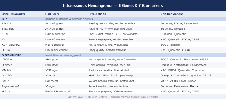
This article is organized around four practical tracks. The first and most detailed covers 7 measurable biomarkers — VEGF-A, D-dimer, MMP-9, hs-CRP, bone-specific alkaline phosphatase, angiopoietin-2, and HIF-1α — explaining what each reveals about lesion activity, how to measure it (with cost ranges), and what to do when results are out of range, with and without supplementation. The second track explores 6 key genes — PIK3CA, TIE2/TEK, KRAS, VHL, KDR/VEGFR2, and HIF1A — explaining how mutations or variants in each may drive abnormal vascular growth, along with targeted lifestyle and nutritional countermeasures. A third section distills ten of the most impactful insights from Eat to Beat Disease by Dr. William Li, whose angiogenesis framework directly challenges conventional thinking about how diet interacts with vascular tumors. Finally, three evidence-informed complementary modalities are presented for those living with symptomatic or monitoring-phase disease. The combination of biomarker tracking, genetic awareness, dietary strategy, and complementary support creates a far more actionable picture than imaging alone.
7 Biomarkers Worth Tracking
Tracking biomarkers for intraosseous hemangioma is not yet standard clinical practice. Most physicians rely on imaging alone to monitor these lesions — and imaging only shows anatomy. It cannot tell you whether a lesion is metabolically active, whether local inflammation is accelerating its growth, or whether your coagulation system is quietly under stress from abnormal vascular channels. Blood-based biomarkers can reveal those dimensions.
The seven markers below are chosen for their direct relevance to hemangioma biology, their practical measurability through commercial laboratories, and the existence of at least some evidence connecting each to vascular tumor activity. None are diagnostic on their own, and none replace imaging surveillance. Tracked together over time, however, they build a picture that scans cannot provide. Always interpret results with a physician familiar with vascular biology.
VEGF-A — The Primary Angiogenic Signal
Why It Matters
VEGF-A (Vascular Endothelial Growth Factor A) is the single most important driver of angiogenesis in hemangiomas. It binds to its primary receptor VEGFR2 on endothelial cells, triggering proliferation, migration, and new vessel formation. In intraosseous hemangiomas, VEGF-A is produced locally within the lesion and, in larger or more active lesions, becomes detectable in systemic circulation. Elevated serum VEGF-A has been associated with more active vascular lesions and higher recurrence rates in related vascular tumors. A rising trend over serial measurements is more informative than any single value. Research on VEGF signaling in bone vascular lesions is indexed at PubMed.
How to Measure It
Serum VEGF-A is available through commercial laboratories such as LabCorp and Quest Diagnostics as a standalone test. A standard blood draw is all that is needed; no fasting is required. Cost range: $40–$120 USD depending on provider and insurance coverage. Direct-to-consumer lab services also offer it. Request the numerical result rather than a simple normal/abnormal flag. Most labs consider values below 500 pg/mL within normal reference range for adults, though optimal target is lower for those with known vascular lesions.
If the Score Is Bad — The Plan Without Supplements
The most powerful non-supplement approach to reducing excessive VEGF-A signaling is dietary. Natural anti-angiogenic foods include green tea (EGCG), cooked tomatoes (lycopene), dark berries (anthocyanins), cruciferous vegetables (sulforaphane), and soy isoflavones — all have demonstrated VEGF-suppressing properties in human and animal studies. Regular moderate aerobic exercise at zone 2 intensity (150–300 minutes per week, conversational pace) normalizes angiogenic signaling over time; acute exercise transiently increases VEGF for healthy vascular remodeling, while chronic exercise training reduces basal pro-angiogenic tone. Reducing visceral adiposity is also critical — fat tissue is a major systemic VEGF source. Intermittent fasting (16:8 protocol) and moderate caloric restriction reduce circulating VEGF and related growth factors.
If the Score Is Bad — The Plan With Supplements or Equipment
- EGCG (green tea extract): 400–800 mg/day of standardized EGCG. Inhibits VEGFR2 kinase activity and reduces VEGF gene expression. Frequency: daily with food. Cycling: 12 weeks on, 4 weeks off. Side effects: potential liver stress at sustained high doses — avoid with liver disease; mild caffeine sensitivity in some; take with food, not on an empty stomach. - Curcumin with piperine: 500–1000 mg curcumin plus 5–10 mg piperine daily for bioavailability. Inhibits VEGF gene expression through NF-κB and HIF-1α suppression. Cycling: 8 weeks on, 2 weeks off. Side effects: GI discomfort, may reduce iron absorption, interacts with blood thinners — caution if anticoagulated. - Resveratrol: 250–500 mg/day trans-resveratrol. Dose-dependently inhibits VEGF transcription. Cycling: continuous with monthly one-week breaks. Side effects: interacts with CYP enzyme-metabolized drugs; possible mild estrogenic effects at very high doses. - Silibinin (milk thistle standardized extract): 250–500 mg/day. Inhibits VEGFR2 activation directly. Cycling: continuous. Side effects: generally well tolerated; mild GI effects in a minority.
D-Dimer — A Window Into Localized Coagulation
Why It Matters
D-dimer is a fibrin degradation product — it rises when coagulation is activated and clots are simultaneously being broken down. Vascular malformations, including hemangiomas, can induce localized intravascular coagulation (LIC) — a condition particularly relevant for larger cavernous or venous-type lesions where blood flow is sluggish within abnormal channels. LIC is not systemic DIC, but it creates persistent clot formation and fibrinolysis within the lesion that chronically elevates D-dimer. This biomarker provides indirect evidence of whether a lesion is hemodynamically active. Research linking D-dimer to vascular malformation activity is indexed at PubMed.
How to Measure It
D-dimer is one of the most widely available and affordable blood markers in clinical medicine. Cost range: $20–$60 USD. Reference ranges are typically expressed in fibrinogen equivalent units (FEU), with values below 500 ng/mL FEU considered normal. Keep in mind that many conditions — infection, recent surgery, pregnancy, active inflammation — can also elevate D-dimer. Trend tracking over time matters more than any isolated value, and context is essential for interpretation.
If the Score Is Bad — The Plan Without Supplements
Reducing vascular stasis and systemic coagulation activation are the primary non-supplement targets. Daily low-impact movement (30–60 minutes of walking) improves venous return and reduces localized clot formation. Adequate hydration (at least 2L water daily) supports blood fluidity. If the hemangioma is in the spine, extended bed rest should be minimized. Addressing systemic inflammatory drivers — excess visceral fat, metabolic syndrome, insulin resistance — reduces background fibrinogen and coagulation activity. The Mediterranean dietary pattern has robust evidence for reducing coagulation markers including D-dimer.
If the Score Is Bad — The Plan With Supplements or Equipment
Important: Any supplement affecting coagulation must be reviewed with your physician before use, particularly if you take anticoagulants or antiplatelet agents.
- Omega-3 fatty acids (EPA + DHA): 2–4 g/day combined EPA/DHA from marine oil or algae-based sources. Reduces fibrinogen and platelet aggregation. Cycling: continuous. Side effects: blood-thinning effect — contraindicated at high dose with warfarin; fishy aftertaste manageable with enteric-coated formulas. - Nattokinase: 2000–4000 FU/day away from meals. Fibrinolytic enzyme from fermented soy that directly breaks down fibrin. Cycling: 8 weeks on, 4 weeks off. Side effects: significant fibrinolytic effect — contraindicated with warfarin, heparin, or aspirin therapy; discontinue at least 2 weeks before surgery. - Serrapeptase: 40,000–120,000 SPU/day on an empty stomach. Proteolytic and fibrinolytic enzyme. Cycling: 6 weeks on, 2 weeks off. Side effects: similar bleeding risk profile to nattokinase; rare reports of pneumonitis with long-term high-dose use. - Graduated compression garments (equipment): For limb-adjacent or spinal lesions where venous stasis is a factor, fitted graduated compression can reduce local vascular stagnation. No cycling needed — use as directed. Obtain proper fit and pressure grading from a vascular specialist.
MMP-9 — The Matrix Degradation Marker
Why It Matters
MMP-9 (Matrix Metalloproteinase-9) is a zinc-dependent enzyme that degrades collagen IV and other extracellular matrix components, physically clearing the path for new blood vessels to expand into surrounding tissue. In bone, elevated MMP-9 allows hemangioma vascular channels to invade and displace the mineralized matrix — a mechanism relevant to lesion growth and local bone destruction. MMP-9 is both a downstream target of VEGF signaling and an independent inflammatory marker. Elevated serum MMP-9 correlates with more aggressive vascular tumor behavior in related conditions and reflects the broader pro-angiogenic inflammatory environment. Research on MMP-9 in bone vascular pathology is indexed at PubMed.
How to Measure It
Serum MMP-9 is available through commercial reference labs, though it is less commonly ordered than the other markers on this list. Some integrative oncology practices include it in vascular tumor monitoring panels. Cost range: $60–$150 USD. Typical adult reference values are below 130 ng/mL, but vary by laboratory. Trend monitoring over 3–6 month intervals is the most useful application.
If the Score Is Bad — The Plan Without Supplements
Reducing visceral adiposity is one of the highest-yield non-pharmacological MMP-9 interventions — adipose tissue is a prolific producer of MMP-9, and meaningful reductions in visceral fat produce measurable MMP-9 decreases. An anti-inflammatory dietary pattern — Mediterranean-style, low glycemic — reduces the cytokine environment (particularly IL-6 and TNF-α) that drives MMP transcription. Limiting alcohol intake is important, as alcohol upregulates MMP-9 in multiple tissue contexts. Consistent, quality sleep (7–9 hours) reduces overnight NF-κB-mediated MMP-9 gene expression.
If the Score Is Bad — The Plan With Supplements or Equipment
- N-Acetylcysteine (NAC): 600–1200 mg/day. Antioxidant that reduces NF-κB activity and downstream MMP-9 expression. Cycling: continuous or 12 weeks on, 4 weeks off. Side effects: GI discomfort in some individuals; may interact with certain chemotherapy agents — disclose to oncologist if relevant. - EGCG (green tea extract): 400–800 mg/day. Directly inhibits MMP-9 expression and activity. (Full dosing in VEGF-A section.) Cycling: 12 weeks on, 4 weeks off. - Quercetin: 500–1000 mg/day. Flavonoid that reduces MMP-9 through NF-κB inhibition. Cycling: continuous. Side effects: generally well tolerated; rare headache; avoid extremely high doses if thyroid disease is present, as quercetin may inhibit thyroid peroxidase at megadoses. - Curcumin with piperine: 500–1000 mg/day. Directly inhibits MMP-9 transcription via NF-κB. (Full dosing in VEGF-A section.) Cycling: 8 weeks on, 2 weeks off.
hs-CRP — The Inflammatory Environment Marker
Why It Matters
High-sensitivity C-reactive protein (hs-CRP) is the most accessible general marker of systemic low-grade inflammation. It is not specific to intraosseous hemangioma, but it reflects the inflammatory microenvironment that fuels angiogenesis. Chronic low-grade inflammation upregulates VEGF, MMP-9, HIF-1α, and NF-κB — all major pro-angiogenic drivers. An hs-CRP consistently above 2–3 mg/L suggests an inflammatory state that likely contributes to vascular lesion activity. The long-term goal for most adults is hs-CRP below 1 mg/L. Research on inflammation and angiogenesis in vascular tumor contexts is indexed at PubMed.
How to Measure It
hs-CRP is one of the cheapest, most widely available blood markers in clinical medicine. Cost range: $15–$40 USD. Many primary care physicians order it routinely for cardiovascular risk assessment. Unlike standard CRP, the high-sensitivity assay detects the lower levels relevant to chronic inflammatory risk. No fasting is required. It is worth testing at baseline and every 3–6 months when actively managing inflammation.
If the Score Is Bad — The Plan Without Supplements
The Mediterranean dietary pattern has the strongest human evidence for lowering hs-CRP — it emphasizes olive oil, fatty fish, legumes, vegetables, nuts, and whole grains while minimizing processed food and refined carbohydrates. Regular moderate aerobic exercise (150+ minutes per week) consistently reduces hs-CRP across most population groups. Sleep quality is significantly underappreciated: even one week of fragmented or shortened sleep can double hs-CRP. Managing body weight — particularly reducing visceral fat — remains the highest-yield single intervention for chronically elevated CRP. Smoking cessation, if relevant, produces some of the fastest CRP reductions of any modifiable factor.
If the Score Is Bad — The Plan With Supplements or Equipment
- Omega-3 fatty acids (EPA + DHA): 2–4 g/day. The single most evidence-backed supplement for hs-CRP reduction across multiple RCTs. Cycling: continuous. Side effects: mild blood-thinning; fishy breath manageable with enteric coating. - Curcumin with piperine: 500 mg/day with piperine. Multiple randomized controlled trials show clinically meaningful hs-CRP reductions. Cycling: 8 weeks on, 2 weeks off. Side effects: GI, iron absorption, anticoagulant interaction — see above. - Magnesium glycinate: 300–400 mg/day before bed. Magnesium deficiency is extremely prevalent and correlates with elevated CRP. The glycinate form is better tolerated and less laxative than oxide forms. Cycling: continuous. Side effects: loose stools at high doses; otherwise very safe. - Vitamin D3 + K2: 2000–4000 IU D3 daily with 100–200 mcg MK-7 K2. Supplementation consistently reduces hs-CRP when serum 25-OH vitamin D is below 40 ng/mL — check before supplementing. Cycling: continuous with periodic 25-OH D monitoring. Side effects: hypercalcemia at excessive D3 doses; K2 may interact with vitamin K-antagonist anticoagulants.
Bone-Specific Alkaline Phosphatase — Tracking Bone Remodeling
Why It Matters
Bone-specific alkaline phosphatase (BALP) is an isoform of alkaline phosphatase produced exclusively by osteoblasts during bone matrix formation. In the context of intraosseous hemangioma, elevated BALP can indicate active bone matrix disruption at the lesion site — the abnormal vascular channels physically displace bone trabeculae, and adjacent bone responds by upregulating remodeling activity. BALP is more specific to bone than total alkaline phosphatase, which also reflects liver enzyme activity. Elevated BALP alongside clinical symptoms or imaging progression warrants closer surveillance. Bone remodeling biomarker research relevant to bone tumors is available at PubMed.
How to Measure It
BALP can be ordered as isoform-specific alkaline phosphatase or bone ALP through commercial reference labs. Cost range: $50–$120 USD. It is sometimes bundled with bone metabolism panels that include vitamin D, PTH, and calcium. Normal adult range is approximately 11–48 mcg/L, varying by laboratory. Context matters — if total ALP is elevated, separating bone and liver contributions requires the isoform test.
If the Score Is Bad — The Plan Without Supplements
When BALP elevation is secondary to hemangioma-driven bone remodeling, the primary target is the lesion's inflammatory and angiogenic environment — addressed through the VEGF-A, hs-CRP, and MMP-9 strategies above. For bone support specifically, weight-bearing exercise (walking, bodyweight resistance training) stimulates healthy osteoblast/osteoclast balance. Adequate dietary protein (1.0–1.6 g/kg/day) is essential for bone matrix synthesis. Eliminating excess alcohol and minimizing corticosteroid use (when medically possible) reduces secondary bone losses that could further elevate compensatory BALP.
If the Score Is Bad — The Plan With Supplements or Equipment
- Vitamin K2 (MK-7 form): 100–200 mcg/day. Activates osteocalcin and directs calcium to bone matrix rather than soft tissues. Cycling: continuous. Side effects: interacts with vitamin K-antagonist anticoagulants (warfarin) — contraindicated without physician oversight in anticoagulated patients. - Vitamin D3: 2000–4000 IU/day, calibrated to achieve serum 25-OH D of 40–60 ng/mL. Supports osteoblast function and calcium metabolism. Cycling: continuous with periodic monitoring. Side effects: hypercalcemia at doses above 10,000 IU/day long-term. - Boron: 3–6 mg/day. Supports vitamin D activation and bone mineral metabolism. Cycling: continuous. Side effects: very safe at these doses; avoid higher doses (>20 mg/day). - Silicon (orthosilicic acid form): 10–25 mg/day. Supports collagen synthesis in bone matrix. Cycling: continuous. Side effects: generally very safe.
Angiopoietin-2 — The Vascular Destabilizer
Why It Matters
Angiopoietin-2 (Ang-2) is a glycoprotein that destabilizes the endothelium by competitively displacing Angiopoietin-1 from the TIE2 receptor. When Ang-2 is elevated, blood vessels become more permeable, more prone to sprouting, and more responsive to VEGF signals. In vascular malformations and hemangiomas, elevated Ang-2 is a marker of active vascular remodeling. Ang-2 acts as a permissive signal — it primes vessels for VEGF-driven expansion — and is both a useful activity marker and a target in clinical trials for vascular tumors. Angiopoietin signaling research in vascular lesions is indexed at PubMed.
How to Measure It
Serum Angiopoietin-2 is available through specialty and reference labs. It is not a routine panel item and is most commonly ordered in oncology, integrative medicine, or vascular anomaly specialty contexts. Cost range: $80–$200 USD. Reference values are typically below 3 ng/mL in healthy adults. This is one of the more advanced markers on this list — for those new to biomarker tracking, start with VEGF-A and hs-CRP first and add Ang-2 as a second-tier measurement.
If the Score Is Bad — The Plan Without Supplements
Ang-2 is strongly associated with cardiometabolic risk factors — obesity, insulin resistance, dyslipidemia, and hypertension all independently elevate it. Addressing these through diet and exercise produces direct Ang-2-lowering effects. Regular zone 2 aerobic exercise performed consistently over 8–12 weeks reduces Ang-2 in metabolically unhealthy individuals. Keeping fasting glucose below 90 mg/dL and fasting insulin below 5 µIU/mL is the most sustainable long-term Ang-2 management strategy. Visceral fat reduction is, again, among the highest-yield interventions.
If the Score Is Bad — The Plan With Supplements or Equipment
- Berberine: 500 mg, 2–3 times daily with meals. Activates AMPK, reducing pro-inflammatory and pro-angiogenic signaling including Ang-2. Cycling: 8 weeks on, 4 weeks off. Side effects: GI discomfort particularly at initiation; lowers blood sugar — caution in diabetics on medication; may interact with metformin and some statins. - Resveratrol: 250–500 mg/day. Inhibits NF-κB and reduces VEGF/Ang-2 co-activation. Cycling: continuous with monthly one-week breaks. Side effects: CYP enzyme interactions; see VEGF-A section. - Alpha-lipoic acid (R-ALA form): 300–600 mg/day. Reduces vascular endothelial inflammation and normalizes endothelial signaling. Cycling: 12 weeks on, 4 weeks off. Side effects: may lower blood glucose; GI sensitivity at higher doses; use R-form rather than racemic mixture for better bioavailability.
HIF-1α — The Hypoxia Master Regulator
Why It Matters
HIF-1α (Hypoxia-Inducible Factor 1-alpha) is the transcriptional master regulator of the cellular hypoxia response. Under low-oxygen conditions, HIF-1α is stabilized and activates hundreds of downstream genes — VEGF foremost among them — as well as angiopoietins, erythropoietin, and glucose transporters. In intraosseous hemangiomas, the structurally abnormal vascular architecture creates localized hypoxic pockets that chronically stabilize HIF-1α and maintain a sustained pro-angiogenic state. Critically, HIF-1α can also be activated by oncogenic mutations (such as PIK3CA or VHL loss) independent of actual oxygen levels, creating a pseudo-hypoxic state. Research on HIF-1α in bone vascular tumors is available at PubMed.
How to Measure It
Direct serum HIF-1α measurement is available through specialty labs but has limited clinical utility as a standalone systemic marker due to protein instability in blood samples. Cost range: $90–$200 USD. A more practical and affordable approach is to measure its downstream proxies: VEGF-A (already on this list), erythropoietin (EPO) (~$40–80 USD), and lactate dehydrogenase (LDH) (~$15–30 USD). Elevated EPO and LDH in the context of elevated VEGF-A together suggest an active HIF response — this triad is more informative than serum HIF-1α alone.
If the Score Is Bad — The Plan Without Supplements
The single most powerful driver of chronic HIF-1α elevation outside of genetic mutations is sleep-disordered breathing — particularly obstructive sleep apnea. Overnight hypoxic episodes repeatedly stabilize HIF-1α and chronically upregulate VEGF. If you snore, wake unrefreshed, or experience daytime sleepiness, a home sleep study is worth pursuing. CPAP therapy for confirmed sleep apnea normalizes nocturnal HIF-1α activation within weeks — arguably the most impactful single HIF-modulating intervention that exists. Beyond sleep, reducing excess body weight (obesity reduces tissue oxygen delivery efficiency) and engaging in regular moderate aerobic exercise to improve VO2max and oxygen utilization are the most impactful systemic interventions.
If the Score Is Bad — The Plan With Supplements or Equipment
- NAC (N-Acetylcysteine): 600–1200 mg/day. Reduces ROS — reactive oxygen species are required cofactors for HIF-1α stabilization in many pathways. Cycling: continuous or 12 weeks on, 4 weeks off. Side effects: GI discomfort; avoid with certain chemotherapy agents — check with oncologist. - Quercetin: 500–1000 mg/day. Directly inhibits HIF-1α accumulation and reduces downstream VEGF transcription. Cycling: continuous. Side effects: generally safe; see MMP-9 section. - EGCG: 400–800 mg/day. Enhances prolyl hydroxylase activity, helping the cell degrade HIF-1α more effectively under normoxic conditions. Cycling: 12 weeks on, 4 weeks off. Side effects: see VEGF-A section. - CPAP / BiPAP device (equipment): For confirmed obstructive sleep apnea, this is the highest-yield HIF-1α-lowering intervention available to most patients. Not optional if sleep apnea is confirmed — its impact on VEGF and angiogenic signaling is substantial.
Taken together, these seven biomarkers form a functional monitoring panel that gives a working picture of what is happening biologically around and within an intraosseous hemangioma. The genetic section that follows explains why some people's panels trend consistently higher than others — and what molecular variants may be at the root.
6 Genes That Shape How These Lesions Behave
The genetics of intraosseous hemangioma operates on two distinct levels. The first is somatic mutations — genetic changes that occur in the lesion's own cells, not inherited from your parents. These are found only in the hemangioma tissue and drive its local biology. The second is germline variants — inherited polymorphisms present in every cell of your body that affect how your entire biology handles angiogenesis, inflammation, and hypoxia. Identifying somatic mutations requires tissue sampling (biopsy or surgical specimen analysis). Germline variants can be identified through consumer genetic testing or clinical genomics panels.
Most intraosseous hemangiomas arise from somatic events in the lesion itself. But germline variants shape the biological environment in which those events occur — they determine the soil's fertility, so to speak. The six genes below span both levels, and the same pathways reappear in the biomarker section, which is exactly the point: genes and biomarkers are two windows into the same molecular story.
PIK3CA — The Most Common Driver Mutation
What It Does
PIK3CA encodes p110α, the catalytic subunit of phosphatidylinositol 3-kinase (PI3K). This enzyme phosphorylates PIP2 to PIP3, activating the AKT/mTOR cascade — one of the most fundamental regulators of cell growth, survival, metabolism, and angiogenesis. Activating somatic mutations in PIK3CA are the most commonly identified driver mutations in vascular malformations, including hemangiomas. Hotspot mutations at E542K, E545K, and H1047R are most frequently reported. When PIK3CA is constitutively active, it drives chronic endothelial cell proliferation, survival, and VEGF secretion — precisely the biology that characterizes active, growing hemangiomas. In germline form, PIK3CA mutations cause the PIK3CA-Related Overgrowth Spectrum (PROS), which includes multiple vascular anomalies. Research on PIK3CA in vascular malformations is indexed at PubMed.
If the Gene Has a Bad Variant — The Plan Without Supplements
The most evidence-supported lifestyle approach to reducing PI3K/AKT/mTOR activity is intermittent fasting and caloric restriction. These interventions robustly suppress mTORC1 — the same downstream target inhibited by rapamycin, which has emerging clinical evidence specifically for PIK3CA-driven vascular malformations. A low-refined-carbohydrate diet with low glycemic load keeps insulin and AKT/mTOR suppressed throughout the day. Regular aerobic exercise activates AMPK, which directly counteracts mTOR signaling. Managing fasting insulin below 8 µIU/mL and HbA1c below 5.4% are the most practical and measurable metabolic targets.
If the Score Is Bad — The Plan With Supplements or Equipment
- Berberine: 500 mg, 2–3 times daily with meals. Activates AMPK and directly inhibits mTORC1, mirroring aspects of rapamycin's mechanism. Cycling: 8 weeks on, 4 weeks off. Side effects: GI discomfort; blood glucose lowering — caution with diabetes medication. - EGCG: 400–800 mg/day. Inhibits PI3K/AKT signaling and reduces VEGF expression downstream. Cycling: 12 weeks on, 4 weeks off. - Resveratrol: 250–500 mg/day. Activates SIRT1 and AMPK, modulating PI3K pathway activity. Cycling: continuous. - Clinical note: Prescription mTOR inhibitors (sirolimus/rapamycin, alpelisib) are in active clinical investigation for PIK3CA-driven vascular malformations. If your hemangioma is symptomatic or demonstrably growing, asking a specialist about current trials in this area is worth pursuing.
TIE2 / TEK — The Endothelial Stability Gene
What It Does
TIE2 (also called TEK) encodes a tyrosine kinase receptor on endothelial cells whose natural ligands are Angiopoietin-1 and Angiopoietin-2. Activating TIE2 mutations cause constitutive AKT and ERK signaling in endothelial cells — independent of ligand binding — resulting in chronically leaky, structurally abnormal, pericyte-poor vessels. These are the hallmarks of venous malformations and some hemangiomas. TIE2 mutations can be somatic (found in isolated lesions) or germline (causing Multiple Cutaneous and Mucosal Venous Malformations, MCMVM — multiple lesions throughout the body). The angiopoietin-2 biomarker described above is directly connected to this pathway. Research on TIE2 mutations in vascular anomalies is indexed at PubMed.
If the Gene Has a Bad Variant — The Plan Without Supplements
TIE2-mediated AKT activation shares downstream pathways with PIK3CA — intermittent fasting, AMPK-activating exercise, and a low-insulin dietary pattern remain the core lifestyle countermeasures. Because TIE2 mutations also create structurally fragile vessels prone to thrombosis, managing the D-dimer biomarker and preventing vascular stasis through regular movement and hydration is especially important in this genetic context. If multiple lesions are present, formal genetic testing and counseling is the first priority.
If the Score Is Bad — The Plan With Supplements or Equipment
- Berberine: as described above — AKT pathway modulation. - Omega-3 fatty acids: 2–4 g/day. Particularly relevant for endothelial protection and anti-thrombotic support given TIE2-associated vascular fragility. Cycling: continuous. Side effects: blood-thinning — discuss with physician if coagulopathy is present. - Clinical note: Low-dose aspirin (81 mg/day) is sometimes used in venous malformation patients with elevated D-dimer risk — a decision to make with your vascular specialist, not independently.
KRAS — The RAS Pathway Activator
What It Does
KRAS encodes a membrane GTPase that transmits growth signals from cell surface receptors to downstream cascades — most importantly RAF/MEK/ERK and PI3K/AKT. Gain-of-function somatic KRAS mutations lock the protein in its active state, constitutively driving endothelial cell proliferation, VEGF production, and vessel formation. These mutations are well established in arteriovenous malformations (AVMs) and are increasingly being identified across a broader spectrum of vascular anomalies including some hemangiomas. Research on KRAS in vascular anomalies is indexed at PubMed.
If the Gene Has a Bad Variant — The Plan Without Supplements
KRAS-driven pathways are among the most difficult to modulate through lifestyle alone, as RAS signaling is deeply embedded in cell biology. The most actionable strategy is reducing upstream drivers: minimizing insulin and IGF-1 (growth signals that activate RAS pathways indirectly), managing systemic inflammation (NF-κB potentiates RAS signaling), and maintaining robust antioxidant status (reactive oxygen species promote RAS pathway activity). A protein-adequate diet with low glycemic load is the dietary foundation. Regular exercise reduces IGF-1 and insulin sensitivity — two of the upstream inputs to RAS.
If the Score Is Bad — The Plan With Supplements or Equipment
- Curcumin: multiple in vitro and early human studies show RAS-pathway modulatory effects. 500–1000 mg/day with piperine. Cycling: 8 weeks on, 2 weeks off. - Quercetin: reduces MEK/ERK downstream signaling. 500–1000 mg/day. Cycling: continuous. - Clinical note: MEK inhibitors (trametinib, cobimetinib) are available by prescription and being studied specifically in KRAS-driven vascular anomalies. Discuss with a vascular anomaly specialist if KRAS mutation is confirmed in your lesion.
VHL — The HIF-1α Gatekeeper
What It Does
VHL encodes the Von Hippel-Lindau tumor suppressor protein, whose primary role is targeting HIF-1α for proteasomal degradation. When VHL is mutated or silenced, HIF-1α accumulates regardless of oxygen levels, driving chronic, massive VEGF expression in a pseudo-hypoxic state. In germline form, VHL mutations cause VHL syndrome — a hereditary condition characterized by hemangioblastomas (brain, spinal cord, retina), renal cell carcinoma, and pheochromocytoma. Somatic VHL variants may also contribute to isolated vascular lesions. Research on VHL and angiogenesis is indexed at PubMed.
If the Gene Has a Bad Variant — The Plan Without Supplements
If multiple hemangiomas, a family history of VHL-associated tumors, or renal lesions are present, formal VHL genetic testing and specialist referral is the first priority — not a supplement. From a lifestyle standpoint, the goal is to reduce HIF-1α stabilization from non-genetic sources: treat sleep apnea, maintain healthy body weight, avoid chronic hypoxic environments, and engage in regular aerobic exercise to improve tissue oxygen delivery. These steps do not reverse the VHL mutation but substantially reduce the compounding hypoxic load.
If the Score Is Bad — The Plan With Supplements or Equipment
- NAC, Quercetin, EGCG: the three HIF-1α-modulating supplements described in biomarker 7 are the most directly relevant here. Same dosing and cycling apply. - CPAP: if sleep apnea is confirmed — the highest-impact equipment intervention for HIF-1α. - Clinical note: Belzutifan (an HIF-2α inhibitor) is FDA-approved specifically for VHL syndrome-associated tumors. This is a specialist conversation, not a self-management decision.
KDR / VEGFR2 — The VEGF Amplifier
What It Does and How to Act on It
KDR encodes VEGFR2, the primary signaling receptor for VEGF-A on endothelial cells. Germline polymorphisms in KDR — particularly the Q472H variant — alter receptor kinase activity and affect how strongly endothelial cells respond to a given VEGF signal. KDR variants that increase VEGFR2 sensitivity amplify the angiogenic response to VEGF, potentially contributing to more active vascular lesion behavior even at normal circulating VEGF levels. Research on KDR polymorphisms and angiogenesis is indexed at PubMed. Since KDR variants amplify the VEGF response, the intervention logic is to reduce the upstream signal — VEGF-A — using the full strategy described in biomarker 1. EGCG is particularly relevant in this context because it acts partly by inhibiting VEGFR2 kinase activity directly. Anti-angiogenic dietary patterns, weight management, and anti-inflammatory lifestyle measures address the problem at the signal level. Silibinin (250–500 mg/day) has specific VEGFR2-inhibitory properties and is worth prioritizing in this genetic context. Cycling: continuous. Side effects: very well tolerated; minor GI effects in some.
HIF1A — The Inherited Hypoxia Sensor
What It Does and How to Act on It
HIF1A is the gene encoding HIF-1α described in biomarker 7. Beyond its role in tumor biology, germline polymorphisms in HIF1A — particularly Pro582Ser (rs11549465) — alter the stability and transcriptional potency of HIF-1α under physiological oxygen conditions. Carriers of the Pro582Ser variant have a constitutively more active HIF-1α protein, meaning their endothelial cells generate stronger angiogenic responses to hypoxic signals even at modest oxygen fluctuations. This amplifies VEGF production and potentially contributes to more active vascular lesion behavior over time. Research on HIF1A polymorphisms is indexed at PubMed. The Pro582Ser variant is identifiable through consumer genetic panels (23andMe, AncestryDNA) or clinical genomics testing. The intervention strategy maps directly to the HIF-1α biomarker section — NAC, quercetin, EGCG, sleep apnea treatment, aerobic exercise for VO2max improvement. What this genetic context adds is the rationale to act proactively and earlier, before biomarkers become clearly abnormal. Carriers have a mechanistic reason to treat sleep quality and metabolic health not as general wellness goals but as biologically specific interventions.
What Dr. William Li's Angiogenesis Research Can Teach You
Eat to Beat Disease: The New Science of How Your Body Can Heal Itself by Dr. William Li — Harvard-trained physician, clinical researcher, and founder of the Angiogenesis Foundation — is one of the most directly relevant works for anyone with a vascular tumor. Dr. Li's central argument is that the body maintains sophisticated angiogenesis defense systems throughout life, and that specific dietary choices can either strengthen or undermine those defenses. He has catalogued hundreds of peer-reviewed studies showing that foods contain natural compounds capable of measurably modulating VEGF, MMP-9, Ang-2, and other pro-angiogenic signals at nutritional doses. His framework directly challenges the conventional medical assumption that diet has no meaningful role in vascular tumor management.
For intraosseous hemangioma, the anti-angiogenic side of his framework is most relevant. Below are ten of the most impactful insights from his research.
Your Body's Anti-Angiogenic Defense System Is More Powerful Than You Think
Li argues that vascular and angiogenesis-dependent tumors are routinely held in check throughout most of our lives by endogenous anti-angiogenic mechanisms. Intraosseous hemangiomas appear in autopsy studies at prevalence rates far higher than clinical incidence — suggesting that the vast majority never become symptomatic because the body's defenses contain them. These defenses include endostatin, angiostatin, thrombospondin-1, and circulating anti-angiogenic peptides. The critical insight is that lifestyle and diet either support or erode these natural mechanisms. When they are intact, the lesion stays contained. When they are chronically undermined, the balance may tip toward growth.
Green Tea Catechins Are Among the Most Studied Natural Anti-Angiogenic Compounds
Dr. Li cites an extensive body of research showing that EGCG from green tea inhibits multiple steps in the angiogenic cascade — VEGFR2 kinase activation, MMP-9 expression, NF-κB signaling, and endothelial cell migration. Population data from Japan and China show that regular consumption of 3–5 cups of green tea daily is associated with reduced rates of angiogenesis-dependent tumors. Mechanistically, EGCG's concentrations in tea are sufficient to achieve measurable plasma levels with direct endothelial effects. Li considers green tea one of the highest-ranking anti-angiogenic dietary choices, and it maps directly to both VEGF-A and MMP-9 in the biomarker panel above.
Cooked Tomatoes With Fat Deliver the Most Bioavailable Anti-Angiogenic Lycopene
Lycopene from cooked tomatoes consumed with fat (such as extra virgin olive oil) is absorbed at levels sufficient to reduce VEGF signaling measurably. Li references prospective studies and mechanistic data showing that two or more servings of cooked tomato products weekly (tomato paste, sauce, roasted tomatoes) produce meaningful reductions in VEGF bioactivity in human blood. The combination of heat processing — which releases lycopene from cell walls — and dietary fat, which facilitates its absorption, doubles effective lycopene exposure compared to raw tomatoes. This is a food-first intervention that is both affordable and palatable.
Dark Berries Provide Anthocyanins That Blunt VEGF-Driven Endothelial Migration
Blueberries, blackberries, tart cherries, and açaí contain anthocyanins — polyphenolic pigments that inhibit VEGF expression, reduce endothelial cell migration, and lower circulating angiogenic markers. Li references studies in which regular berry consumption measurably modulated angiogenic markers in human blood within weeks. The dose is realistic: one serving (80–100g) of mixed dark berries daily achieves anthocyanin concentrations with meaningful anti-angiogenic effects. This is among the easiest dietary shifts to implement and maintain long-term.
Soy Isoflavones Inhibit Angiogenesis at Food-Level Doses
Genistein, soy's primary isoflavone, inhibits VEGF-driven tube formation in endothelial cells and reduces VEGF gene expression in multiple in vitro and human study contexts. Li addresses the estrogenic concern associated with soy directly — at food-level consumption (one to two servings of whole soy foods daily, not megadose supplements), the anti-angiogenic benefit appears to predominate in most populations. He distinguishes fermented soy products (miso, tempeh, natto) as particularly favorable due to increased isoflavone bioavailability, with natto having the additional benefit of delivering nattokinase (the fibrinolytic enzyme discussed in the D-dimer section above).
Extra Virgin Olive Oil — A Polyphenol-Rich Anti-Angiogenic Fat
Li identifies high-quality extra virgin olive oil as one of the foundational dietary elements with consistent anti-angiogenic evidence. Its polyphenols — particularly oleocanthal, hydroxytyrosol, and oleuropein — inhibit VEGF and reduce MMP activity through NF-κB suppression. He emphasizes the importance of distinguishing quality EVOO (high polyphenol content, recent harvest, properly stored in dark glass) from commodity refined olive oil, which has been processed to the point where most active compounds are stripped. Practically, 2–4 tablespoons of high-quality EVOO daily, used as a cooking base and dressing, provides the relevant polyphenol dose.
Visceral Fat Is a Pro-Angiogenic Organ — Not Just a Risk Factor
One of Li's most practically impactful insights is that excess visceral adipose tissue functions as an active pro-angiogenic organ — not merely a cardiovascular risk marker. Visceral fat produces VEGF, Ang-2, inflammatory cytokines, and matrix metalloproteinases at rates that directly fuel vascular tumor activity systemically. Reducing visceral fat through diet and exercise is, in his framing, a direct anti-angiogenic intervention. This reframes weight management from a general wellness recommendation into a specific therapeutic target for vascular lesion management — a conceptual shift that motivates the effort differently.
Fatty Fish as a Weekly Cornerstone of Anti-Angiogenic Diet
EPA and DHA from fatty fish (salmon, sardines, mackerel, anchovies) reduce arachidonic acid-derived eicosanoids that stimulate VEGF and NF-κB. Li references prospective cohort studies linking two or more servings of oily fish weekly to reduced rates of angiogenesis-dependent disease. He prioritizes whole food consumption over supplements in most cases, noting that fish also provides astaxanthin — a carotenoid with independent anti-angiogenic and antioxidant properties. For those who cannot regularly consume fatty fish, algae-based EPA/DHA supplements are the most bioavailable alternative.
Resveratrol's Evidence Is Real but Dose-Limited in Food Form
Li covers resveratrol carefully and honestly. The in vitro and animal evidence for resveratrol's anti-angiogenic effects is robust — it inhibits VEGF transcription, suppresses MMP-9, and activates SIRT1. However, the concentrations achievable through wine consumption alone are likely too low to reproduce these effects directly. He recommends consuming resveratrol-rich whole foods (red grapes, peanuts, mulberries, dark cocoa) regularly, while acknowledging that supplementation at 250–500 mg/day trans-resveratrol provides more pharmacologically relevant doses. He avoids overclaiming for either form, which makes his recommendation more trustworthy.
The Cumulative Anti-Angiogenic Diet Score Matters More Than Any Single Food
Perhaps Li's most practically useful finding for daily decision-making: it is not any single food but the cumulative anti-angiogenic profile of the entire diet that produces measurable, sustained effects. A diet consistently rich in green tea, cooked tomatoes, dark berries, cruciferous vegetables, fatty fish, and extra virgin olive oil — while minimizing refined carbohydrates, processed foods, and excess red meat — creates a sustained anti-angiogenic metabolic environment. No individual food substitutes for the overall pattern. The daily implementation question is not "did I take my anti-angiogenic supplement?" but "is my diet as a whole anti-angiogenic today?"
Complementary Approaches Worth Considering
The evidence base for complementary approaches specific to intraosseous hemangioma is limited — this condition is relatively uncommon, and few modalities have been studied in this precise population. The following three options are included because they have meaningful human clinical evidence for the broader biological targets most relevant to this condition: chronic pain management in symptomatic lesions, systemic inflammatory marker reduction, and normalization of the vascular and autonomic environment. None are curative; all carry low risk when applied appropriately.
Low-Level Laser Therapy / Photobiomodulation
Photobiomodulation (PBM) uses specific near-infrared and red wavelengths (typically 630–1000 nm) to stimulate mitochondrial energy production (via cytochrome c oxidase activation), reduce local inflammation, and modulate vascular biology. For intraosseous hemangioma, its primary clinical application is pain management for symptomatic spinal lesions, where localized bone pain from vertebral expansion or periosteal involvement may be present. Secondarily, PBM's anti-inflammatory effects — reduction in prostaglandins, IL-1β, and TNF-α at the tissue level — are relevant to reducing the inflammatory microenvironment that sustains lesion activity.
A systematic review of PBM in musculoskeletal pain — indexed in the literature at PubMed — demonstrates consistent, clinically meaningful benefit for localized bone and soft tissue pain across multiple randomized trials. PBM's effects on VEGF and angiogenic signaling in pathological contexts remain under active investigation, with the evidence base still largely experimental rather than clinical for this application specifically.
In practice, PBM for spinal pain associated with vertebral hemangioma involves treatment by a licensed physiotherapist or rehabilitation physician using a class III or IV laser device. Typical protocols use 810–830 nm wavelength at 3–5 sessions per week for 4–6 weeks, applied at the symptomatic spinal level. Home-use devices (panel-type red light therapy or targeted near-infrared devices) exist for ongoing maintenance. Contraindication: do not apply directly over an uncharacterized or actively growing lesion without medical clearance — consult your physician before initiating treatment near the lesion site.
Mindfulness-Based Stress Reduction (MBSR)
MBSR is an 8-week structured program combining formal meditation, body scan, and mindful movement practices. Developed by Jon Kabat-Zinn at the University of Massachusetts Medical Center, it has accumulated one of the largest evidence bases of any mind-body intervention in medicine. Its relevance to intraosseous hemangioma is twofold: chronic pain management for patients with symptomatic spinal or skull-base lesions, and systemic inflammatory biomarker reduction — mindfulness practice measurably reduces hs-CRP, IL-6, and cortisol, all markers directly relevant to the biomarker panel above.
A well-cited randomized controlled trial by Cherkin and colleagues (JAMA, 2016) demonstrated that MBSR was superior to usual care for chronic low back pain at both 8 weeks and 52 weeks. For inflammatory biomarkers specifically, multiple RCTs examining MBSR's effects on CRP and IL-6 are indexed at PubMed, consistently showing reductions in inflammatory markers after completing the 8-week program. Chronic psychological stress activates the HPA axis, elevating cortisol and downstream NF-κB signaling — the same inflammatory cascade that drives VEGF and MMP-9. MBSR directly interrupts this pathway.
MBSR is accessible in multiple formats: in-person 8-week group programs (common at hospitals and cancer centers), app-based guided programs, and online courses certified to the original curriculum. The core practice is 20–45 minutes of formal meditation daily. For patients with spinal hemangioma-related pain, body scan and gentle mindful movement modified to avoid pain-provoking positions are the most relevant techniques. Risk is exceptionally low — it is one of the safest complementary interventions available.
Breathing-Based Therapies
Structured breathing practices — including diaphragmatic breathing, box breathing (4-4-4-4 pattern), and nasal breathing training — modulate the autonomic nervous system and blood gas physiology in ways that directly reduce systemic inflammation, normalize HIF pathway activity, and improve tissue oxygenation. Chronic sympathetic nervous system activation (the stress response) elevates VEGF, CRP, cortisol, and NF-κB signaling. Controlled, slow breathing reliably activates parasympathetic tone, downregulating these same signals. The relevance to HIF-1α is mechanistically direct: improving respiratory efficiency reduces nocturnal and daytime hypoxic episodes that chronically stabilize HIF-1α and drive angiogenesis.
Human evidence for breathing practices in inflammatory marker and pain management is growing. Research on diaphragmatic breathing and autonomic regulation is indexed at PubMed, and a 2017 study in Frontiers in Psychology demonstrated measurable reductions in salivary cortisol after controlled breathing training. Multiple studies show reductions in CRP and IL-6 with sustained practice.
The most practical starting protocol is slow diaphragmatic box breathing: inhale through the nose for 4–5 seconds, hold for 4 seconds, exhale slowly through the nose or pursed lips for 6–8 seconds, hold 4 seconds. Practice for 10–15 minutes twice daily. Nasal breathing is strongly preferred over mouth breathing — nasal airflow contributes to nitric oxide production and more efficient oxygen extraction. Wim Hof-style hyperventilation protocols are not recommended for vascular anomaly patients, as rapid CO2 fluctuations and transient blood pressure changes may stress structurally abnormal vasculature. Start conservatively and discuss with your physician if you have spinal cord involvement from the lesion.
Conclusion
Intraosseous hemangioma is a condition that does not demand panic — but it does reward a more informed approach than most patients receive at diagnosis. The seven biomarkers covered here — VEGF-A, D-dimer, MMP-9, hs-CRP, bone-specific alkaline phosphatase, angiopoietin-2, and HIF-1α — give you a practical, serial monitoring panel that imaging alone cannot provide. The six genetic factors — PIK3CA, TIE2/TEK, KRAS, VHL, KDR/VEGFR2, and HIF1A — help explain why some individuals' biology tilts toward more active lesion behavior and point to specific molecular targets for lifestyle and nutritional intervention. Dr. William Li's anti-angiogenic dietary framework translates laboratory science into daily food choices that are both accessible and evidence-grounded. And the three complementary approaches offer low-risk additions for managing pain and systemic inflammation.
The most important next step is not to start every supplement on this list simultaneously. Begin with the most accessible and informative tests — VEGF-A, hs-CRP, and D-dimer are affordable starting points — establish your baseline, and review the results with a physician open to a functional and integrative perspective. Address the most out-of-range markers first through lifestyle changes before adding supplements. Document, track, and reassess at three to six month intervals. Small, consistent, evidence-based steps — not dramatic interventions — are what produce durable biological change over time.
Musculoskeletal: Bone Conditions Spine Conditions
Cardiovascular: Vascular Conditions
Respiratory: Sleep & Breathing Disorders
Autoimmune: Inflammatory Conditions