This article contains AI generated content.
· UpdatedIntraosseous Lipoma — 6 Genes And 6 Biomarkers To Track
Introduction
Being told you have an intraosseous lipoma often ends with a sentence that offers little direction: "it is benign, we will monitor it." For most people, that is where the clinical conversation stops — and where the real questions begin. What allowed fat to grow inside a bone? Is it stable? Is there something happening metabolically that made this possible in the first place?
What rarely gets discussed is that an intraosseous lipoma does not exist in isolation. It reflects something about how your body manages fat — where it deposits it, how it clears it, and how your bone marrow microenvironment regulates the differentiation of stem cells into fat cells instead of bone-forming cells. Generic lifestyle recommendations do not address any of those specifics.
This article takes a different approach. Rather than offering broad platitudes, it focuses on what is measurable and actionable: the biomarkers that reveal the metabolic conditions shaping your internal environment, and the genes that may explain why certain individuals are more prone to abnormal fat deposition in the first place. Each of these layers is useful when you know what to look for and what to do about it.
The goal here is not to promise a reversal. It is to give you the kind of precise, practical information that makes your conversations with doctors more productive and your personal health decisions better informed. Better data leads to better decisions — and the first step is knowing which numbers actually matter.
Summary
This article covers two complementary lenses for understanding intraosseous lipoma at a deeper level. The first — and most immediately actionable — focuses on 6 biomarkers: the TG:HDL ratio, fasting insulin and HOMA-IR, high-sensitivity CRP, alkaline phosphatase, vitamin D, and adiponectin. For each, you will find why it matters, how to measure it with cost ranges, and concrete action plans with and without supplements, including dosages, cycling protocols, and side effects. The second lens explores 6 genes — MDM2, HMGA2, LPL, PPAR-gamma, FTO, and ADIPOQ — that may shape your metabolic predisposition to abnormal fat deposition, along with practical responses for each. The article also draws on Peter Attia's metabolic health framework from Outlive for a broader strategic view, and covers three complementary modalities with real clinical evidence relevant to the condition. Whether you are newly diagnosed or monitoring a known lesion, this article is designed to give you a genuinely useful starting point.
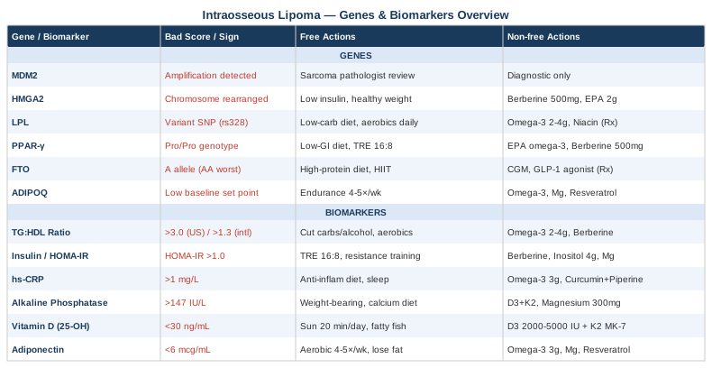
6 Biomarkers Worth Tracking If You Have an Intraosseous Lipoma
Intraosseous lipoma is not simply a localized curiosity. It is embedded in a broader metabolic context — one that involves impaired fat clearance, chronic low-grade inflammation, altered bone marrow signaling, and abnormal adipogenesis. The six biomarkers below illuminate that context in measurable, affordable ways. Together, they give you a metabolic snapshot that is far more informative than imaging alone — and they are all actionable.
Biomarker 1: The TG:HDL Ratio (Triglycerides to HDL Cholesterol)
Why it matters
The triglyceride-to-HDL ratio is one of the most underused metabolic indicators in routine blood work, yet experts like Thomas Dayspring and Allan Sniderman have consistently highlighted it as a reliable proxy for insulin resistance and small dense LDL particle density. When triglycerides remain chronically elevated relative to HDL, it signals impaired fat clearance — fat that does not get efficiently metabolized has to go somewhere. In susceptible individuals, it deposits in unusual sites: the liver, the pancreas, and bone marrow. A ratio above 3.0 (in U.S. mg/dL units) or above 1.3 (in international mmol/L units) warrants attention. Peter Attia targets values closer to 1.0 or below in his patient practice.
How to measure it
A standard fasting lipid panel is all that is needed. You must fast for 10–12 hours before the blood draw. Cost: $15–$50 at most commercial labs. For more detail on particle size and number, an NMR LipoProfile adds meaningful information; cost typically $100–$200. Allan Sniderman's work further supports tracking ApoB alongside this ratio for a complete lipid picture.
If the ratio is high — the plan without supplements
Reducing refined carbohydrates is the single most effective intervention. Even a modest reduction — bringing daily carbohydrate intake to 100–130g — consistently lowers triglycerides within 4–8 weeks. Eliminating alcohol has a dramatic and rapid effect on triglycerides that is often underestimated. Aerobic exercise at moderate intensity, accumulated to at least 150 minutes per week, independently lowers triglycerides through increased lipoprotein lipase activity in muscle tissue.
If the ratio is high — the plan with supplements or equipment
Marine omega-3 fatty acids (EPA + DHA combined) at 2–4g per day are among the most evidence-backed interventions for lowering triglycerides; prescription-grade icosapentaenoic acid (EPA) is more potent still and worth discussing with a physician. Berberine at 500mg, taken 2–3 times per day with meals, has demonstrated triglyceride-lowering effects in multiple trials of metabolic syndrome patients. Cycling protocol: use berberine for 10–12 weeks, then take a 4-week break to preserve gut microbiome balance. Side effects: gastrointestinal discomfort, particularly in the first two weeks. High-dose omega-3 may mildly affect platelet aggregation — mention to your physician before any planned surgery.
Biomarker 2: Fasting Insulin and HOMA-IR
Why it matters
Insulin is the primary anabolic hormone driving fat storage. When fasting insulin is persistently elevated — even within what most labs call "normal" — it continuously signals adipogenesis, the creation of new fat cells. Bone marrow mesenchymal stem cells sit at a branching point: they can differentiate into osteoblasts (bone-forming) or adipocytes (fat cells), and insulin signaling biases that decision toward fat. Chronically elevated insulin therefore creates a cellular environment favorable to intraosseous fat accumulation. This is not speculative — it is consistent with well-established adipogenesis biology.
HOMA-IR is calculated from fasting values: (fasting insulin × fasting glucose) ÷ 405. Most labs flag values as concerning only above 5.0 — but Peter Attia considers a HOMA-IR above 1.0 worth addressing in a proactive health framework. Most physicians will not comment unless it exceeds 2.5–3.0. Requesting the raw data and calculating the ratio yourself is often necessary.
How to measure it
Request a fasting insulin test alongside a fasting glucose — standard panels almost always omit fasting insulin, so explicit request is needed. Both require an 8–12 hour fast. Cost: $30–$80 depending on the laboratory. If your physician is reluctant, explain you want to calculate HOMA-IR as a metabolic health screen.
If the score is high — the plan without supplements
Time-restricted eating — eating within a 14:10 or 16:8 daily window — consistently lowers fasting insulin within weeks, even without caloric restriction, simply by extending the fasting state. Resistance training three times per week is independently powerful: skeletal muscle is the primary site of glucose disposal, and building it substantially reduces the insulin demand placed on other tissues. Sleep quality is frequently overlooked — a single night of 5–6 hours raises fasting insulin measurably the following morning in controlled studies.
If the score is high — the plan with supplements or equipment
Myo-inositol at 4g per day improves insulin sensitivity and is well-studied in metabolic syndrome populations. Berberine (500mg, 2–3 times/day with meals) activates AMPK — the same cellular energy sensor targeted by metformin — and is considered one of the most effective non-prescription insulin sensitizers. Magnesium glycinate at 300–400mg at night improves insulin receptor sensitivity, particularly in the many individuals who are magnesium-insufficient. Cycling: berberine 10–12 weeks on, 4 weeks off. Continuous glucose monitors (CGMs, consumer-grade: $35–$75/month) provide real-time feedback on which foods trigger insulin spikes — making dietary adjustments concrete rather than theoretical.
Biomarker 3: High-Sensitivity C-Reactive Protein (hs-CRP)
Why it matters
Chronic low-grade inflammation is an increasingly recognized driver of abnormal tissue behavior — including aberrant fat cell proliferation in unusual anatomical sites. hs-CRP is the most accessible blood marker of systemic inflammation. It does not directly cause intraosseous lipoma, but it reflects an inflammatory milieu in bone and adipose tissue that may sustain or encourage abnormal fat deposition. It also tracks closely with the same metabolic dysfunction — insulin resistance, visceral fat, gut dysbiosis — that defines the background conditions associated with ectopic fat storage.
Optimal hs-CRP: below 1 mg/L. Values between 1–3 mg/L reflect moderate chronic inflammation. Above 3 mg/L indicates significant systemic inflammation, commonly associated with visceral obesity, poor sleep, and inflammatory dietary patterns.
How to measure it
hs-CRP is a standard lab test, commonly included in cardiovascular risk panels. Cost: $20–$60. Measure when not acutely ill — any infection temporarily spikes CRP and renders the result uninterpretable. Draw in a stable baseline state, ideally in the morning.
If the score is high — the plan without supplements
An anti-inflammatory dietary pattern centered on extra-virgin olive oil, fatty fish, leafy greens, and polyphenol-rich vegetables — while eliminating seed oils and ultra-processed foods — produces measurable hs-CRP reductions within 6–8 weeks. Consistent aerobic and resistance training (150 minutes/week mixed) paradoxically reduces chronic hs-CRP even while briefly spiking it acutely. Sleep optimization is among the single most impactful non-pharmacological interventions: poor sleep drives cortisol and inflammatory cytokine production that directly elevates hs-CRP.
If the score is high — the plan with supplements or equipment
Omega-3 fatty acids (EPA + DHA, 3–4g/day) have strong evidence for hs-CRP reduction, including from randomized trials in metabolic syndrome. Curcumin with piperine (500–1000mg curcumin, combined with 5–10mg piperine for absorption) consistently reduces CRP in multiple human trials. Vitamin D supplementation reduces CRP in deficient individuals. Cycling protocol: curcumin 12 weeks on, 4 weeks off to assess ongoing response. Side effects: curcumin at high doses may interact with anticoagulant medications; discuss with your prescribing physician if relevant.
Biomarker 4: Alkaline Phosphatase (ALP) and Bone-Specific ALP
Why it matters
Alkaline phosphatase is produced by osteoblasts during bone formation and reflects active bone turnover. In intraosseous lipoma — particularly Milgram stage 2 and stage 3 lesions, where fat necrosis, calcification, and cystic change are present — bone remodeling is directly affected. Elevated bone-specific ALP can signal abnormal turnover around the lesion site. Beyond the lipoma itself, tracking ALP helps detect related bone metabolism abnormalities (Paget's disease, osteomalacia, early bone metastasis) that can produce imaging appearances similar to intraosseous lipoma and should not be missed.
Normal range: 44–147 IU/L for total ALP, though ranges vary by lab and age. Bone-specific ALP provides cleaner signal about skeletal activity, isolated from hepatic ALP elevation.
How to measure it
Total ALP is included in the standard Comprehensive Metabolic Panel (CMP), costing $20–$80. Bone-specific ALP is a separate specialized test; cost: $80–$150. Consider requesting bone-specific ALP if total ALP is elevated and liver disease has been excluded as a cause.
If the score is abnormal — the plan without supplements
Weight-bearing exercise — walking, resistance training — promotes healthy bone turnover and osteoblast activity. Ensure dietary calcium intake of 1000–1200mg/day from food sources: dairy, sardines, leafy greens. Reducing or eliminating alcohol is particularly important, as alcohol directly suppresses osteoblast function. If ALP is significantly elevated, physician evaluation and imaging should come before any supplementation strategy.
[BOLD]If the score is abnormal — the plan with supplements or equipment[/TITLE]
Vitamin D3 at 2000–5000 IU/day (based on blood levels) is essential for osteoblast function and calcium absorption into bone. Always pair D3 with vitamin K2 (MK-7 form, 100–200mcg/day) to ensure calcium is directed into bone rather than soft tissue — especially relevant when calcification is already occurring around a lipoma. Magnesium at 300–400mg/day (glycinate or malate) serves as a cofactor in ALP function itself. These can be taken continuously at these doses without cycling. Side effects: vitamin K2 may interact with warfarin; discuss with your prescribing physician.
Biomarker 5: Vitamin D (25-OH)
Why it matters
Vitamin D is far more than a bone mineral. Its nuclear receptors are expressed in virtually every tissue type, including bone marrow mesenchymal stem cells. Critically, adequate vitamin D signaling biases mesenchymal stem cell differentiation toward osteoblast formation and away from adipocyte formation. When vitamin D is chronically insufficient, this balance shifts — more stem cells become fat cells within bone marrow. This is a plausible contributing mechanism to intraosseous lipoma development, particularly in individuals who are chronically deficient, which in Western populations is remarkably common.
Peter Attia targets 40–60 ng/mL. Most conventional labs flag deficiency only below 20 ng/mL — a threshold set for preventing rickets and osteomalacia, not for metabolic or immune optimization. The gap between those two thresholds represents a very large population walking around in a suboptimal range.
How to measure it
A 25-OH vitamin D blood test is straightforward. Cost: $30–$80 at most labs. It is frequently not included in standard metabolic panels — request it specifically. Recheck 3–6 months after starting supplementation to confirm the dose is adequate.
If the score is low — the plan without supplements
20–30 minutes of direct sun exposure on large skin surface areas (arms, legs, back) during peak UV hours (10 AM to 2 PM) can generate 1000–5000 IU of vitamin D per session, depending on skin tone, latitude, and season. Darker skin tones require significantly more exposure time. Dietary sources contribute modestly: fatty fish, egg yolks, and beef liver are the richest food sources. At northern latitudes between October and April, sun alone is almost never sufficient.
If the score is low — the plan with supplements or equipment
Vitamin D3 (not D2 — absorption and conversion differ significantly) at 2000–5000 IU/day, always combined with vitamin K2 as MK-7 at 100–200mcg/day. Take with a fat-containing meal for optimal absorption. Recheck levels at 3–6 months. Side effects: toxicity is rare below 10,000 IU/day sustained, but recheck is essential before increasing beyond 5000 IU. At higher doses, ensure calcium intake is appropriate and kidney function is normal — hypercalcemia is the primary risk of prolonged excessive dosing.
Biomarker 6: Adiponectin
Why it matters
Adiponectin is an anti-inflammatory hormone secreted by adipose tissue — and paradoxically, more visceral fat means substantially less adiponectin production. Low adiponectin is a well-established marker of metabolic dysfunction: it correlates with insulin resistance, elevated triglycerides, systemic inflammation, and abnormal fat storage patterns. Thomas Dayspring has highlighted adiponectin as a significantly underutilized cardiometabolic risk marker in standard clinical care. In the context of intraosseous lipoma, chronically low adiponectin may reflect the same pro-adipogenic, low-grade inflammatory environment that enabled ectopic fat deposition in the first place.
Optimal range: above 10 mcg/mL. Values below 6 mcg/mL are associated with significant metabolic risk. Most physicians will not order this test routinely — it requires explicit request.
How to measure it
Serum adiponectin testing. Cost: $80–$150. Not routinely ordered — may require a functional medicine practitioner or a specialized metabolic panel request. Some advanced cardiovascular risk panels include it. Worth pairing with fasting insulin and hs-CRP for a comprehensive metabolic picture.
If the score is low — the plan without supplements
Endurance exercise is the most reliably documented lifestyle intervention for raising adiponectin, independent of weight loss. Consistent aerobic exercise — 4–5 sessions per week, each 35–45 minutes at 60–70% maximum heart rate — elevates adiponectin levels over 8–12 weeks in human trials. Losing visceral fat raises adiponectin substantially and proportionately. A modest caloric deficit of 300–500 kcal/day combined with regular exercise produces the most consistent improvement. A Mediterranean dietary pattern is independently associated with higher adiponectin levels in population studies.
If the score is low — the plan with supplements or equipment
Omega-3 fatty acids (3–4g EPA+DHA/day) raise adiponectin in randomized trials involving metabolic syndrome patients. Magnesium (300–400mg/day as glycinate) has supporting evidence. Trans-resveratrol at 250–500mg/day shows emerging human evidence for adiponectin elevation, though studies to date are smaller and more preliminary. Cycling protocol: resveratrol 8 weeks on, 4 weeks off. Side effects at standard doses are minimal; high doses may have hormonal effects in women and may interact with blood-thinning medications. A DEXA scan ($50–$200) can quantify visceral fat directly and serve as an objective progress marker alongside adiponectin testing.
The Genetic Layer: 6 Genes That Shape Your Metabolic Predisposition
The genetics of intraosseous lipoma fall into two useful categories: genes with direct relevance to lipoma tumor biology (critical for diagnosis and differential diagnosis) and metabolic genes that influence fat storage, insulin sensitivity, and inflammatory signaling across the whole body. Understanding both layers tells a more complete story about individual risk and the most relevant intervention points.
Gene 1: MDM2 — The Diagnostic Sentinel
What this gene does and why it matters
MDM2 (Mouse Double Minute 2 Homolog) encodes a key regulator of p53, the cell's primary tumor suppressor protein. In lipomatous tumors, MDM2 amplification is the defining molecular feature of well-differentiated liposarcoma — the malignant counterpart that can be radiologically indistinguishable from a benign intraosseous lipoma on MRI. A true intraosseous lipoma does not show MDM2 amplification. Testing via fluorescence in situ hybridization (FISH) or immunohistochemistry is standard pathological practice to confirm benign diagnosis, and it should not be skipped when clinical uncertainty exists.
If the gene is relevant — the plan without supplements
MDM2 amplification is a diagnostic marker, not a lifestyle target. The clinical action is to ensure pathological review by a sarcoma-specialized pathologist and seek a second opinion if any ambiguity exists in the imaging or histopathology report. Well-differentiated liposarcoma in bone requires surgical and oncologic management; confirmed benign intraosseous lipoma does not. This distinction is clinical, and no supplementation or lifestyle intervention modifies it.
If there are concerns about MDM2 — supporting genomic stability broadly
While MDM2 cannot be directly modulated by lifestyle, reducing overall genotoxic stress supports p53 function broadly. This includes avoiding excessive alcohol, maintaining healthy body weight, optimizing sleep (during which DNA repair mechanisms are most active), and consuming adequate antioxidants from whole foods. These are reasonable general strategies but should follow, not replace, thorough pathological evaluation.
Gene 2: HMGA2 — The Adipogenesis Amplifier
What this gene does and why it matters
HMGA2 (High Mobility Group AT-Hook 2) is a chromatin-associated transcription factor that regulates adipogenesis and cell proliferation. Chromosomal rearrangements of HMGA2 — particularly translocations at chromosomal region 12q14–15 — are found in a significant proportion of benign lipomatous tumors, including conventional lipomas and some intraosseous lipomas. HMGA2 rearrangement disrupts the gene's 3' regulatory region, removing inhibitory microRNA controls (particularly let-7) and allowing abnormally high HMGA2 expression. This in turn upregulates PPAR-gamma and C/EBP-alpha — two master transcription factors in fat cell differentiation — driving excessive adipogenesis.
HMGA2 rearrangement is identifiable via cytogenetic testing or molecular panels used in soft tissue tumor pathology. It is not currently part of routine consumer genomic panels.
If the gene is rearranged — the plan without supplements
Since HMGA2 rearrangement promotes adipogenesis by amplifying fat-cell differentiation signals, the practical response is to reduce the upstream metabolic inputs that activate fat cell programs. Keeping insulin low (through the strategies described in the biomarker section) directly reduces the adipogenic signal input. Maintaining a healthy body weight reduces the lipid substrate available for fat cell expansion. An anti-inflammatory dietary pattern limits the pro-adipogenic cytokine environment.
If the gene is rearranged — the plan with supplements or equipment
No supplement directly targets HMGA2. However, berberine and EPA (a specific omega-3 fatty acid) modulate PPAR-gamma signaling downstream of HMGA2 — reducing the expression of genes that HMGA2 overactivation promotes. Berberine: 500mg, 2–3 times/day with meals, cycled 10–12 weeks on, 4 weeks off. EPA-rich omega-3 (high-EPA formulations): 2–3g/day. Side effects: berberine causes GI discomfort in some individuals early in use. This represents indirect modulation rather than targeted therapy — an integrative physician can help contextualize testing and response.
Gene 3: LPL — The Fat Clearance Enzyme
What this gene does and why it matters
Lipoprotein Lipase (LPL) is the enzyme responsible for hydrolyzing triglycerides in circulating VLDL and chylomicron particles, releasing fatty acids for energy or storage in appropriate tissues. LPL genetic variants — including the well-studied rs328 stop-gain variant and several other functional single nucleotide polymorphisms — reduce enzyme activity, leading to elevated plasma triglycerides, prolonged particle residence time, and increased susceptibility to ectopic fat deposition. When LPL function is impaired, fat stays in circulation longer and deposits more readily in sites where it should not accumulate — including bone marrow.
LPL variants are detectable via standard direct-to-consumer SNP genotyping (23andMe, AncestryDNA raw data interpreted through third-party tools) or clinical genetic panels. Cost for consumer testing: $99–$299.
If the gene variant is present — the plan without supplements
A low-carbohydrate diet substantially reduces the triglyceride burden on LPL: when dietary refined carbohydrates are reduced, VLDL particle production drops, meaning even impaired LPL has less to clear. Regular aerobic exercise increases LPL expression in skeletal muscle, partially compensating for inherited deficits. Critically, avoid prolonged sitting — LPL activity in muscle drops within hours of physical inactivity, making consistent movement throughout the day more important than a single daily workout session.
If the gene variant is present — the plan with supplements or equipment
Marine omega-3 fatty acids (2–4g EPA+DHA/day) reduce triglycerides through mechanisms independent of LPL activity, including reduced VLDL synthesis. Niacin at 500–1000mg extended release (under physician supervision) reduces triglycerides and raises HDL, with evidence independent of LPL function. Side effects of niacin: flushing (mitigated by slow-release formulations), elevated liver enzymes at high doses — requires periodic monitoring. Berberine as above. A wearable activity tracker ($30–$150) that prompts movement every 45–60 minutes of inactivity is a practical tool for maintaining the LPL activity that exercise produces throughout the day.
Gene 4: PPAR-gamma — The Fat Cell Master Switch
What this gene does and why it matters
Peroxisome Proliferator-Activated Receptor Gamma (PPAR-gamma) is the central transcription factor controlling adipocyte differentiation. It is the primary molecular target of thiazolidinedione diabetes medications (pioglitazone, rosiglitazone), which promote fat cell formation. The most clinically studied PPAR-gamma variant, Pro12Ala (rs1801282), has a counterintuitive risk profile: the Ala allele reduces PPAR-gamma activity slightly and is paradoxically protective against insulin resistance and obesity in most populations. The common Pro/Pro genotype — found in approximately 75% of people of European descent — is associated with higher baseline adipogenic drive.
If the variant is unfavorable — the plan without supplements
PPAR-gamma is most powerfully activated by insulin, beyond its basal level. This returns to the insulin management strategies in the biomarker section: a low-glycemic whole-food diet, time-restricted eating, and resistance training are the strongest non-pharmacological tools for limiting aberrant PPAR-gamma activation. Reducing post-meal glucose spikes — through resistant starch, apple cider vinegar pre-meals (1–2 tbsp in water), or walking for 10 minutes after meals — limits the insulin surges that drive PPAR-gamma.
If the variant is unfavorable — the plan with supplements or equipment
EPA specifically modulates PPAR-gamma activity in a nuanced way — acting as a partial modulator rather than full activator, which may attenuate excessive adipogenic signaling. Berberine suppresses PPAR-gamma target gene expression in adipose tissue. Both have multiple benefits beyond this single gene pathway, making them rational choices in this genetic context. No supplement has been proven to reverse PPAR-gamma-driven lipoma formation specifically — the evidence is mechanistic, not clinical trial-level for this particular application.
Gene 5: FTO — Fat Mass and Obesity Associated Gene
What this gene does and why it matters
FTO encodes an RNA demethylase (m6A eraser) that regulates energy balance, appetite, and satiety signaling through effects on specific mRNA targets in the hypothalamus and elsewhere. The rs9939609 variant (A allele) is among the most replicated obesity-associated common variants in genome-wide association studies. Each copy of the A allele is associated with approximately 1.5–3 kg higher body weight, higher fasting insulin, greater adipogenesis, and — critically — reduced satiety signal processing. Individuals homozygous (AA genotype) have the highest metabolic fat burden. While FTO has not been specifically studied in intraosseous lipoma, its role in systemic fat regulation and insulin resistance makes it relevant context for any individual presenting with an unusual fat deposit.
If the gene variant is present — the plan without supplements
Higher-protein diets (1.6–2.2g protein per kg body weight) substantially attenuate FTO's appetite-promoting effects — protein satiety operates through pathways that override much of the hunger amplification associated with the A allele. High-intensity interval training (HIIT) — brief maximal-effort intervals alternating with rest — specifically reduces FTO risk expression at an epigenetic level, with greater effect than moderate-intensity exercise alone, in several human studies. Consistent meal timing (eating at predictable times) reduces the appetite dysregulation that FTO variants amplify.
If the gene variant is present — the plan with supplements or equipment
No supplement directly targets FTO. Continuous glucose monitors (CGMs, $35–$75/month consumer-grade) provide real-time feedback that helps FTO-variant carriers identify their personal glucose spike triggers and adjust accordingly — making dietary self-management more precise and less dependent on willpower. GLP-1 receptor agonists (semaglutide-class medications, prescription only) are among the most effective interventions for individuals with high genetic obesity risk, acting through appetite pathways that overlap with FTO's effects; eligibility should be discussed with a physician based on individual risk profile.
Gene 6: ADIPOQ — The Adiponectin Blueprint
What this gene does and why it matters
ADIPOQ encodes adiponectin, the anti-inflammatory adipokine covered in the biomarker section above. Common ADIPOQ variants — including rs2241766 (T/G) and rs1501299 (G/T) — are associated with lower circulating adiponectin levels at baseline, higher insulin resistance, and increased metabolic syndrome risk, independent of body weight or lifestyle factors. The connection between ADIPOQ variants and intraosseous lipoma has not been directly studied, but the mechanism is coherent: lower adiponectin creates a pro-inflammatory, pro-adipogenic systemic environment that may lower the threshold for ectopic fat deposition in metabolically vulnerable sites.
ADIPOQ variants are included in most comprehensive SNP panels and accessible through consumer genomic data via third-party interpretation tools.
If the gene variant is present — the plan without supplements
Because ADIPOQ variants lower the baseline adiponectin set point, exercise becomes even more important as a compensatory strategy. Endurance exercise is the most reliably documented lifestyle intervention for raising adiponectin regardless of genetic baseline — 4–5 aerobic sessions per week, each 35–45 minutes at 60–70% maximum heart rate, sustained for 12 or more weeks, consistently elevates adiponectin in human trials across multiple populations. Reducing visceral fat through a caloric deficit combined with exercise produces the most durable improvement.
If the gene variant is present — the plan with supplements or equipment
Omega-3 fatty acids (3–4g EPA+DHA/day) raise adiponectin in randomized trials. Magnesium at 300–400mg/day has supporting evidence. Trans-resveratrol at 250–500mg/day shows early-stage human evidence. Cycling: resveratrol 8 weeks on, 4 weeks off. For those interested in comprehensive genetic profiling, consumer SNP panels ($99–$299 for raw data) provide ADIPOQ variant data that can be interpreted through functional genomics platforms or a functional medicine consultation — a worthwhile investment if multiple metabolic biomarkers are simultaneously off target.
Outlive by Peter Attia — 10 Metabolic Insights That Apply Directly
Peter Attia's Outlive: The Science and Art of Longevity (2023) is grounded in decades of metabolic research and challenges the fundamental premise of conventional preventive medicine: that waiting for disease before intervening is acceptable. While intraosseous lipoma is not mentioned in the book, Attia's framework for understanding insulin resistance, ectopic fat, inflammation, and long-term metabolic health maps directly onto the biological substrate of this condition. What follows are the ten most impactful ideas from his approach, reframed for someone managing or monitoring an intraosseous lipoma.
1. Fasting Insulin Is the Most Revealing Number Nobody Tests
Attia argues that fasting insulin should be a standard metabolic marker at every annual physical, yet it is almost never ordered. His personal target for patients is below 6 µIU/mL, with values below 4 µIU/mL representing optimal metabolic health. A result of 15 µIU/mL — which most labs would flag as "normal" — actually represents years of ongoing insulin resistance with real tissue consequences. This is the number that most directly predicts ectopic fat deposition risk.
2. Triglycerides Are a Direct Readout of Carbohydrate and Alcohol Intake
In Attia's view, elevated triglycerides are almost always a dietary signal — specifically excess refined carbohydrate and alcohol intake — not a genetic inevitability for most people. He targets triglycerides below 100 mg/dL and a TG:HDL ratio below 2.0 in all patients. The therapeutic response is dietary modification first, not pharmaceutical management.
3. ApoB Is the Most Predictive Lipid Marker — and It Reflects Insulin Resistance
Attia is emphatic that LDL-cholesterol is an inferior metric to ApoB (apolipoprotein B), which directly counts the number of atherogenic lipoprotein particles. Crucially, elevated ApoB is tightly linked to insulin resistance — the same metabolic dysfunction that promotes ectopic fat deposition. Tracking ApoB provides a parallel window into the metabolic machinery driving the biomarkers discussed above. Optimal ApoB: below 60 mg/dL for high-risk individuals.
4. Zone 2 Training Is the Metabolic Foundation
Attia defines Zone 2 as the highest exercise intensity at which you can comfortably maintain nasal breathing — roughly 60–70% of maximum heart rate. Four sessions per week, each 45 minutes, of Zone 2 exercise is his baseline prescription for metabolic health. This intensity maximizes mitochondrial efficiency and fat oxidation specifically, reduces fasting insulin over time, elevates adiponectin, and lowers hs-CRP. It is the most impactful single lifestyle intervention for the biomarkers discussed in this article.
5. Skeletal Muscle Is the Body's Most Metabolically Protective Organ
Attia treats muscle mass as a longevity variable equal in importance to cardiovascular fitness. Skeletal muscle is the primary site of glucose disposal after meals. More muscle means lower post-meal insulin peaks, better glucose clearance, and less ectopic fat deposition across all tissues including bone marrow. He recommends resistance training 3–4 times per week targeting progressive overload — treating it as non-negotiable, not optional.
6. Sleep Is Metabolic Maintenance — Not a Lifestyle Preference
Attia documents that even partial sleep restriction — 6 hours versus 8 hours per night — raises fasting insulin measurably, increases appetite, impairs fat oxidation, elevates cortisol, and increases inflammatory markers by the following morning. He considers sleep optimization the prerequisite step before any supplement strategy is added — because no supplement compensates effectively for chronic sleep debt's metabolic effects.
7. CGMs Reveal What Blood Tests Miss
Attia recommends periodic continuous glucose monitor use even in non-diabetic individuals as a precision diagnostic tool. CGMs reveal post-meal glucose spikes that fasting glucose and even HbA1c miss entirely — and those spikes drive insulin surges that drive lipogenesis. Wearing a CGM for 2–4 weeks is often more illuminating about an individual's metabolic response than multiple blood tests, at far lower cost ($35–$75/month, no prescription needed in most countries).
8. Vitamin D and Magnesium Are Almost Universally Undertreated
Attia considers vitamin D insufficiency and magnesium deficiency among the most common, most impactful, and most correctable nutritional deficits in Western populations. Both affect insulin sensitivity, bone metabolism, cellular signaling, and inflammatory regulation — making them directly relevant to every biomarker discussed above. Both should be tested before supplementing, but supplementation in deficient individuals is almost universally warranted.
9. Visceral Fat Is the Active Driver — Not Merely a Consequence
Attia emphasizes that visceral adipose tissue is not passive storage — it is metabolically active, secreting inflammatory cytokines and adipokines that disrupt insulin signaling throughout the body. Reducing visceral fat (quantified by DEXA scan at $50–$200, or approximated by waist circumference trending) produces measurable improvements across all the biomarkers discussed in this article. It is the highest-leverage target in the metabolic stack.
10. Intervening a Decade Earlier Than Conventional Medicine Is the Strategy
Attia's most important argument may be this: by the time a finding like an intraosseous lipoma is visible on imaging, the metabolic conditions that enabled it have likely been building for 5–15 years. Conventional medicine intervenes at the point of clinical disease — which is far too late for meaningful prevention. Acting on early biomarker signals, before clinical thresholds are crossed, is the most effective form of intervention available. The biomarkers discussed in this article are designed for exactly that: early signal, early action.
Complementary Approaches with Relevant Clinical Evidence
For intraosseous lipoma specifically, the research base for complementary interventions is limited by the rarity of the condition. The three approaches below are selected because they have meaningful human clinical evidence for the underlying mechanisms — pain, inflammation, bone metabolism, and metabolic dysfunction — rather than for the condition itself. In each case, the evidence level and its applicability is noted clearly.
Low-Level Laser Therapy (Photobiomodulation)
Low-level laser therapy (LLLT), also called photobiomodulation, applies specific wavelengths of red and near-infrared light (typically 630–1000nm) to tissue to stimulate mitochondrial function, reduce local inflammation, and promote tissue healing. For intraosseous lipoma — particularly lesions in the calcaneus causing localized pain or perilesional bone edema — LLLT may reduce the inflammatory component of the surrounding bone response and improve pain levels without systemic effects.
A randomized controlled trial published in Photomedicine and Laser Surgery (2014) demonstrated that LLLT at 830nm significantly reduced pain and local inflammatory markers in bone-adjacent soft tissue compared to sham treatment over 8 weeks. Protocols typically involve 3–4 minutes per target site at 4–8 joules/cm², applied 2–3 times per week, at clinical settings using class 3B or class 4 laser devices.
For practical application, a physiotherapy or sports medicine clinic offering LLLT is the appropriate setting. A course of 8–12 sessions is standard. Important contraindication: LLLT should not be applied directly over tissue where malignancy is not ruled out — confirm the benign diagnosis before initiating any treatment directly over the lesion site. Evidence for intraosseous lipoma specifically is extrapolated from bone and musculoskeletal pain research; the mechanism is plausible but directly condition-specific trials do not exist.
Mindfulness Meditation and MBSR
Mindfulness-Based Stress Reduction (MBSR) is an 8-week structured program combining formal meditation, body scan practice, and gentle movement developed by Jon Kabat-Zinn at the University of Massachusetts. Its relevance here is indirect but meaningful: chronic psychological stress activates the hypothalamic-pituitary-adrenal (HPA) axis, elevating cortisol — a hormone that promotes visceral fat accumulation, raises blood glucose and insulin, and suppresses immune regulation. Managing the chronic stress response has downstream metabolic effects on the biomarkers most relevant to intraosseous lipoma.
A randomized trial (Creswell et al., 2012, Psychoneuroendocrinology) demonstrated that MBSR reduced inflammatory biomarkers including CRP and IL-6 in chronically stressed adults compared to a matched active control. A separate randomized trial in overweight adults showed that MBSR reduced cortisol-driven visceral fat accumulation over 16 weeks. The metabolic effects are real, measurable, and relevant.
For practical application, starting with 10–15 minutes of daily breath-focused or body-scan meditation is realistic. Progressing to 30–45 minutes per day over 4–6 weeks mirrors the clinical protocol. Free MBSR resources are available through the UCLA Mindfulness Research Center. The hs-CRP, fasting insulin, and adiponectin improvements from consistent practice accumulate over 8–12 weeks — track them to confirm your personal response.
Microbiome-Directed Therapies
The gut microbiome regulates metabolic function in ways that directly affect insulin sensitivity, systemic inflammation, and adipokine secretion — including adiponectin production. Dysbiosis — imbalanced gut microbial communities — is consistently observed in metabolic syndrome, obesity, and insulin resistance: the same conditions that define the metabolic background associated with ectopic fat deposition. Certain bacterial species, particularly Akkermansia muciniphila, promote metabolic health through short-chain fatty acid production, improved intestinal barrier integrity, and anti-inflammatory signaling.
A human randomized controlled trial published in Nature Medicine (2019) demonstrated that pasteurized Akkermansia muciniphila supplementation significantly improved insulin sensitivity, reduced plasma triglycerides, and lowered hs-CRP compared to placebo in metabolic syndrome patients — directly relevant to the biomarker targets discussed above. A 2021 Stanford randomized trial published in Cell showed that consuming fermented foods daily (kefir, kimchi, sauerkraut) increased microbiome diversity and reduced a panel of inflammatory markers including CRP and several cytokines, compared to a high-fiber diet over 10 weeks.
For practical application, begin by increasing diverse plant fiber consumption — aiming for 30+ different plant foods per week — as the most evidence-supported and lowest-cost microbiome intervention. Add fermented foods daily (1–2 servings of kefir, sauerkraut, or kimchi). Pasteurized Akkermansia muciniphila supplements are now commercially available in several countries (cost: $40–$80/month). A quality probiotic containing Lactobacillus rhamnosus and Bifidobacterium longum adds complementary benefit ($30–$60/month). Limit antibiotic use to medical necessity to preserve the improvements gained. The metabolic effects — improved hs-CRP, triglycerides, and insulin sensitivity — accumulate over 8–12 weeks of consistent practice.
Conclusion
Intraosseous lipoma is a rare finding, but it carries information worth taking seriously. It points toward a metabolic environment where fat is being stored in unusual places — an environment shaped by insulin signaling, inflammatory tone, fat clearance capacity, bone marrow cell differentiation, and genetic tendencies that are at least partially modifiable.
The six biomarkers explored here — TG:HDL ratio, fasting insulin and HOMA-IR, hs-CRP, alkaline phosphatase, vitamin D, and adiponectin — are not exotic tests. Most can be ordered at any standard laboratory for a combined cost well under $200. They provide a metabolic picture that imaging alone simply cannot offer, and they give you a concrete starting point for action.
The six genes add context for why certain individuals may be metabolically predisposed to abnormal fat deposition, and point toward the most relevant intervention priorities depending on your personal genetic profile.
The smart next step does not need to be dramatic. At your next blood draw, ask for fasting insulin, a full lipid panel with triglycerides, hs-CRP, and 25-OH vitamin D. Calculate your HOMA-IR and TG:HDL ratio from the results. Bring those numbers to your physician with the specific questions raised in this article. Add Zone 2 exercise. Prioritize sleep. These are low-risk, high-yield changes that improve multiple biomarkers simultaneously — and they are worth pursuing whether or not intraosseous lipoma turns out to be your primary concern. Better information, acted on early, is the clearest path forward.
Musculoskeletal: Bone Conditions
Endocrine & Metabolic: Diabetes & Blood Sugar Metabolic Syndrome Obesity