This article contains AI generated content.
· UpdatedJuvenile Dermatomyositis — 5 Genes And 6 Biomarkers To Track
Introduction
Juvenile dermatomyositis is one of the rarest and most biologically complex autoimmune conditions affecting children. If your child has been diagnosed — or if you spent months trying to get a diagnosis confirmed — you already know how disorienting this experience can be. The rash, the muscle weakness, the fatigue: these are real, measurable phenomena with a biological logic underneath. Yet most of the information available describes JDM in the broadest possible terms, rarely addressing what is actually driving the disease at the level of genes, immune signaling, and blood chemistry.
Most health resources approach JDM through symptoms and standard treatment protocols. Far fewer explain which immune pathways are chronically overactivated, which genetic predispositions helped set the stage, or which measurable markers can be used to track disease activity between clinic visits. That is where the most actionable information lives — and it is exactly where this article focuses.
The approach here is precise and practical. No cures are promised. What is offered instead is a clear map of the biological signals that matter most in JDM: the six biomarkers with the most clinical relevance, the five genes most consistently associated with JDM susceptibility, and realistic strategies for addressing problematic results with or without supplements and equipment. It draws on current research in pediatric rheumatology, genomics, immunology, and integrative medicine.
Two core strategies are developed in depth below. The first — and the most immediately actionable — focuses on six key biomarkers that track JDM activity from multiple angles, explaining how each is measured, what a poor result actually signals biologically, and what can be done about it. The second examines five genetic variants associated with JDM susceptibility and what the research suggests about compensating for each one. Together, these strategies offer a more complete foundation for medical conversations and day-to-day decision-making than symptoms alone ever could.
6 Biomarkers That Reveal What Is Actually Happening in Juvenile Dermatomyositis
Tracking biomarkers in JDM is not simply about confirming a diagnosis. It is about understanding disease activity in real time, detecting flares before they escalate, assessing whether treatment is genuinely working, and catching serious complications early. The six markers below represent the most clinically meaningful targets currently available, each offering a distinct and complementary window into the disease.
Biomarker 1: Myositis-Specific Autoantibodies
Why it matters: Myositis-specific autoantibodies (MSAs) are proteins produced by the immune system that mistakenly target the body's own cellular machinery. In JDM, the three most clinically significant are anti-MDA5, anti-TIF1γ, and anti-NXP2. These are not purely diagnostic — they are predictive. Anti-MDA5 positivity is strongly associated with rapidly progressive interstitial lung disease and carries the most urgent prognostic weight. Anti-TIF1γ correlates with more severe and persistent skin involvement. Anti-NXP2 is linked to calcinosis and severe muscle disease. Knowing which antibody is present reshapes the entire monitoring and treatment strategy — the clinical management of an anti-MDA5-positive child looks very different from that of an anti-NXP2-positive child.
How to measure it: MSA panels are performed through specialized immunofluorescence or line-blot assays. They are not included in standard autoimmune screens and require referral to a rheumatology center or a specialized reference laboratory. Costs typically range from $200 to $600 depending on panel breadth and insurance coverage. Testing should be completed at diagnosis. Repeat testing is generally not necessary unless the clinical picture changes significantly or a new complication emerges.
If the score is bad — the plan without supplements: Anti-MDA5 positivity requires proactive respiratory monitoring — pulmonary function tests and high-resolution CT of the chest should be scheduled at defined intervals, not triggered only by symptoms. For anti-TIF1γ positivity, rigorous and consistent sun protection is particularly critical, since UV exposure perpetuates skin inflammation through keratinocyte stress and innate immune activation. For all MSA-positive children, a strict anti-inflammatory lifestyle — eliminating ultra-processed foods, optimizing sleep quality, and reducing environmental immune triggers — provides a meaningful foundation. Frequency: pulmonary function testing every six months for anti-MDA5-positive children; skin assessment every three months for all MSA-positive children.
If the score is bad — the plan with supplements or equipment: No supplement directly clears autoantibodies. However, omega-3 fatty acids (EPA and DHA combined at 2–4 g per day from a high-quality fish oil) have demonstrated anti-inflammatory effects in human trials and may reduce downstream immune activation by modulating eicosanoid signaling. Vitamin D3 supplementation, titrated to maintain serum 25(OH)D between 50 and 80 ng/mL, is associated with improved immune regulation across multiple autoimmune conditions and has specific mechanistic relevance to JDM through vitamin D receptor-mediated dendritic cell modulation. Both omega-3s and Vitamin D can be taken continuously without cycling. Side effects of high-dose fish oil include mild GI discomfort and a slight blood-thinning effect — discuss with the treating physician. Phototherapy devices should be avoided in JDM, as UV exposure can worsen skin disease.
Biomarker 2: Creatine Kinase
Why it matters: Creatine kinase (CK) is an enzyme released when muscle cells are damaged. Elevated CK is a direct indicator of active myositis. In JDM, levels can range from mildly elevated to dramatically high, and they correlate reasonably well with disease activity and treatment response. However, an important caveat exists: some children with active JDM — particularly those with predominant skin involvement or interstitial lung disease — maintain normal CK throughout their disease course. CK should never be interpreted in isolation. Normalized CK during treatment is a reassuring sign; re-elevation after normalization is an early and useful flare signal.
How to measure it: CK is included in standard metabolic and muscle enzyme panels, available at any laboratory for approximately $20–$60. It should be measured at diagnosis, at every significant treatment change, and at minimum every three months during active disease or during medication tapering. Morning draws are preferred, as physical activity can transiently raise CK and confound interpretation.
If the score is bad — the plan without supplements: Significantly elevated CK in confirmed active myositis requires medical management — typically corticosteroids and steroid-sparing agents. From a lifestyle standpoint, strict rest during acute inflammatory phases reduces further mechanical muscle damage. Once disease is brought under control, graded physical therapy — not unstructured exercise — is the appropriate path to muscle recovery. High-intensity unsupervised activity during active disease can worsen CK elevation and delay recovery. Frequency: CK should begin normalizing within weeks of effective treatment. Persistent elevation despite treatment is a clear signal to reassess the therapeutic strategy with the treating rheumatologist.
If the score is bad — the plan with supplements or equipment: Coenzyme Q10 (100–300 mg per day) supports mitochondrial function in muscle tissue and has shown benefit in some inflammatory myopathy contexts, though evidence specific to JDM remains limited. Magnesium glycinate (150–300 mg per day) supports muscle function and reduces cramping. Creatine monohydrate (3–5 g per day) has demonstrated muscle-protective properties in adult inflammatory myopathies — pediatric-specific evidence is emerging. Hydrotherapy equipment (pool access for water-based physical therapy) provides the lowest-impact path to muscle rehabilitation. No cycling is needed for magnesium or CoQ10; creatine can be taken continuously. Monitor CK response at six-week intervals following any intervention change.
Biomarker 3: Ferritin
Why it matters: Ferritin is both an iron-storage protein and a powerful acute-phase reactant — it rises significantly in the presence of systemic inflammation. In JDM, markedly elevated ferritin (particularly above 500 ng/mL) signals more severe, systemic disease. At extreme elevations above 10,000 ng/mL, ferritin becomes a critical danger signal for macrophage activation syndrome (MAS), a potentially life-threatening complication of JDM that requires urgent intervention. This dual role makes ferritin both a disease-activity marker and a safety marker that must be followed consistently throughout the JDM disease course.
How to measure it: Ferritin is a standard blood test available at any laboratory for approximately $20–$50. During active JDM, monthly monitoring is appropriate. In stable, well-controlled disease, quarterly checks are sufficient. Any sharp, unexplained rise in ferritin — regardless of other markers — should trigger urgent evaluation for MAS, as this complication can evolve rapidly.
If the score is bad — the plan without supplements: Persistently elevated ferritin signals uncontrolled systemic inflammation. Reducing dietary sources of inflammatory load — refined sugars, trans fats, ultra-processed foods — has documented effects on ferritin in systemic inflammatory conditions. Sleep architecture is particularly relevant: IL-1β and IL-6 (the cytokines that most directly drive ferritin production) are substantially regulated by sleep quality and circadian timing. A consistent sleep schedule, dark sleep environment, and low evening light exposure directly modulate these cytokine pathways. Anti-inflammatory dietary patterns such as the Mediterranean diet or the Autoimmune Protocol have shown ferritin-lowering effects in inflammatory disease populations.
If the score is bad — the plan with supplements or equipment: Curcumin in a bioavailable form (BCM-95 or liposomal, 500–1000 mg daily) has been shown across multiple human trials to reduce IL-6 — the primary cytokine driver of ferritin elevation. N-Acetylcysteine (NAC, 600 mg twice daily with food) supports glutathione production and has broad antioxidant effects that reduce inflammatory amplification. Neither requires cycling, but both should be used with physician awareness in a child with active autoimmune disease. Infrared sauna, used cautiously and only in remission with physician clearance, may support systemic inflammatory load reduction over time — JDM-specific evidence is lacking, and this should not be used during active skin disease.
Biomarker 4: Type I Interferon Score
Why it matters: Type I interferons — specifically IFN-α and IFN-β — are antiviral signaling molecules that, in JDM, are chronically and inappropriately overactivated. The interferon signature, measured as the expression level of interferon-stimulated genes in whole blood, is one of the most consistently and significantly elevated findings across JDM patient populations. It correlates strongly with disease activity and often remains elevated even when surface-level markers such as CK appear controlled. Landmark research by Virginia Pascual and colleagues at the Texas Scottish Rite Hospital for Children established the type I IFN pathway as a central and upstream driver of JDM pathology — not merely a bystander. Tracking the IFN score provides a window that other biomarkers simply cannot.
How to measure it: The type I IFN score is measured via a gene expression assay from whole blood (examining the upregulation of interferon-stimulated genes such as IFI27, IFI44L, IFIT1, RSAD2, and SIGLEC1). It is not yet part of routine clinical practice but is available at academic medical centers and through specialized research laboratories. Cost ranges from $150 to $500. Some pediatric rheumatology research centers include IFN scoring in observational study protocols at no cost to patients — ask the treating rheumatologist about availability and whether enrollment in a monitoring protocol is possible.
If the score is bad — the plan without supplements: The IFN pathway is activated in part by nucleic acid sensing — the innate immune system detecting double-stranded RNA or DNA fragments released during cellular stress or viral infection. Consistent, broad-spectrum sun protection (SPF 50+ with UVA blocking, applied daily and reapplied every two hours during outdoor activity) is one of the most evidence-supported free interventions in JDM. UV radiation directly activates nucleic acid sensing in keratinocytes, making photosensitivity not merely a cosmetic concern but a mechanistic one. Viral infection prevention through hand hygiene and avoiding sick contacts during active disease phases reduces the most potent external IFN trigger. Keeping vaccinations current (where medically appropriate and discussed with the treating rheumatologist) is also relevant.
If the score is bad — the plan with supplements or equipment: Vitamin D3 at doses targeting serum levels of 60–80 ng/mL has documented IFN-modulating effects through downregulation of TLR7 and TLR9 signaling — the innate sensing pathways that drive IFN production. Hydroxychloroquine (a prescription antimalarial agent) specifically suppresses endosomal TLR-mediated IFN production and is a first-line immunomodulatory agent in JDM — its mechanism of action directly targets IFN pathway overactivation. Low-dose naltrexone (LDN) has emerging evidence as an IFN and immune modulator in autoimmune conditions; it is not yet standard in pediatric JDM but may be worth discussing at specialized academic centers. Frequency: Vitamin D daily, with blood level monitoring every six months.
Biomarker 5: Aldolase
Why it matters: Aldolase is a glycolytic enzyme found in muscle tissue that, like CK, is released into circulation when muscle cells are damaged. Its specific clinical value in JDM lies in a critical gap: some children with active, ongoing muscle inflammation maintain entirely normal CK throughout their disease course. In these cases, aldolase may be the only blood-based muscle damage marker that is elevated, making it an essential complement to CK rather than a redundant test. It is particularly informative when CK normalizes during treatment but clinical symptoms persist — a discordance that can signal ongoing subclinical myositis.
How to measure it: Aldolase is measured through a standard blood test available at most reference laboratories for $30–$80. Pediatric-specific reference ranges must be applied, as normal values in children differ meaningfully from adult reference ranges. It should be measured alongside CK at diagnosis and monitored every three months during active disease. In remission, biannual monitoring is appropriate.
If the score is bad — the plan without supplements: The same principles that apply to elevated CK are relevant here: strict rest during active inflammatory phases, followed by carefully graded rehabilitation through supervised physical therapy as disease comes under control. Water-based physical therapy (hydrotherapy) is particularly well-suited to JDM rehabilitation — it allows meaningful muscle engagement while dramatically reducing joint and muscle stress compared to land-based exercise.
If the score is bad — the plan with supplements or equipment: B vitamins — particularly riboflavin (B2) and niacin (B3) — support mitochondrial energy metabolism in muscle cells. A high-quality B-complex supplement taken daily is low-risk and broadly supportive of oxidative energy production in recovering muscle tissue. L-carnitine (500–1000 mg per day) supports fatty acid transport into mitochondria and has been studied in some myopathy contexts with generally favorable results. No cycling is required for either. Avoid high-dose niacin supplementation without physician monitoring, as it can cause flushing and, at very high doses, hepatic effects.
Biomarker 6: Interleukin-6
Why it matters: Interleukin-6 (IL-6) is a pro-inflammatory cytokine that is produced in excess in JDM and drives much of the systemic inflammatory burden: elevated ferritin, elevated CRP and ESR, muscle catabolism, fatigue, and pain all have IL-6 as a significant upstream contributor. Critically, IL-6 may remain elevated even when surface-level markers appear controlled under treatment, suggesting ongoing smoldering inflammation beneath apparent remission. The therapeutic relevance is direct — tocilizumab, an IL-6 receptor blocker used in rheumatoid arthritis, is being explored in refractory JDM, reflecting how central this cytokine is to disease maintenance.
How to measure it: IL-6 is measured via ELISA from serum or plasma, available through most reference laboratories at $50–$150 per draw. Because IL-6 has a relatively short half-life and can fluctuate substantially, consistent methodology matters: same time of day (morning, fasting), same laboratory, and serial measurement at three-month intervals alongside other inflammatory markers are all important for interpretability.
If the score is bad — the plan without supplements: Sleep is among the most powerful and underused modulators of IL-6. Even a single night of partial sleep deprivation produces measurable increases in IL-6 that persist into the following day. Maintaining a consistent sleep and wake schedule, reducing screen-based light exposure before bed, and keeping the sleep environment dark and thermally cool are all directly relevant biological interventions. Brief cold exposure — one to two minutes of cold water at the end of a shower — activates norepinephrine release, which has documented IL-6 suppressing effects; this is appropriate for older children and adolescents in remission, not during active disease. Target frequency: daily sleep hygiene practices; cold exposure three to five times per week when clinically appropriate.
If the score is bad — the plan with supplements or equipment: Quercetin (500 mg daily) is a flavonoid with documented IL-6-lowering effects in human randomized trials, acting through NF-κB inhibition. Berberine (250–500 mg twice daily with meals) also modulates NF-κB — the key transcription factor driving IL-6 production — and has growing evidence across inflammatory conditions. Resveratrol (200–500 mg daily) activates SIRT1, producing complementary anti-inflammatory effects. None require cycling, though berberine can cause GI discomfort in sensitive individuals, particularly when first introduced. As always, discuss with the treating physician before adding any supplement to a child with an active autoimmune condition.
Tracking these six biomarkers longitudinally — not just at one isolated time point — transforms them from individual numbers into a narrative about how the disease is evolving. That narrative is what enables early, precise adjustments rather than reactive crisis management.
The Genetic Architecture Behind Juvenile Dermatomyositis
Genetics does not determine destiny in JDM. But understanding which genetic variants are associated with susceptibility, immune dysregulation, and disease course offers important context: it helps explain why children respond differently to the same treatments, why certain complications are more likely in some patients, and which biological compensations are most mechanistically relevant. Five genetic factors have the strongest and most consistent evidence in JDM.
Gene 1: HLA-DRB1*03:01 and HLA-DQA1*05:01
HLA (human leukocyte antigen) genes encode proteins responsible for presenting antigens to T cells. Specific alleles — particularly HLA-DRB1*03:01 and HLA-DQA1*05:01 — represent the strongest genetic risk factor for JDM identified to date, replicated across multiple ancestral populations and cohorts. When these alleles are present, the threshold for immune recognition of self-antigens may be lowered, contributing to the breakdown of self-tolerance that initiates autoimmunity.
If the gene is present — the plan without supplements: HLA alleles cannot be modified, but their downstream consequences can be modulated. The most important strategy is reducing the immune activation events that provoke a break in self-tolerance: minimizing unnecessary antibiotic courses, limiting environmental toxin exposure, and supporting gut barrier integrity. Intestinal permeability is one pathway through which microbial antigens inappropriately engage HLA-mediated immune responses — and dietary diversity, fermented foods, and prebiotic fiber intake all support gut barrier function without requiring supplements. Frequency: these are lifestyle-level commitments, implemented consistently.
If the gene is present — the plan with supplements or equipment: Vitamin D3 (maintaining 60–80 ng/mL) directly modulates HLA-dependent antigen presentation by regulating dendritic cell maturation and reducing the co-stimulatory signals that drive autoreactive T cell activation. Multi-strain probiotic supplementation (10–50 billion CFU, combining Lactobacillus and Bifidobacterium strains) supports microbiome diversity, which modulates systemic immune tone through the gut-immune axis. Both can be taken daily without cycling. Minor GI adjustment may occur with probiotics in the first one to two weeks.
Gene 2: PTPN22 (rs2476601)
PTPN22 encodes a phosphatase that normally functions as a brake on T cell and B cell receptor signaling. The rs2476601 risk variant reduces the enzyme's inhibitory activity, leaving lymphocytes in a state of lower activation threshold — more likely to respond to weak or ambiguous signals, including self-antigens. This variant is shared across multiple autoimmune conditions including rheumatoid arthritis, lupus, type 1 diabetes, and JDM, making it one of the most widely replicated autoimmune susceptibility variants in genetic research. Its presence in JDM patients helps explain the shared immune dysregulation architecture underlying these conditions.
If the gene is bad — the plan without supplements: Since PTPN22 risk acts through lowered T cell and B cell activation thresholds, interventions that support immune regulation are most mechanistically logical. In the pediatric context, prolonged breastfeeding, diverse early-life microbiome exposure, and reduced unnecessary antibiotic courses represent preventive factors. For already-diagnosed children, psychological stress reduction is particularly relevant — cortisol dysregulation worsens the T cell regulatory deficits already present in PTPN22 risk carriers. Structured mindfulness practice, consistent sleep timing, and actively reducing household stress responsivity all support immune regulatory pathways through HPA axis normalization.
If the gene is bad — the plan with supplements or equipment: Omega-3 supplementation (EPA and DHA, 2–4 g per day from fish oil) modulates T cell receptor signaling and downstream cytokine production through lipid raft modification and resolvin generation — reasonable mechanistic rationale and some human evidence supports omega-3s as gentle, broad-spectrum immune modulators. Green tea extract standardized to EGCG (200–400 mg daily) has immunomodulatory properties relevant to T cell function, including Treg enhancement. No cycling is required; monitor liver enzymes with long-term EGCG use.
Gene 3: IFIH1 (Encoding the MDA5 Protein)
IFIH1 encodes MDA5 (melanoma differentiation-associated gene 5), an innate immune sensor that detects double-stranded RNA — a molecular pattern associated with viral infection. Gain-of-function variants in IFIH1 lead to heightened IFN-α production even in the absence of actual viral infection, because the sensor is constitutively too sensitive. This gene has a unique dual relevance in JDM: gain-of-function IFIH1 variants increase disease susceptibility through excessive IFN production, while the anti-MDA5 autoantibody — one of the most prognostically significant in JDM — is directed against the protein this gene encodes.
If the gene is bad — the plan without supplements: Given that IFIH1 gain-of-function variants lower the threshold for IFN activation through double-stranded RNA sensing, viral trigger avoidance becomes particularly important. Rigorous infection prevention (hand washing, avoiding sick contacts during disease flares) and consistent broad-spectrum sun protection (as UV light activates innate sensing pathways analogous to dsRNA detection in keratinocytes) are high-priority, cost-free interventions. Anti-inflammatory dietary patterns reduce baseline immune activation and may limit the amplitude of IFN flares triggered by environmental signals.
If the gene is bad — the plan with supplements or equipment: Vitamin D3 (target 60–80 ng/mL) downregulates TLR and RIG-I-like receptor signaling — the pathway through which MDA5 drives IFN production — through vitamin D response element-mediated gene regulation. NAC (600 mg twice daily) reduces oxidative stress that amplifies MDA5-driven IFN signaling. Hydroxychloroquine (prescription only) specifically inhibits endosomal nucleic acid sensing pathways and is a first-line agent in JDM — its mechanism is directly relevant to IFIH1 gain-of-function risk. Frequency: Vitamin D and NAC daily; hydroxychloroquine timing to be discussed with the treating physician.
Gene 4: STAT4
STAT4 is a transcription factor that mediates downstream signaling from IL-12 and type I interferons. Risk variants in STAT4 are associated with enhanced responsiveness to IFN-α — meaning that the IFN signals already elevated in JDM are amplified further in STAT4 risk carriers. These variants are found across multiple autoimmune conditions and represent a shared pathway of immune dysregulation downstream of the type I IFN axis. In JDM, STAT4 risk contributes to the intensity of the interferon signature rather than initiating it independently.
If the gene is bad — the plan without supplements: Since STAT4 amplifies IFN signals rather than generating them, the most logical strategy is reducing IFN stimulation upstream — the same measures applicable to IFIH1 above: consistent sun protection, infection avoidance, stress management, and anti-inflammatory dietary patterns. These reduce the amplitude of the IFN signal that STAT4 then magnifies.
If the gene is bad — the plan with supplements or equipment: Curcumin in bioavailable form (500–1000 mg daily) has STAT3 and STAT4 inhibitory properties documented in human immune cell studies, acting through JAK-STAT pathway modulation. Resveratrol similarly modulates STAT pathway activation through SIRT1-dependent mechanisms. Both can be taken daily without cycling; combine curcumin with piperine (if not using a liposomal formulation) to ensure adequate absorption.
Gene 5: IRF5
IRF5 (Interferon Regulatory Factor 5) is a transcription factor that, when risk variants are present, drives increased production of multiple pro-inflammatory cytokines simultaneously — IFN-α, IL-6, and TNF-α among them. IRF5 risk alleles have been associated with JDM and several other systemic autoimmune diseases, and they contribute to the multi-cytokine inflammatory environment characteristic of severe JDM. Because IRF5 drives several inflammatory arms in parallel, its effects are broader than single-cytokine dysregulation, and the lifestyle strategies that address it have multi-pathway reach.
If the gene is bad — the plan without supplements: Sleep quality, dietary anti-inflammatory patterns, UV protection, stress reduction — these apply here with particular relevance because IRF5 responds to activation signals from multiple upstream sources. Optimizing all of these simultaneously is more impactful than addressing any one in isolation.
If the gene is bad — the plan with supplements or equipment: Quercetin (500 mg daily) has documented IRF5-pathway inhibitory effects through NF-κB suppression. Fish oil (EPA and DHA, 2–4 g daily) modulates cytokine production at multiple levels including IRF5-downstream targets through resolvin and protectin generation. Both are taken daily without cycling and are low-risk in the context of a pediatric autoimmune management plan, used alongside — not instead of — medical treatment.
Genetic testing for these variants is available through direct-to-consumer platforms (23andMe provides some HLA and PTPN22 data) or through clinical genetic panels ordered by a physician. Understanding which variants are present shapes which biological strategies are most relevant — and provides a more rational basis for prioritizing interventions.
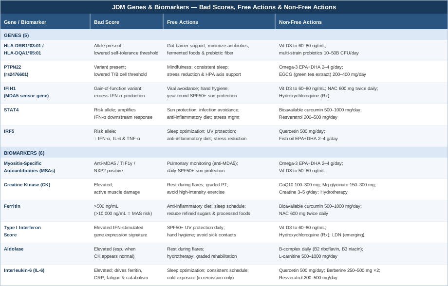
Understanding the biology at the level of individual genes and markers is valuable — but it becomes even more powerful when framed within the broader scientific narrative that has transformed how JDM is understood over the past two decades.
The Interferon Hypothesis: 10 Research Insights That Reshape How JDM Is Understood
The past twenty years of JDM research have converged on a single dominant biological theme: type I interferon overactivation as the central upstream driver of disease. This is not a fringe perspective — it is now the dominant model in pediatric rheumatology, supported by gene expression studies, genetic data, treatment response evidence, and mechanistic immunology. Below are ten of the most impactful findings from this body of research, each representing a meaningful shift in how JDM can be understood and managed.
1. JDM Is Primarily an Interferon Disease, Not Simply a Muscle Disease
Early models of JDM centered on T cell infiltration of muscle as the primary mechanism. More recent transcriptomic and immunological research has demonstrated that chronically elevated type I interferon signaling is the upstream driver, and that muscle inflammation is largely a downstream consequence of this systemic immune dysregulation. This reframing has direct implications for treatment strategy: targeting the IFN pathway directly — as newer biological agents including JAK inhibitors do — may be more effective in refractory cases than solely suppressing downstream inflammation.
2. The Interferon Signature Often Predicts Disease Activity More Reliably Than CK
Multiple research cohorts have demonstrated that the type I IFN gene expression score tracks disease activity more reliably than CK in some patient subgroups — particularly those with predominantly skin or pulmonary involvement and normal CK. In these patients, CK alone provides false reassurance. The IFN score provides a truer picture of ongoing immune activation even when muscle enzymes appear controlled.
3. UV Light Is a Direct Disease Trigger Through a Specific Biological Mechanism
UV radiation activates nucleic acid sensing in skin keratinocytes — the same innate immune pathway that drives IFN overproduction in JDM. This is why JDM often first presents or flares during summer months, and why photosensitivity is not merely a cosmetic concern but a mechanistic one. Rigorous, year-round sun protection is a direct biological intervention, not optional adjunctive care.
4. Viral Infections Precede Many JDM Diagnoses and Relapses
Retrospective studies and international patient registry data have documented that a substantial proportion of JDM diagnoses and disease relapses are temporally associated with prior viral infections. This supports a model of viral-triggered innate immune activation in genetically susceptible individuals as the proximate environmental cause of both JDM onset and flares — and explains why infection prevention during vulnerable periods matters.
5. Gut Microbiome Dysbiosis Is Measurably and Consistently Different in JDM
Emerging research has documented significantly altered gut microbiome composition in children with JDM compared to healthy controls, with consistent reductions in butyrate-producing bacteria including Faecalibacterium prausnitzii. Since butyrate is essential for regulatory T cell development in the gut lamina propria, this dysbiosis may perpetuate the immune dysregulation underlying JDM — and microbiome-directed dietary interventions represent a rational complementary strategy.
6. Calcinosis Has a Predictable Biological Logic and Is Not Random
Calcinosis — calcium deposition in soft tissues — occurs most consistently in areas of ongoing microvasculature damage and correlates strongly with delayed or inadequate treatment. Research has identified anti-NXP2 antibody positivity and prolonged active disease duration as the strongest predictors. Early, aggressive treatment with sufficient immunosuppression is the most evidence-supported strategy for calcinosis prevention, underscoring why tight disease control matters beyond symptom management.
7. Skin Disease Activity Is Often the Earliest Flare Signal
Clinical research using validated assessment tools — including the Cutaneous Assessment Tool (CAT) and the modified Dermatomyositis Skin Severity Index — has shown that changes in cutaneous disease activity frequently precede upcoming muscle flares before CK rises or other markers shift. Regular, systematic skin assessment is therefore not only cosmetically relevant but a practical early warning system for disease re-activation.
8. Anti-MDA5 Positivity Mandates a Distinct Monitoring Protocol
Multi-center data from Japan, the United Kingdom, North America, and Europe have consistently documented that anti-MDA5-positive JDM patients face meaningfully higher risk of rapidly progressive interstitial lung disease — a complication that can escalate from absent to severe within weeks. Standard respiratory symptom monitoring alone is insufficient in this subgroup. Proactive pulmonary function testing and high-resolution CT of the chest must be built into the care plan from the time of antibody identification.
9. Sleep Quality Directly Amplifies IFN Signaling — Making It a Biological Intervention
Human experimental studies have documented that partial sleep deprivation — even a single night — produces measurable increases in type I interferon-related gene expression and pro-inflammatory cytokine levels. In a disease driven by IFN overactivation, sleep optimization is not supportive care; it is a direct intervention on the core disease mechanism. Consistent sleep timing, darkness, and thermal regulation of the sleep environment are all modifiable variables with biological relevance in JDM.
10. JAK Inhibitors Validate the Interferon Model and Are Reshaping Refractory JDM Management
JAK (Janus kinase) inhibitors — including baricitinib, tofacitinib, and ruxolitinib — block the intracellular signaling cascade through which type I interferons drive gene expression and immune activation. Their emerging efficacy in refractory JDM, documented in case series and early clinical trials, represents the strongest possible validation of the IFN hypothesis: the drugs work precisely because they interrupt the pathway the research identified as central. Families managing refractory JDM should be aware of this therapeutic direction in their discussions with academic pediatric rheumatologists.
Complementary Approaches With Relevant Evidence in JDM
The following approaches do not replace medical treatment. They represent adjunctive strategies with meaningful human evidence — or strong mechanistic rationale — that may reduce disease burden, support quality of life, or address specific aspects of JDM biology. Each is worth discussing with the treating team before implementation.
The Autoimmune Protocol — Sarah Ballantyne
The Autoimmune Protocol (AIP) was developed by Dr. Sarah Ballantyne, a research scientist and author of The Paleo Approach, specifically for autoimmune conditions. It is a structured dietary elimination and reintroduction framework that removes foods associated with gut permeability and immune activation — grains, legumes, dairy, nightshades, eggs, nuts, seeds, alcohol, and refined sugars — while emphasizing nutrient-dense whole foods, organ meats, diverse vegetables, bone broths, and fermented foods. The core rationale is that gut barrier disruption is a shared mechanism across autoimmune diseases, and that removing dietary triggers allows the intestinal lining to heal, reducing the inappropriate presentation of microbial and dietary antigens to the systemic immune system.
A clinical trial published in Inflammatory Bowel Diseases (Konijeti et al.) demonstrated measurable reductions in inflammatory biomarkers and significant clinical improvement in patients with inflammatory bowel disease following AIP. While JDM-specific trial data does not yet exist, the shared mechanisms of gut permeability and systemic immune dysregulation across autoimmune conditions make AIP a scientifically logical adjunct for JDM — particularly given the microbiome dysbiosis findings described in the research section above. Sarah Ballantyne's protocol is the most systematically documented dietary framework for autoimmune management currently available.
In practice, AIP for a child with JDM involves a 4–6 week strict elimination phase followed by systematic, single-food reintroduction to identify personal triggers. Parents should work with a registered dietitian experienced in AIP to ensure nutritional adequacy — particularly for calcium, protein, and total caloric intake — since muscle wasting is already a risk in JDM. The protocol requires genuine commitment, and ongoing monitoring of growth metrics is essential in pediatric patients.
Mindfulness Meditation and MBSR
Mindfulness-Based Stress Reduction (MBSR), developed by Jon Kabat-Zinn at the University of Massachusetts Medical School, is an eight-week structured program combining mindfulness meditation, body scanning, and gentle yoga. Its relevance in JDM has two distinct dimensions: psychological stress is a documented driver of immune dysregulation and cytokine elevation — including IL-6 and IFN-α — and children with chronic illness face significant emotional burden that compounds the physiological disease process in measurable biological ways.
A randomized controlled trial published in Brain, Behavior, and Immunity demonstrated that MBSR produced measurable reductions in inflammatory markers including IL-6 in adults with chronic inflammatory conditions. A systematic review in Frontiers in Immunology confirmed immune-modulating effects of mindfulness practices across autoimmune and inflammatory conditions, with consistent effects on HPA axis function and pro-inflammatory cytokine profiles.
For children with JDM, adapted mindfulness programs exist — including Mindfulness-Based Cognitive Therapy for Children (MBCT-C) and child-specific MBSR formats. Sessions of 15–20 minutes daily, guided initially by a trained instructor or age-appropriate app (Headspace for Kids, Calm for Kids), are a realistic and low-risk starting point. Even parental mindfulness practice can reduce household stress reactivity, which has downstream effects on the child's cortisol environment. Evidence in JDM specifically is indirect but the biological rationale is sound and the risk is negligible.
Microbiome-Directed Therapies
Research consistently identifies gut microbiome dysbiosis as a feature of JDM and related pediatric autoimmune conditions. Reduced microbial diversity, lower abundance of short-chain fatty acid (SCFA)-producing bacteria — particularly Faecalibacterium prausnitzii and Roseburia species — and increased intestinal permeability have been documented in pediatric autoimmune populations. SCFAs, especially butyrate, are essential for regulatory T cell development in the gut — and Tregs are the immune cells responsible for suppressing aberrant self-reactive responses. Their depletion in JDM is not incidental: it may be a sustaining mechanism of immune dysregulation.
Research published in Nature Medicine demonstrated that microbiome composition predicted autoimmune relapse risk in pediatric patients, and that specific bacterial taxa were associated with immune regulatory gene expression patterns. Fecal microbiota transplantation (FMT) remains experimental for autoimmune indications, but targeted dietary and probiotic approaches are evidence-informed and immediately accessible.
Practically, microbiome-directed therapy in JDM begins with dietary diversity: aiming for 30 or more different plant foods per week, incorporating daily fermented foods (yogurt, kefir, sauerkraut, kimchi — ensuring compatibility with any elimination protocol in use), and emphasizing prebiotic-rich foods (garlic, leeks, asparagus, Jerusalem artichokes, green banana). Multi-strain probiotic supplementation (10–50 billion CFU daily, including Lactobacillus rhamnosus GG and Bifidobacterium longum) complements the dietary foundation. Butyrate supplementation (sodium butyrate or tributyrin, 300–600 mg daily) represents an emerging option with growing mechanistic support for Treg induction. These are adjunctive measures — they modulate immune tone over time rather than treating active disease acutely.
Gentle Yoga
Yoga is among the best-studied complementary practices in inflammatory musculoskeletal conditions. Its relevance in JDM lies in its ability to support muscle function and flexibility without the high-impact mechanical load that can worsen active muscle inflammation — while simultaneously delivering documented stress-reduction and anti-inflammatory effects through HPA axis modulation and vagal nerve activation.
A randomized trial published in Arthritis Care and Research demonstrated that a structured gentle yoga program reduced inflammatory biomarkers including IL-6 and CRP while improving functional capacity in adults with inflammatory arthritis. In pediatric rheumatic disease, yoga-based physical therapy components have been incorporated at several academic centers with positive outcomes in fatigue, quality of life, and functional measures.
For children with JDM, yoga must be matched strictly to disease phase. During active flares, yoga is contraindicated — muscle protection is the priority. In the recovery and remission phases, restorative and gentle yoga — not hot yoga, not vinyasa flow, not any form of high-intensity yoga — can begin under physical therapist guidance, progressing from 15 to 30-minute sessions three to four times per week. Restorative poses, gentle range-of-motion work, and breathing exercises are the appropriate foundation. Inversions and poses stressing recovering neck and shoulder muscles should be avoided until strength is clearly and verifiably restored.
Low-Level Laser Therapy and Photobiomodulation
Photobiomodulation (PBM), also called low-level laser therapy (LLLT), uses red and near-infrared light (630–1064 nm) to stimulate cellular energy production through cytochrome c oxidase in the mitochondrial electron transport chain. The result is increased ATP synthesis, reduced oxidative stress, and modulation of pro-inflammatory cytokines including TNF-α and IL-6 at the cellular level. In JDM, PBM is relevant primarily as a potential adjunct for muscle repair and local inflammation management during recovery phases.
A 2020 systematic review in Lasers in Medical Science found significant effects of PBM on muscle recovery, oxidative stress markers, and inflammatory cytokines in musculoskeletal conditions. A randomized controlled trial in rheumatoid arthritis demonstrated reduced joint inflammation and IL-6 levels following PBM treatment. JDM-specific trials do not yet exist, but the shared mechanisms of inflammatory myopathy and the favorable safety profile of near-infrared PBM make it a biologically plausible adjunct.
In practice, home-use red light panels emitting 630–850 nm at 30–100 mW/cm² are available at $200–$800. Clinical-grade LLLT is available at physical therapy centers and some academic rheumatology facilities. For JDM, PBM should never be applied to areas of active skin rash — photosensitivity in JDM represents a real contraindication, and UV or intense visible light over inflamed skin can worsen local immune activation. Application to affected muscle groups (thighs, proximal upper extremities) during documented remission, 10–20 minutes per session, three to five times per week, is a reasonable protocol with physician clearance. This is an adjunctive strategy in remission — not an active-disease intervention.
Conclusion
Juvenile dermatomyositis is biologically complex, but it is increasingly well-mapped. The combination of myositis-specific autoantibody profiling, muscle enzyme monitoring, ferritin and IL-6 tracking, and emerging interferon scoring provides a far more precise picture of disease activity than general inflammatory markers alone. Understanding the genetic architecture — particularly HLA, PTPN22, IFIH1, STAT4, and IRF5 — adds context for why the immune system is responding the way it is, and which biological compensations have the strongest mechanistic rationale for any given child.
The most practical next step is not to implement everything at once. It is to review the current monitoring plan with the treating rheumatologist and ask which of these biomarkers are being tracked and which are not. Confirm that MSA subtyping has been completed and that its implications for monitoring frequency have been discussed. Ask about the availability of interferon scoring through research protocols. From there, the lifestyle and supplementary strategies outlined here can be introduced systematically — in dialogue with the clinical team — to complement medical management with precision. Better information, applied carefully and collaboratively, is how better long-term outcomes are built.
Musculoskeletal: Muscle Conditions
Autoimmune: Inflammatory Conditions Autoimmune Skin Conditions