This article contains AI generated content.
Juvenile Tibial Osteochondrosis: 5 Genes and 7 Biomarkers to Track
When Shin and Knee Pain Doesn't Fit the Standard Explanation
A growing child with persistent pain below the kneecap, a bowed lower leg that doesn't straighten with age, or a knee that locks and swells after sport rarely gets a satisfying explanation. Most families are told some version of "it's growing pains, give it rest and ice," and while that advice isn't wrong, it's rarely enough. It doesn't explain why one sibling develops Osgood-Schlatter disease during a growth spurt and another doesn't, why some cases of tibia vara resolve with bracing and others need surgery, or why a well-nourished, active teenager can still end up with a growth-plate problem.
Generic guidance is broad because it has to work for every child. It doesn't account for the fact that a child's vitamin D status, growth velocity, body composition, and inherited cartilage biology all interact differently in each case. Juvenile tibial osteochondrosis is not one disease — it's an umbrella term covering conditions like Osgood-Schlatter disease (tibial tuberosity apophysitis), Blount's disease (tibia vara from disrupted proximal tibial growth), and osteochondritis dissecans of the tibial plateau. Treating them all with the same rest-and-stretch script misses the more specific, more useful question: what, in this particular child, is putting extra stress on a still-forming growth plate, and what can realistically be measured and adjusted?
That's the deeper approach this article takes. Rather than repeating "reduce activity and wait it out," it looks at the biomarkers that reflect what's actually happening in a growing tibia — vitamin D and mineral status, growth-axis activity, bone turnover, body composition — and at the early genetic research that may explain why some children are more vulnerable to begin with. Neither approach offers a guaranteed fix; growth-plate biology is complex and much of the pediatric-specific evidence is still developing. But better information, measured rather than assumed, tends to lead to better decisions: a more targeted conversation with a pediatrician, a more sensible training adjustment, a supplement decision based on an actual blood result instead of a guess.
Summary
This article works through two complementary lenses on juvenile tibial osteochondrosis. The main section covers seven biomarkers worth tracking — from vitamin D and the calcium-phosphate-PTH axis to bone turnover markers and body composition — explaining what each one reveals about growth-plate stress, how it's measured, typical cost, and what a realistic improvement plan looks like both with and without supplements. A shorter section afterward looks at five genes tied to cartilage structure and growth signaling (COL2A1, ACAN, GDF5, VDR, and IGF1) and what the still-early human evidence actually supports. There's also a breakdown of ten mechanotransduction and bone-remodeling concepts drawn from a leading longevity physician's deep dive on bone health, applied specifically to growing tibias, and a review of complementary approaches — from targeted stretching to photobiomodulation — with an honest look at where the evidence is solid and where it's thin. Read together, these sections aim to replace "just rest it" with something more specific: what to test, what it means, and what to actually do next.
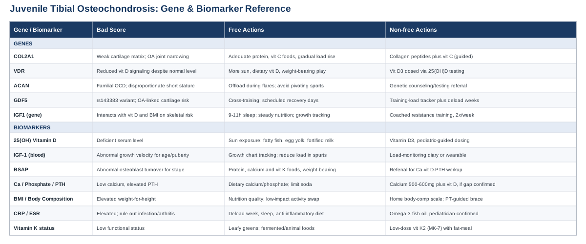
7 Biomarkers Worth Tracking in Juvenile Tibial Osteochondrosis
Before getting into individual markers, it's worth borrowing a principle popularized by physicians like Peter Attia: measure the thing directly rather than relying on a generic reference range or a visual impression. A knee X-ray shows structure, but it doesn't show whether a child's mineralization is on track, whether their growth axis is unusually active right now, or whether their inflammatory markers suggest something other than mechanical overuse. The biomarkers below are the practical, mostly blood-based measurements that give that additional layer of information — some are cheap and available at any pediatric visit, others require a specialized lab.
1. Serum 25-Hydroxyvitamin D
This is the standard measure of the body's vitamin D reserves, and it matters here because vitamin D governs calcium absorption and the mineralization of growth-plate cartilage. Deficiency disorganizes the growth plate in classic rickets, and a retrospective study of children at a pediatric obesity clinic found those with vitamin D deficiency were substantially more likely to carry a Blount's disease diagnosis, although another study found deficiency rates similar to healthy children — so the link is plausible but not settled. The mechanical disruption vitamin D deficiency causes at the growth plate is well described independent of any single orthopedic diagnosis.
How to Measure It
A simple blood draw for 25(OH)D, costing roughly 40 to 100 dollars out of pocket (often less or covered through a pediatric visit). It's frequently bundled into broader metabolic or bone-health panels.
If the Score Is Low, the Plan Without Supplements
Ten to twenty minutes of midday sun exposure on arms and legs several times a week (adjusted for skin tone and season), regular outdoor weight-bearing play, and dietary sources like fatty fish, egg yolks, and fortified milk or cereal. Retest after about three months rather than assuming improvement.
If the Score Is Low, the Plan With Supplements or Equipment
Vitamin D3 at a maintenance dose of roughly 600 to 1,000 IU per day for most children, or a higher repletion dose (often 1,000 to 2,000 IU per day for 8 to 12 weeks) for a confirmed deficiency, under pediatric guidance rather than self-directed. Retest every three to six months during active correction. Side effects are rare at these doses, but unsupervised high-dose regimens risk hypercalcemia — this is not a "more is better" nutrient in children.
2. IGF-1 (Growth Axis Activity)
Insulin-like growth factor 1 mediates growth hormone's effect on linear bone growth and the proliferation of growth-plate chondrocytes. It's relevant here because rapid growth velocity — being unusually tall or heavy for age during a growth spurt — is a recognized risk pattern for Osgood-Schlatter disease, and IGF-1 is the biological signal driving that velocity.
How to Measure It
A blood test costing roughly 50 to 150 dollars. Interpretation requires age-, sex-, and pubertal-stage-adjusted reference ranges — a result that looks "high" for an adult may be entirely normal for a child mid-growth-spurt, so this is a value to interpret with a pediatrician or pediatric endocrinologist rather than in isolation.
If the Pattern Looks Unusual, the Plan Without Supplements
Track height on a growth chart and temporarily reduce training volume or intensity during the steepest phase of a growth spurt, which is the known window of highest risk for traction apophysitis at the tibial tuberosity. A simple paper or app-based training log that notes symptom flares against growth measurements over a few months often reveals the pattern clearly.
If the Pattern Looks Unusual, the Plan With Supplements or Equipment
No supplement safely or appropriately raises or lowers IGF-1 in a healthy child, and none should be attempted. The useful "equipment" here is a load-monitoring tool — a basic training diary or a low-cost wearable that logs weekly activity hours — used to correlate symptom flares with growth timing. A genuinely abnormal IGF-1 result belongs in front of a pediatric endocrinologist, not a supplement plan.
3. Bone-Specific Alkaline Phosphatase (BSAP)
BSAP is an enzyme marker of osteoblast activity — it tells you how much active bone formation is happening. It naturally runs high in infancy and again during puberty, tracking the physiological surges of longitudinal growth, so unusually high or low values relative to age and pubertal stage can flag abnormal bone turnover alongside growth-plate stress.
How to Measure It
A blood test, typically 50 to 120 dollars, usually only available through larger reference labs as part of a bone turnover panel. The result must be compared against pediatric, pubertal-stage-specific reference intervals — adult ranges are not applicable and will misread a normal pubertal surge as abnormal.
If the Score Is Off, the Plan Without Supplements
Consistent protein intake, calcium- and vitamin-K-rich foods, and regular moderate weight-bearing activity all support normal osteoblast function without any product purchase involved.
If the Score Is Off, the Plan With Supplements or Equipment
There's no supplement that targets BSAP directly. If a turnover pattern suggests a metabolic bone condition rather than simple mechanical overuse, the correct next step is referral for evaluation of the calcium-vitamin D-PTH axis (below), not self-supplementation. Retest in three to six months given the natural pubertal swing in this marker.
4. Calcium, Phosphate, and Parathyroid Hormone (PTH) Panel
This trio governs the mineralization of growth-plate cartilage. When vitamin D is low, calcium absorption drops, PTH rises to compensate, and that compensatory rise disrupts the normal breakdown of hypertrophic chondrocytes at the growth plate — the same mechanism seen in nutritional rickets, and a plausible contributor across the osteochondrosis spectrum even outside classic rickets.
How to Measure It
A basic metabolic panel covers calcium and phosphate for roughly 30 to 80 dollars; intact PTH is a separate order, typically 40 to 90 dollars.
If the Panel Is Off, the Plan Without Supplements
Dietary calcium and phosphate from dairy, fortified plant milks, fish eaten with bones, and legumes, while moderating processed foods and sodas that skew the phosphate-to-calcium ratio.
If the Panel Is Off, the Plan With Supplements or Equipment
Calcium supplementation (roughly 500 to 600 mg elemental calcium in split doses, paired with vitamin D) only after confirming a genuine dietary gap — most children who eat any dairy or fortified food don't need it. Side effects of oversupplementing include constipation and, in excess over time, kidney stone risk, so this is a fix-the-gap intervention, not a blanket recommendation.
5. BMI and Body Composition Trend
Excess weight-for-height is the single most consistently proven risk factor for Blount's disease, and higher BMI is also linked to Osgood-Schlatter disease. This is essentially a mechanical-load biomarker: more body mass means more compressive and shear force on the medial proximal tibial physis and on the tibial tuberosity during quadriceps loading.
How to Measure It
BMI percentile is tracked for free at any routine pediatric visit. For a more precise read, a home bioelectrical impedance scale (30 to 150 dollars) or a DEXA body composition scan (100 to 250 dollars) separates fat mass from lean mass more accurately than BMI alone.
If the Trend Is Unfavorable, the Plan Without Supplements
Sustainable nutrition quality improvements rather than calorie restriction in a still-growing child, plus a shift toward lower-impact activity — swimming, cycling — when higher-impact sport is currently aggravating symptoms. This is best done with a pediatrician or dietitian setting age-appropriate targets; adult weight-loss approaches don't transfer safely to children.
If the Trend Is Unfavorable, the Plan With Supplements or Equipment
No supplement belongs in pediatric weight management. The useful equipment is a home body composition scale checked monthly (daily weight fluctuates too much to be meaningful) and, during active flares, a physical-therapist-guided offloading brace or activity modification rather than a product-based fix.
6. CRP and ESR (Inflammatory Screen)
These don't diagnose osteochondrosis, but they're an important filter. Persistent joint or shin pain with elevated CRP or ESR should prompt evaluation for infection or juvenile inflammatory arthritis rather than an assumption of mechanical overuse — a distinction diagnostic reviews of both Osgood-Schlatter disease and osteochondritis dissecans specifically flag as a differential to rule out.
How to Measure It
A simple blood draw, usually ordered together: CRP around 15 to 40 dollars, ESR around 15 to 30 dollars.
If the Score Is Elevated, the Plan Without Supplements
If the workup points to overtraining-related inflammation rather than infection or arthritis, standard load management — a deload week, improved sleep, and an anti-inflammatory eating pattern rich in fish, fruit, and vegetables — is the appropriate response.
If the Score Is Elevated, the Plan With Supplements or Equipment
Omega-3 fish oil at a pediatric-appropriate dose (confirmed with a pediatrician) may modestly support general inflammatory tone during a recovery block. It is not a treatment for elevation caused by infection or arthritis — a persistently elevated CRP or ESR needs medical workup first, full stop.
7. Vitamin K Status
Vitamin K is the often-overlooked cofactor for carboxylating osteocalcin and other bone matrix proteins, which is what allows those proteins to actually bind calcium. It complements vitamin D's role in absorption by supporting proper matrix mineralization once calcium is available.
How to Measure It
This isn't routinely tested. Specialized labs offer undercarboxylated osteocalcin or PIVKA-II as indirect functional markers, at roughly 80 to 150 dollars — an advanced option worth considering only if the more standard markers above are already optimized and questions remain. Most families can reasonably skip this and focus on dietary sufficiency.
If Intake Looks Low, the Plan Without Supplements
Regular leafy greens (vitamin K1) and some fermented or animal-based foods (vitamin K2) are sufficient for most children eating a varied diet — no special protocol needed.
If Intake Looks Low, the Plan With Supplements or Equipment
Routine supplementation isn't necessary for a healthy, varied diet. If pursued based on a specific test result, a low-dose vitamin K2 (as MK-7) taken with a fat-containing meal, under pediatric guidance, is reasonable; cycling isn't typically required. The one real caution is drug interaction with anticoagulant medications, which is rarely relevant in an otherwise healthy child but worth flagging.
Taken together, these seven markers turn "just rest it" into something closer to "here's specifically what your child's biology is doing right now." That measurement-first mindset extends naturally into the genetic side of the picture, where the evidence is earlier but the underlying logic is similar.
What Genetic Research Suggests About Tibial Growth Plate Vulnerability
Genetic research into juvenile tibial osteochondrosis specifically is thin — most of what exists comes from adjacent fields like adult osteoarthritis, rare familial skeletal disorders, and general pediatric growth research, extrapolated to this context. That's worth stating plainly rather than overselling: none of the genes below have been validated in a dedicated genetic study of Osgood-Schlatter disease or Blount's disease. They're biologically plausible contributors based on what these genes are known to do in cartilage and growth-plate biology, in the same spirit that researchers like Ali Torkamani have pushed for connecting genomic variants to actionable, everyday health decisions rather than leaving them as abstract risk scores.
1. COL2A1 (Collagen Type II)
COL2A1 encodes type II collagen, the main structural protein of the growth plate's hyaline cartilage. Variants here affect how well that cartilage matrix holds together under repetitive load. The clearest human evidence comes from adult knee osteoarthritis research, where COL2A1 genotype tracked specifically with joint space narrowing — a structural cartilage effect, not a pediatric osteochondrosis finding, but a plausible upstream mechanism.
If the Gene Looks Unfavorable, the Plan Without Supplements
Distribute adequate protein across the day (roughly 1.0 to 1.2 g/kg/day for an active child), include vitamin-C-rich foods that support collagen synthesis, avoid secondhand smoke exposure, and increase training load gradually rather than in sudden jumps — a standard "10 percent rule" applied to weekly volume.
If the Gene Looks Unfavorable, the Plan With Supplements or Equipment
Hydrolyzed collagen peptides (5 to 15 g) with about 50 mg of vitamin C, taken roughly an hour before loading activity, has adult connective-tissue evidence behind it, though pediatric-specific trials are lacking — use only with pediatric provider input. Cycle it through active training blocks and skip it during off-season. Side effects are minimal; mild GI upset is possible, and those with fish or bovine allergies should check the source.
2. VDR (Vitamin D Receptor)
VDR determines how sensitively cells respond to vitamin D, independent of blood vitamin D levels themselves. Common variants (FokI, BsmI, TaqI, ApaI) have been studied in bone density and osteoarthritis research, sometimes alongside COL2A1 in the same studies, but evidence is mixed and no pediatric osteochondrosis data exists yet.
If the Gene Looks Unfavorable, the Plan Without Supplements
Because receptor sensitivity can't be changed through lifestyle, the practical lever is ensuring vitamin D supply is generous rather than borderline: regular sun exposure, dietary sources, and weight-bearing activity to amplify the downstream signaling that does occur.
If the Gene Looks Unfavorable, the Plan With Supplements or Equipment
The same vitamin D3 approach described in the biomarker section applies, guided by actual 25(OH)D testing rather than blind dosing — a less sensitive receptor is an argument for testing more diligently, not for taking more vitamin D without a number to guide it.
3. ACAN (Aggrecan)
ACAN encodes aggrecan, the main proteoglycan giving cartilage its compressive resistance. This is the one gene on this list with genuinely strong human evidence — but only for a rare, distinct subset of cases. Autosomal dominant ACAN mutations cause a documented familial form of osteochondritis dissecans, often paired with disproportionate short stature, running in families across generations.
If Family History Suggests This, the Plan Without Supplements
For the common, non-familial presentation, mechanical protection during flares (offloading, temporary activity modification) and avoiding high-impact pivoting sports while symptomatic are the reasonable defaults. For a child with both short stature and a strong family history of early joint disease, the useful step is a referral conversation, not a home protocol.
If Family History Suggests This, the Plan With Supplements or Equipment
Genetic counseling and testing through a pediatric orthopedic genetics service (typically several hundred dollars) is the appropriate "equipment" here if familial ACAN-related disease is suspected. Glucosamine and chondroitin are sometimes tried for general cartilage support; adult osteoarthritis evidence for them is weak and there's no pediatric OCD trial data, so this should be framed honestly as low-confidence and optional, not a genetic fix.
4. GDF5 (Growth Differentiation Factor 5)
GDF5 guides joint formation and cartilage development at the growth plate. A specific variant, rs143383, has been linked to knee osteoarthritis risk in meta-analyses of Caucasian populations. It hasn't been studied in juvenile osteochondrosis directly, but its known role in joint cartilage development makes it a reasonable candidate pathway.
If the Gene Looks Unfavorable, the Plan Without Supplements
Cross-training instead of single-sport specialization — a factor specifically noted as protective in systematic reviews of Osgood-Schlatter risk factors — plus built-in recovery days between high-load sessions.
If the Gene Looks Unfavorable, the Plan With Supplements or Equipment
There's no GDF5-targeted supplement. The more useful equipment is a simple training-load tracker (a log or low-cost wearable) paired with deload weeks every four to six weeks, since load management is the actionable variable regardless of genotype.
5. IGF1 (Growth Axis Genotype)
Beyond blood IGF-1 levels, the IGF1 gene itself has variants studied in children that interact with vitamin D status and BMI — meaning genotype may shape how growth velocity, weight, and vitamin D sufficiency combine to affect a child's skeletal development. This is human pediatric evidence, though it hasn't been tested against osteochondrosis diagnoses specifically.
If the Gene Looks Unfavorable, the Plan Without Supplements
Consistent, age-appropriate sleep (9 to 11 hours for school-age children and young teens, since most growth hormone pulses occur in deep sleep), steady nutrition that avoids rapid excess weight gain, and routine growth-velocity and BMI-percentile tracking with a pediatrician.
If the Gene Looks Unfavorable, the Plan With Supplements or Equipment
No supplement should be used to influence the GH/IGF-1 axis in a healthy growing child outside a diagnosed deficiency managed by endocrinology — that line shouldn't be crossed. The legitimate equipment lever is qualified, age-appropriate resistance training (bodyweight or light-load, coached), roughly twice a week, which supports musculoskeletal development through mechanical stimulus rather than hormonal manipulation.
The genetic picture, in short, explains predisposition better than it prescribes treatment — which is exactly why the mechanotransduction principles in the next section, borrowed from a physician who has spent years studying how bone actually responds to load, are worth understanding regardless of what any single gene shows.
10 Lessons From a Leading Longevity Physician's Deep Dive on Bone Health
Peter Attia's long-form episode on bone health, Navigating bone health: early life influences and advanced strategies for improvement and injury prevention, is framed mostly around adult osteoporosis prevention. But its central argument — that bone is a mechanosensory tissue that remodels in direct proportion to the load placed on it — maps directly onto how a growing tibia responds to training, rest, and repair. Here are the ten points from that discussion most relevant to a child dealing with tibial osteochondrosis.
1. Bone Is Living Tissue, Not a Fixed Structure
Bone is vascularized, constantly remodeling tissue, not an inert scaffold. A growth plate under repetitive stress isn't a static weak point — it's actively responding, for better or worse, to whatever load pattern it's exposed to week after week.
2. Two Cell Types Are Always in a Tug-of-War
Osteoblasts build bone, osteoclasts break it down, and the two are in continuous balance. Overuse injuries like Osgood-Schlatter disease happen when the loading side of that equation outpaces the repair side for long enough that the tissue can't keep up.
3. "Lifting Heavy Stuff Matters"
Attia's own phrase for mechanotransduction: bone and connective tissue respond most strongly to progressive resistance loading, not just repetitive impact. For a young athlete stuck in a cycle of running-related shin and knee pain, this is an argument for adding qualified strength work rather than only accumulating more mileage.
4. Imaging Measures Structure, Not Just Symptoms
DEXA scanning is the gold standard for adult bone mineral density precisely because it gives an objective number instead of a guess. The same logic supports using the biomarkers and, where warranted, imaging described earlier in this article rather than relying on how a child's leg looks or how much pain they report on a given day.
5. Deficiency Exists on a Spectrum
Osteopenia and osteoporosis aren't a single on/off switch — they're gradations of an ongoing process. Growth-plate stress is similar: it builds gradually before becoming a diagnosed condition, which is the argument for catching trends early through tracking rather than waiting for a clear injury.
6. Hormonal Timing Shapes the Whole Trajectory
Estrogen dramatically shapes bone density trajectories in adults; in growing children, the pubertal hormonal shift similarly governs when growth spurts happen and when growth-plate vulnerability peaks. Timing, not just total load, matters.
7. Nutrition Supports the Structure, It Doesn't Replace Loading
Attia is clear that nutrition and supplementation (calcium, vitamin D) support bone health but don't substitute for the mechanical stimulus of activity. The same applies here: correcting a vitamin D deficiency matters, but it won't offset a training load a growing tibia genuinely can't tolerate right now.
8. Complete Immobility Has Its Own Cost
Prolonged bed rest or forced sedentary periods cause rapid bone loss. This is a useful counterpoint to blanket "just rest completely" advice for tibial osteochondrosis — relative rest and activity modification, rather than total immobilization, is generally the better-supported approach in the clinical literature on Osgood-Schlatter disease specifically.
9. The Early Years Are a Skeletal Investment Window
Peak bone mass is substantially determined by childhood and adolescent habits. The years when tibial osteochondrosis typically shows up are the same years that set the trajectory for skeletal health decades later — a reason to manage these conditions thoughtfully rather than simply waiting for a child to "grow out of it."
10. Proactive Measurement Beats Reactive Treatment
Attia's broader case for earlier, more frequent bone density screening in adults reflects a mindset worth borrowing here: check the relevant markers and load patterns before a problem becomes a diagnosis, rather than only responding once a child is already limping.
These principles reinforce a theme running through the biomarker and gene sections alike: growth-plate health responds to measurable inputs — load, minerals, hormones, sleep — more than to guesswork. That same logic extends to a set of complementary approaches that, while not substitutes for medical evaluation, have some genuine evidence behind them for this condition.
Complementary Approaches Worth Considering
None of the following replace a pediatric orthopedic evaluation, imaging when indicated, or the biomarker-guided steps above. But for the muscle tightness, training-load, and pain-coping components of juvenile tibial osteochondrosis, a few complementary approaches have real, condition-relevant evidence.
Targeted Stretching and Flexibility Work (Yoga-Based)
Reduced hamstring and quadriceps flexibility is a specific, biomechanically documented risk factor for Osgood-Schlatter disease — tighter hamstrings increase quadriceps force transmitted through the patellar tendon into the tibial tuberosity, and developmental-stage flexibility deficits predict who develops symptoms among adolescent athletes. A structured, yoga-style flexibility routine is a reasonable, low-cost way to work on exactly this deficit.
A biomechanical modeling study quantified this directly: shortening hamstring optimal length by 30 percent increased accumulated quadriceps force by nearly 22 percent, concentrated in early stance and terminal swing phases of gait, and the same research group's broader work on adolescent soccer players found flexibility deficits predictive of who went on to develop Osgood-Schlatter disease (The Biomechanics Effect of Hamstring Flexibility on the Risk of Osgood-Schlatter Disease).
In practice, this means a daily 10 to 15 minute quadriceps and hamstring flexibility sequence, done consistently rather than only during flare-ups, ideally guided initially by a physical therapist or athletic trainer to confirm technique. It's a genuinely low-risk addition; the main caution is avoiding aggressive stretching directly over an acutely inflamed tibial tuberosity.
Massage Therapy and Manual Soft-Tissue Work
Because quadriceps and surrounding muscle tightness is directly implicated in traction stress on the tibial tuberosity, manual therapy aimed at reducing that tightness is a logical complementary step, and it's commonly used alongside standard physical therapy in adolescent sports medicine settings.
A retrospective cohort study of adolescent athletes with Osgood-Schlatter disease found that adding quadriceps release therapy to standard local treatment led to significantly shorter recovery time and fewer treatment sessions than local treatment alone (The impact of quadriceps release therapy on rehabilitation of adolescent athletes with Osgood-Schlatter disease), suggesting a real, if modest, added benefit rather than a purely relaxing effect.
Realistically, this looks like periodic sessions with a therapist experienced in pediatric sports injuries, focused specifically on the quadriceps and surrounding hip musculature rather than generic massage, used as an adjunct to — not a replacement for — activity modification and the flexibility work above.
Low-Level Laser Therapy (Photobiomodulation)
Photobiomodulation is a light-based therapy that's been studied for various tendinopathies, and there's biological plausibility for extending it to the tendon-attachment stress seen in Osgood-Schlatter disease, since both involve overloaded connective tissue trying to heal under continued mechanical demand.
The evidence base is encouraging but not condition-specific yet: a systematic review and meta-analysis of low-level red and near-infrared photobiomodulation for tendinopathy generally found superior pain relief compared to minimal intervention (The effect of low-level red and near-infrared photobiomodulation on pain and function in tendinopathy), and a dedicated randomized trial testing it specifically for Osgood-Schlatter and Sever's disease in youth athletes is currently registered and underway, meaning direct evidence for this exact condition is still emerging rather than established.
Given that, this is reasonable to try under the guidance of a sports medicine or physical therapy provider using a proper clinical device, but it shouldn't be treated as a proven fix, and it doesn't replace load management or flexibility work — it's a pain-modulation adjunct at most, with a favorable safety profile and minimal side effects reported in the tendinopathy literature.
Mindfulness-Based Pain Coping
Chronic or recurring pain during a growth spurt, even when the underlying mechanical issue is being managed appropriately, can affect a young athlete's sleep, mood, and relationship with their sport. Mindfulness-based stress reduction has been studied specifically for chronic musculoskeletal pain coping, which is relevant to the frustration and activity anxiety that often accompanies a drawn-out case of tibial osteochondrosis.
Evidence here is genuinely mixed rather than strongly positive: pilot randomized trials of online mindfulness-based stress reduction for chronic musculoskeletal pain show promise and feasibility (The feasibility and acceptability of an online mindfulness-based stress reduction program for chronic musculoskeletal pain), while other trials in different chronic pain populations have not found significant benefit — this is a supportive tool for coping, not a treatment for the underlying growth-plate issue.
Applied here, this means a short, age-appropriate mindfulness or breathing practice (10 minutes, a few times a week) aimed at pain coping and training-related stress, best introduced through a pediatric psychologist or a reputable app designed for adolescents, and framed honestly to the child as a coping tool rather than a cure.
Putting It All Together
Juvenile tibial osteochondrosis rarely has one clean cause, which is exactly why one generic answer never quite satisfies. The biomarker panel above — vitamin D, IGF-1, bone-specific alkaline phosphatase, the calcium-phosphate-PTH axis, body composition, inflammatory markers, and vitamin K status — gives a concrete way to see what's actually driving stress on a specific child's growth plate, and each one comes with a realistic next step, with or without supplementation. The genetic research is earlier and less conclusive, but genes like COL2A1, ACAN, GDF5, VDR, and IGF1 at least explain why identical training loads don't affect every child the same way. Layered on top, the mechanotransduction principles from bone health research and the complementary approaches with real condition-specific evidence round out a plan that's about load management and measured biology, not guesswork.
None of this replaces a pediatric orthopedic evaluation, especially when there's swelling, locking, night pain, or a limb deformity that isn't improving — those findings need clinical assessment, not a supplement plan. But for the more common, milder presentations, the next smart step is usually simple: track symptoms against training load and growth spurts for a few weeks, ask a pediatrician about the biomarkers most relevant to your child's specific presentation, and treat any supplement or equipment decision as something to revisit with an actual number, not a guess.
Musculoskeletal: Joint Conditions Tendon & Ligament Conditions