This article contains AI generated content.
· UpdatedKashin-Beck Disease Genes and Biomarkers: 4 Genes and 6 Biomarkers to Track
Introduction
Kashin-Beck disease occupies an unusual position in medicine. It is geographically concentrated — endemic across parts of Tibet, rural China, and Siberian Russia — yet the biological processes driving it touch on mechanisms that are relevant far beyond those borders: selenium metabolism, selenoprotein function, oxidative stress in cartilage, and mycotoxin exposure from stored grain. For anyone navigating a diagnosis, or trying to make sense of their risk in an affected region, the information available in most clinical settings stops at "selenium deficiency" and leaves the harder, more specific questions unanswered.
The problem with the simple version is that it does not account for why two people with near-identical diets in the same village can have dramatically different outcomes. Genetic variants in the genes that encode selenoprotein-synthesizing enzymes mean that antioxidant capacity in joint cartilage can vary significantly from person to person — even when dietary intake looks the same on paper. Mycotoxin contamination of grain, often underweighted in clinical discussions, adds an independent layer of oxidative stress that selenium supplementation alone does not fully address. The real picture is layered, and working with it requires more than one data point.
This article focuses on what can actually be measured and acted upon. The primary section covers six biomarkers — spanning functional selenium status, direct indicators of cartilage breakdown, mycotoxin burden, and oxidative damage — that together provide a far more granular picture of what is happening than a standard workup. A shorter companion section covers four genetic variants with meaningful evidence in the context of KBD and selenoprotein biology, with practical protocols for each.
Better information does not promise better outcomes on its own, but it creates a foundation for smarter clinical decisions. Understanding which biomarkers are elevated, and whether a genetic variant is blunting the effectiveness of otherwise reasonable interventions, changes what the intervention should look like. That shift — from generic to targeted — is what this article is designed to support.
Summary
This article covers the six most actionable biomarkers for tracking Kashin-Beck disease — including serum selenium, functional GPx enzyme activity, two direct cartilage degradation markers (CTX-II and COMP), a mycotoxin burden panel that most clinicians never order, and an oxidative stress panel that shows whether antioxidant defenses are actually working. For each, you will find what the marker reveals, how to test it with cost estimates, and a concrete action plan — with and without supplements — if results come back outside the optimal range.
The genetics section that follows examines four gene variants — GPX1, SELENOP, TXNRD1, and COL2A1 — that influence how effectively the body synthesizes antioxidant selenoproteins and maintains cartilage matrix integrity. These variants help explain why some people are more vulnerable than others, and what a genetically informed protocol might look like.
Beyond the core biomarker and genetics content, the article covers ten research-grounded insights from Dr. Ben Lynch's work on antioxidant gene variants, five complementary modalities with meaningful clinical evidence for joint-related conditions, and practical guidance throughout on cycling, dosing, and when to involve a specialist.
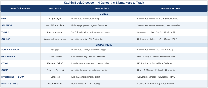
6 Biomarkers Worth Tracking in Kashin-Beck Disease
Tracking biomarkers turns a vague clinical picture into something workable. The six markers below map onto the core processes in KBD: selenium metabolism, cartilage destruction, oxidative burden, and mycotoxin exposure. None of them is sufficient alone, but together they give a comprehensive view — and each one leads to specific, actionable steps if results fall outside the optimal range.
1. Serum Selenium
Selenium is the nutritional foundation of every meaningful intervention in KBD. It is the raw material for the selenoproteins — glutathione peroxidases, thioredoxin reductases, selenoprotein P — that serve as the primary antioxidant defense system for chondrocytes, the cells that build and maintain cartilage. Studies from endemic Chinese and Tibetan populations consistently find that affected individuals have serum selenium levels below 20–30 μg/L, compared to a healthy reference range of approximately 80–150 μg/L. These are not mild deficiencies; they represent near-total depletion of the substrate that antioxidant enzymes depend on to function.
What makes serum selenium particularly important as a tracking marker is that it reflects both recent dietary intake and longer-term bioavailability, and it can change meaningfully within weeks of dietary or supplemental intervention. It is also cheap, widely available, and a reasonable starting point before ordering more specialized tests. The limitation is that serum selenium alone does not tell you whether the selenium is being converted efficiently into active selenoproteins — that requires functional testing, covered in the next marker.
How to Measure It
A standard blood draw, ordered by most primary care physicians or self-ordered through direct-to-consumer labs. LabCorp and Quest Diagnostics both offer standalone serum selenium tests. Cost ranges from $30 to $80. The NIH Office of Dietary Supplements selenium fact sheet provides a detailed clinical reference on interpretation and toxicity thresholds. For people monitoring disease activity or response to intervention, testing every 90 days initially, then every six months once stable, is a reasonable cadence. Whole-blood selenium (as opposed to serum) reflects longer-term status and can be ordered simultaneously for a more complete picture.
If the Score Is Low: Plan Without Supplements
Brazil nuts are the most practical dietary intervention: a single nut provides 70–90 mcg of selenium — roughly a full day's requirement. Two nuts per day, five days a week (avoiding daily use to prevent gradual accumulation toward toxicity over time), is a sustainable protocol. Beyond Brazil nuts, sardines, tuna, eggs, chicken liver, sunflower seeds, and shellfish are meaningful secondary sources. People living in low-selenium-soil regions — which overlap significantly with KBD-endemic zones — cannot rely on grain as a selenium source regardless of quantity consumed, since soil selenium content determines how much crops accumulate. Cooking methods matter: avoiding prolonged boiling of protein foods helps retain selenium that would otherwise leach into cooking water. Retest after 8–12 weeks of sustained dietary change.
If the Score Is Low: Plan With Supplements or Equipment
Selenomethionine is the preferred supplemental form — the organic form found in food, absorbed and retained in tissue more effectively than inorganic sodium selenite. A typical correction dose is 100–200 mcg/day. The tolerable upper intake level set by the Institute of Medicine is 400 mcg/day for adults; exceeding this over weeks risks selenosis, presenting as hair loss, nail changes, and in severe cases neurological symptoms. A practical protocol for deficiency correction: 200 mcg/day for 60 days, then reduce to 100 mcg/day as a maintenance dose, retesting every 90 days. Selenium supplementation trials conducted in KBD-endemic Chinese populations — published in journals including the European Journal of Epidemiology — have shown reductions in disease activity markers when baseline levels were severely depleted. The benefit is most pronounced at severe deficiency; supplementing into an already-adequate range yields diminishing returns and carries toxicity risk.
2. Erythrocyte Glutathione Peroxidase (GPx) Activity
Serum selenium tells you what substrate is available; erythrocyte GPx activity tells you what the cell is actually doing with it. Glutathione peroxidase 1 (GPx1) is the main intracellular antioxidant enzyme in chondrocytes — it neutralizes hydrogen peroxide and lipid hydroperoxides that would otherwise trigger apoptosis and matrix breakdown. GPx1 requires selenium as a structural component of its active site (as selenocysteine), but it also requires glutathione as its reducing substrate and is subject to genetic variation in efficiency regardless of selenium availability. In KBD patients, erythrocyte GPx activity is consistently and severely depressed — often 50–70% below healthy controls — and this functional impairment is a more direct reflection of cellular antioxidant capacity than the selenium level alone.
The clinical significance is this: two people can have comparable serum selenium yet profoundly different GPx activity, because GPx1 function depends on both substrate availability and the genetic coding of the enzyme itself. This is why testing selenium alone can miss the picture, particularly in people who have selenium in the normal range but still show signs of oxidative-stress-related cartilage damage.
How to Measure It
Erythrocyte GPx activity is measured from a standard blood draw and reported as units per gram of hemoglobin (U/g Hb). It is available through specialty functional medicine and research labs, including Doctor's Data. Cost ranges from $50 to $150. Testing both serum selenium and GPx activity together is the most informative approach — it allows you to distinguish between selenium deficiency as the bottleneck (serum selenium low, GPx activity low) and genetic or cofactor limitations (serum selenium adequate but GPx activity still low). Results in children should be interpreted against age-adjusted norms, since enzyme activity varies by developmental stage.
If the Score Is Low: Plan Without Supplements
GPx1 depends on two inputs: selenium (the enzyme's active site) and glutathione (the reducing substrate it uses to neutralize peroxides). Dietary strategies to raise glutathione without supplementation center on providing its precursor amino acids — cysteine (from eggs, meat, poultry, garlic, onions, whey protein), glycine (from bone broth, gelatin, and skin-on meat), and glutamate (from fermented foods and most dietary proteins). Cruciferous vegetables — broccoli, Brussels sprouts, kale, cauliflower — activate the Nrf2 transcription factor, which upregulates endogenous GPx1 expression regardless of selenium status. Regular moderate-intensity aerobic exercise (three to five sessions per week) independently activates Nrf2 and raises antioxidant enzyme capacity over weeks, including GPx1 and superoxide dismutase.
If the Score Is Low: Plan With Supplements or Equipment
N-acetylcysteine (NAC) at 600–1200 mg/day is the most evidence-supported glutathione precursor supplement. It raises intracellular cysteine availability and, through it, drives glutathione synthesis — directly feeding the substrate side of the GPx1 reaction. Combining selenomethionine (100–200 mcg/day) with NAC (600 mg/day twice daily) addresses both sides simultaneously: the enzyme's active site and its reducing substrate. Riboflavin (B2) at 25–50 mg/day is required for glutathione reductase, the enzyme that recycles oxidized glutathione back to its active form — it is an overlooked cofactor that significantly affects GPx cycle efficiency. Alpha-lipoic acid at 300–600 mg/day regenerates glutathione and provides dual aqueous and lipid-phase antioxidant protection. Start NAC at 300 mg twice daily and build over two to four weeks to reduce GI side effects. Retest GPx activity after 90 days of the combined protocol.
3. CTX-II (Urinary Type II Collagen Telopeptide)
CTX-II is one of the most specific measurable signals of ongoing cartilage destruction. Type II collagen is the main structural protein in articular and epiphyseal cartilage; when it is degraded by matrix metalloproteinases (MMPs), characteristic fragments are released and appear in urine. CTX-II (C-terminal crosslinking telopeptide of type II collagen) is the most validated of these fragments. It rises before structural damage becomes visible on imaging, making it a leading indicator of cartilage breakdown rather than a trailing one. In KBD, cartilage degradation is the central pathological event — the loss of articular and growth plate cartilage is what causes joint deformities, growth retardation, and disability. Tracking CTX-II gives a real-time window into whether that destructive process is active, stable, or responding to intervention.
Peter Attia has discussed urinary CTX-II in the context of musculoskeletal longevity, noting its usefulness as a leading marker for cartilage health that can change years before a clinician would otherwise detect joint deterioration on imaging. In the context of KBD, where early intervention in the disease process is far more consequential than late-stage management, this leading quality makes CTX-II especially valuable.
How to Measure It
Urinary CTX-II is measured from a second-morning urine sample, normalized to creatinine to control for dilution effects. ELISA-based assays are standard; clinical testing is available through specialty labs. Cost ranges from $80 to $200. Interpretation in children and adolescents requires extra care: CTX-II is naturally elevated during growth phases as type II collagen turnover accelerates in growth plates, which overlaps mechanistically with KBD's typical age of onset. The ordering clinician should be aware of this when interpreting results in young patients. In adults, the focus should be on trend over time rather than single-point values — declining CTX-II over a 90–120-day intervention period is a meaningful signal that cartilage degradation is slowing.
If the Score Is High: Plan Without Supplements
Reducing mechanical overload on damaged cartilage is the structural intervention that matters most. For people with KBD-related joint deformities, this means strategically redistributing joint stress: custom orthotics for lower limb misalignment, appropriate footwear with cushioning and alignment support, and replacing high-impact activities with low-impact alternatives (swimming, cycling, water aerobics). Cartilage has no blood supply and depends on cyclic compression and decompression — the kind that happens during controlled, low-load movement — to exchange nutrients and waste products through the synovial fluid. Total immobilization is therefore harmful; the goal is controlled, low-stress movement, not rest. An anti-inflammatory dietary pattern — rich in long-chain omega-3 fatty acids from fatty fish, colored vegetables, olive oil, and low in refined carbohydrates — reduces the IL-1β and TNF-α signaling that activates the MMPs responsible for collagen degradation and elevated CTX-II.
If the Score Is High: Plan With Supplements or Equipment
Undenatured type II collagen (UC-II) at 40 mg/day is one of the most targeted interventions for elevated CTX-II. Clinical trials in osteoarthritis patients have demonstrated reductions in joint pain and collagen degradation markers compared to glucosamine/chondroitin, with the proposed mechanism involving oral tolerance — training immune tolerance to type II collagen and reducing autoimmune contributions to cartilage breakdown, a mechanism that may be particularly relevant in KBD. Hydrolyzed collagen peptides at 10 g/day provide the glycine, proline, and hydroxyproline substrate for new collagen synthesis. Vitamin C at 500–1000 mg/day is essential as the cofactor for hydroxylation of proline and lysine residues in collagen — without it, newly synthesized collagen chains cannot cross-link into functional fibers. Boswellia serrata extract (standardized to AKBA fraction, 100–200 mg/day) has demonstrated direct MMP-inhibitory activity in human cartilage tissue studies, addressing the upstream driver of collagen fragmentation. Retest CTX-II every 90–120 days on the protocol.
4. COMP (Cartilage Oligomeric Matrix Protein)
COMP is a large pentameric glycoprotein embedded in the cartilage extracellular matrix. Under normal conditions it stays there. When chondrocytes are under significant mechanical stress, or when cartilage matrix integrity is being actively disrupted, COMP is released — first into synovial fluid, then into serum. In KBD research, serum COMP is significantly elevated in affected individuals compared to healthy controls from the same geographic areas, and levels correlate with disease severity. Unlike CTX-II, which measures breakdown of the collagen scaffold specifically, COMP reflects damage to the non-collagenous components of cartilage, making the two markers complementary — one tells you about collagen, the other about the broader matrix.
Serum COMP can rise in the early stages of KBD before significant structural damage has accumulated. This early-signal quality, combined with its direct relationship to chondrocyte stress (rather than just collagen fragment levels), makes it particularly valuable when trying to detect disease activity or response to treatment in the earlier stages of the condition.
How to Measure It
Serum COMP is measured by ELISA from a standard blood draw. It is primarily available through specialty or research labs. Cost ranges from $100 to $300 depending on availability. Like CTX-II, COMP results are most useful as a trend rather than a single-point measure — a falling COMP on retesting after 90 days of intervention is more meaningful than any absolute value. A clinician with rheumatology or sports medicine expertise will be best positioned to interpret COMP in the context of KBD rather than in the osteoarthritis framework where it is more commonly discussed.
If the Score Is High: Plan Without Supplements
Reducing peak chondrocyte stress is the primary physical intervention for elevated COMP. Aquatic therapy — strength, mobility, and cardiovascular work performed in water — reduces joint loading by 40–60% while maintaining all the muscular benefits of exercise, making it particularly appropriate for people with KBD-related deformities. Progressive strengthening of the periarticular muscles — quadriceps and hip abductors for knee involvement, rotator cuff for shoulder, paraspinal muscles for spine — provides a shock-absorbing effect that reduces peak cartilage contact forces during movement. Sleep quality is relevant here in a specific way: IGF-1, the principal anabolic growth factor for cartilage matrix maintenance, peaks during deep sleep. Poor sleep quality reduces both IGF-1 output and the window during which cartilage matrix can be actively replenished.
If the Score Is High: Plan With Supplements or Equipment
Oral hyaluronic acid (HA) at 200 mg/day has demonstrated measurable reductions in COMP in people with early joint disease across several randomized trials, likely through effects on synovial inflammation and joint lubrication that reduce ongoing chondrocyte stress. Fish oil at 2–4 g EPA+DHA per day reduces IL-1β and TNF-α signaling — the inflammatory cytokines that drive the chondrocyte stress response behind COMP release. Curcumin (as a high-bioavailability phytosomal formulation, 500 mg/day) has been evaluated in multiple randomized trials on joint disease biomarkers and pain, consistently showing reductions in inflammatory and cartilage degradation markers. Collagen peptides at 10 g/day support structural matrix integrity from the supply side. All protocols targeting COMP require at least 90 days of consistent use before retesting, given the slow turnover rate of cartilage matrix components.
5. Urinary Mycotoxin Panel (T-2 Toxin and Deoxynivalenol)
This is the biomarker most commonly absent from KBD management plans — and one of the most informative. The mycotoxin hypothesis of KBD holds that Fusarium-derived trichothecene mycotoxins — particularly T-2 toxin and deoxynivalenol (DON) — contaminate grain stored under cold, humid conditions typical of KBD-endemic regions, and that consuming this grain directly damages chondrocytes and amplifies selenium-deficiency-driven oxidative stress. T-2 toxin is a potent inhibitor of protein synthesis and a direct mitochondrial toxin; its effects in chondrocytes mirror many of the cellular features of KBD. Research groups from Shaanxi and Sichuan provinces have detected both T-2 and DON in grain samples from KBD-endemic villages. Animal studies using grain from endemic areas — and using T-2 toxin directly — have reproduced KBD-like cartilage lesions. The relationship is not proven to be exclusive, but it is substantial enough that knowing a person's mycotoxin burden matters for designing an effective plan.
How to Measure It
Urinary mycotoxin testing is available through specialty functional and environmental medicine labs. Mosaic Diagnostics (formerly the Great Plains Laboratory) and RealTime Laboratories both offer comprehensive trichothecene panels including T-2 toxin and DON. These are urine-based tests, typically using a first-morning sample. Cost ranges from $200 to $500 for a full mycotoxin panel. This is not available through standard primary care channels; a functional medicine, integrative, or environmental medicine specialist will typically need to facilitate ordering or guide a self-pay option. It is worth noting that trichothecene mycotoxins are volatile and can be inhaled as well as ingested — mold-contaminated housing in cold, damp climates is a secondary exposure route worth investigating if grain exposure has been controlled.
If the Score Is High: Plan Without Supplements
Source elimination is the first and most impactful step. Identifying and substantially reducing grain-based foods — particularly wheat, corn, and sorghum from sources with uncertain storage conditions — removes the primary exposure pathway. Switching to grain from well-regulated, commercial sources with documented drying and low-humidity storage conditions (international commercial grain generally carries lower mycotoxin loads than small-village-stored grain in endemic areas) can meaningfully reduce the body burden. Within endemic areas, grain diversification — reducing dependence on a single stored crop — has historically correlated with declining KBD rates alongside selenium interventions. On a dietary level: rotating toward animal proteins, legumes, and root vegetables reduces grain dependency; ensuring that any grain used is from fresh, low-humidity sources reduces ongoing exposure even when grain cannot be fully eliminated.
If the Score Is High: Plan With Supplements or Equipment
Food-grade activated charcoal at 500–1000 mg, taken 1–2 hours away from meals and any supplements (not simultaneously, as it will bind nutrients), can adsorb trichothecene mycotoxins in the gastrointestinal tract before systemic absorption. A cycling protocol — 5 days on, 2 days off — prevents any issue with sustained nutrient binding. Cholestyramine, a prescription bile acid sequestrant, is used by some functional medicine physicians as a more potent mycotoxin binder in cases of significant exposure; this requires medical supervision and monitoring. Silymarin (milk thistle) at 400–600 mg/day of standardized extract supports hepatic phase II detoxification of mycotoxin metabolites. NAC and liposomal glutathione support the phase II detoxification pathways more broadly. Bentonite clay at 1/2 teaspoon in water, 30 minutes before a meal, is used by some practitioners for gut-level mycotoxin binding; evidence for trichothecenes specifically is preliminary. Retest urinary mycotoxins after 60–90 days of environmental and supplemental intervention.
6. Oxidative Stress Panel: Malondialdehyde (MDA) and 8-OHdG
MDA and 8-OHdG are two of the most directly informative biomarkers of oxidative damage at the tissue level. Malondialdehyde (MDA) is an end-product of lipid peroxidation — when reactive oxygen species attack polyunsaturated fatty acids in cell membranes — and elevated plasma MDA is evidence that antioxidant defenses are being overwhelmed. 8-hydroxydeoxyguanosine (8-OHdG) is formed when hydroxyl radicals damage DNA, and its urinary presence is one of the most sensitive markers of ongoing oxidative DNA damage. In published research on KBD populations, both MDA and 8-OHdG are significantly elevated compared to matched controls from non-endemic areas. These are not abstract research findings — they are measurable, and tracking them provides a direct readout of whether antioxidant interventions are actually reducing oxidative damage in practice.
The reason to track both together is that they reflect different aspects of oxidative stress: MDA is primarily a membrane-damage signal, while 8-OHdG is a nuclear damage signal. An antioxidant protocol that addresses one but not the other is incompletely effective. Together, they tell a complete story about whether the interventions discussed in the sections above — selenium, GPx support, mycotoxin reduction — are actually working at the cellular level.
How to Measure It
Plasma MDA is measured via the TBARS assay (thiobarbituric acid reactive substances). Urinary 8-OHdG is measured by ELISA. Both are available from specialty labs including Doctor's Data and Genova Diagnostics. The cost for the pair typically ranges from $100 to $300. Neither is standard in most primary care settings. Morning, fasted samples provide the most reproducible baseline; both markers fluctuate significantly with acute exercise, illness, and dietary factors. Because of this variability, always retest under the same conditions — same time of day, same fasting duration — as the baseline measurement.
If the Score Is High: Plan Without Supplements
A polyphenol-rich dietary pattern is the most evidence-supported dietary intervention for reducing oxidative damage markers. Berries (blueberries, strawberries, raspberries), dark leafy greens, green tea, and extra-virgin olive oil consistently reduce circulating oxidative stress markers in human trials, primarily through Nrf2 pathway activation that upregulates endogenous antioxidant enzymes. Removing major dietary pro-oxidant sources matters equally: refined industrial seed oils (corn, soybean, sunflower) are high in easily-oxidized linoleic acid and elevate plasma MDA in controlled feeding studies. Reducing processed meat (heme iron is a gut-level pro-oxidant) and eliminating alcohol (which depletes glutathione) helps from the other direction. Intermittent fasting — 12–16 hour overnight fasting — activates autophagy and reduces circulating MDA measurably; this is accessible without any cost or supplementation.
If the Score Is High: Plan With Supplements or Equipment
Coenzyme Q10 (CoQ10) as ubiquinol at 100–300 mg/day is one of the most evidence-supported mitochondrial antioxidants for reducing lipid peroxidation; it works within the inner mitochondrial membrane, directly limiting the ROS production that leads to downstream MDA formation. Mixed tocopherol vitamin E (not synthetic dl-alpha-tocopherol) at 200–400 IU/day protects cell membrane fatty acids from peroxidation. Astaxanthin at 4–12 mg/day is among the most potent lipid-phase antioxidants studied in humans; it has been shown to reduce both MDA and 8-OHdG in clinical trials, with a particularly strong effect on membrane protection. Combined with the selenium plus NAC protocol described above, these create layered antioxidant defense across both aqueous (intracellular) and lipid (membrane) compartments — which is precisely where the dual MDA/8-OHdG damage signal originates. Retest the oxidative stress panel every 90–120 days of consistent supplementation.
Building this biomarker picture over time — baseline, then quarterly for the first year — provides something most standard KBD management plans lack: evidence that the chosen interventions are working at the level where the disease is actually occurring. With those measurements in hand, the genetic context below helps explain what may be limiting response and what protocol adjustments are warranted.
Genes That Shape Kashin-Beck Disease Susceptibility
Biomarkers show what is happening; genetics help explain why the same environment produces different outcomes in different people. The four gene variants covered here influence antioxidant enzyme efficiency, selenium transport, and cartilage matrix integrity. None of these variants determines fate, but each creates a meaningful biological tendency — and knowing which applies changes the intervention priorities.
Consumer genetic testing through services like 23andMe provides raw data that can be analyzed for many of the variants below using interpretation tools like Strategene or similar SNP analysis platforms. For those without genetic data, the functional biomarkers above — particularly GPx activity and serum selenium — often provide indirect evidence of which genetic bottlenecks may be active.
GPX1 — The Frontline Antioxidant Gene
GPX1 encodes glutathione peroxidase 1, the main intracellular antioxidant enzyme in chondrocytes and most other cell types. It is a selenoprotein — selenium is incorporated as selenocysteine at the catalytic center. The most studied functional variant is rs1050450 (Pro198Leu), a C-to-T substitution that results in a leucine-for-proline substitution at position 198 of the enzyme. The TT genotype reduces GPx1 enzyme activity by approximately 20–40% compared to the wild-type CC genotype. Carriers of the T allele do not necessarily have low serum selenium, but their GPx1 enzyme converts available selenium into antioxidant protection less efficiently. In the context of KBD — where cartilage cells are already under severe oxidative stress from selenium deficiency and possible mycotoxin exposure — reduced GPx1 efficiency from a genetic variant compounds the deficit.
The rs1050450 T allele is common in general populations (roughly 30–40% of individuals carry at least one T allele), and it has been associated in research with increased susceptibility to cancers where oxidative stress plays a role, consistent with a genuine functional effect on antioxidant capacity.
If the Gene Is Unfavorable: Plan Without Supplements
The primary dietary strategy for GPX1 rs1050450 T-allele carriers is to compensate for reduced enzyme efficiency with higher dietary selenium provision — giving the less-efficient enzyme more substrate to work with. Target dietary selenium at the upper end of the adequate intake range, not just the minimum. Brazil nuts (1–2 per day, not daily over long periods), organ meats weekly, and regular consumption of selenium-rich seafood (tuna, sardines, shrimp) are the practical anchors. Nrf2-activating foods — cruciferous vegetables, garlic, turmeric — stimulate GPX1 gene expression, partially compensating for reduced per-enzyme efficiency by making more enzyme molecules. Avoiding prolonged high-intensity exercise (which temporarily depletes glutathione and overwhelms GPx1 capacity) while maintaining regular moderate exercise (which builds GPx capacity over time) is the right balance.
If the Gene Is Unfavorable: Plan With Supplements or Equipment
For TT genotype carriers or those with confirmed low GPx activity, the supplementation target should be the higher end of the therapeutic selenium range: selenomethionine at 150–200 mcg/day (below the 400 mcg upper limit), combined with NAC at 600 mg twice daily. The logic is simple: if each GPx1 molecule is less efficient, more substrate (selenium, glutathione) is needed to achieve the same protective throughput. Sulforaphane supplementation (from broccoli sprout extract, 30–60 mg/day of active sulforaphane) is a validated Nrf2 activator that upregulates GPX1 transcription — increasing the number of enzyme molecules expressed even if each is less active per molecule. This combination addresses enzyme efficiency from two angles simultaneously. Monitor with GPx activity testing every 90 days.
SELENOP — The Selenium Transport Gene
SELENOP (also called SEPP1) encodes selenoprotein P, the main selenium-transport protein in blood. The liver synthesizes SELENOP and secretes it into circulation, where it delivers selenium to peripheral tissues — including cartilage. SELENOP carries approximately 50–60% of the selenium in plasma. Variants in SELENOP affect how much selenium actually reaches peripheral tissues, independent of dietary intake or serum selenium levels. The key SNP rs3877899 (Ala234Thr) reduces SELENOP secretion efficiency, meaning that peripheral tissues may receive less selenium than serum levels suggest is available.
This is the genetic basis for a counterintuitive clinical picture: a person with adequate serum selenium who nonetheless shows low GPx activity in peripheral blood cells and low selenium in hair or nail tissue. SELENOP variants explain this distribution defect — the supply is adequate but the delivery network is impaired.
If the Gene Is Unfavorable: Plan Without Supplements
Dietary strategies that favor organic selenium forms (selenomethionine, selenocysteine) over inorganic forms are more relevant for SELENOP variant carriers. Organic selenium is absorbed more readily and enters tissues through amino acid transport pathways that are independent of SELENOP — reducing dependence on SELENOP as a delivery mechanism. Foods naturally high in selenomethionine (Brazil nuts, eggs, fish, poultry) are thus preferentially useful. Adequate protein intake overall supports tissue delivery of organic selenium through albumin-bound transport pathways that can partially bypass SELENOP deficiency. Ensuring adequate iodine status (through iodized salt or seafood) is also relevant: iodine deficiency occurs alongside selenium deficiency in many KBD-endemic regions and is itself an independent risk factor for impaired selenoprotein synthesis.
If the Gene Is Unfavorable: Plan With Supplements or Equipment
For SELENOP variant carriers, selenomethionine is particularly preferred over sodium selenite, because selenomethionine is transported by amino acid carriers rather than relying solely on SELENOP for tissue delivery. The dosing rationale is the same as for GPX1 variants: supplement toward the higher end of the safe range (150–200 mcg/day) to ensure that even with reduced SELENOP-mediated delivery, enough reaches peripheral tissues. Some practitioners use selenium-enriched yeast as an alternative organic form, which has been studied in European populations with SELENOP variants. A key practical step is testing selenium in multiple compartments — serum, whole blood, and ideally hair or nails — to assess whether tissue delivery is actually occurring. If serum selenium rises appropriately with supplementation but hair/nail selenium remains low, SELENOP-related delivery impairment is strongly suggested.
TXNRD1 — The Thioredoxin Reductase Gene
TXNRD1 encodes thioredoxin reductase 1, the enzyme at the heart of the thioredoxin antioxidant system — a parallel and complementary antioxidant network to the glutathione/GPx system. Like GPx1, TXNRD1 is a selenoprotein: it contains a selenocysteine residue in its catalytic C-terminal sequence, making its activity directly selenium-dependent. TXNRD1 reduces thioredoxin, which in turn maintains protein disulfide balance, supports ribonucleotide reductase (required for DNA repair), and regenerates ascorbic acid. In KBD, where oxidative stress is both severe and chronic, having a normally functioning thioredoxin system alongside the glutathione system provides redundant antioxidant protection for chondrocytes. Variants that reduce TXNRD1 expression or activity narrow this redundancy.
Research on TXNRD1 polymorphisms has primarily come from oncology, where variants have been associated with altered cancer risk — consistent with a genuine role in cellular oxidative defense. Direct evidence for TXNRD1 variants specifically in KBD susceptibility is limited; the connection is mechanistic rather than proven by KBD-specific genetic association studies at this stage.
If the Gene Is Unfavorable: Plan Without Supplements
Because TXNRD1 is also selenium-dependent, dietary selenium adequacy is the foundational dietary strategy here as well. Beyond selenium, vitamin C (ascorbic acid) is a direct substrate of the thioredoxin system — TXNRD1 regenerates dehydroascorbate back to ascorbic acid. High dietary vitamin C from fruits and vegetables supports the thioredoxin cycle biochemistry independently of supplementation. Foods rich in zinc (oysters, pumpkin seeds, red meat) support TXNRD1 structure. The broader principle of reducing the demand on the antioxidant systems — by reducing dietary pro-oxidants, controlling chronic inflammation, and maintaining aerobic fitness — reduces the load on a potentially less-efficient TXNRD1.
If the Gene Is Unfavorable: Plan With Supplements or Equipment
The combined selenium plus NAC protocol described for GPX1 is equally appropriate here, since both antioxidant systems share selenoprotein dependence on selenium. Adding vitamin C at 500–1000 mg/day specifically supports the thioredoxin substrate cycle. Lipoic acid at 300–600 mg/day is both a direct antioxidant and a substrate for the thioredoxin system, providing additional redundancy. Some practitioners also use CoQ10 and mitochondria-targeted antioxidants in TXNRD1 variant carriers, since mitochondrial oxidative stress is a primary driver of TXNRD1 demand. There is no well-validated specific cycling protocol for TXNRD1 support; following the selenium protocol cadence (90 days on at therapeutic dose, retest, then maintenance dose) is reasonable.
COL2A1 — The Cartilage Collagen Gene
COL2A1 encodes type II collagen, the primary structural protein of articular and growth plate cartilage. Rare mutations in COL2A1 cause a spectrum of severe chondrodysplasias (including Stickler syndrome and spondyloepiphyseal dysplasia), while common variants in the general population may modulate susceptibility to cartilage damage without causing disease on their own. In the context of KBD, the hypothesis is that individuals with COL2A1 variants that produce a structurally more vulnerable collagen matrix may be more susceptible to the cartilage damage driven by oxidative stress and MMP activation when selenium deficiency and mycotoxin exposure are present. Direct genetic association studies linking common COL2A1 variants to KBD risk are limited; this relationship is mechanistically plausible but less proven than the selenoprotein gene associations above.
It is worth flagging honestly: if genetic testing reveals a COL2A1 variant of uncertain significance, interpreting its clinical relevance in the KBD context requires caution and ideally consultation with a geneticist or specialist familiar with cartilage biology.
If the Gene Is Unfavorable: Plan Without Supplements
Strategies that reduce both the oxidative drivers of MMP activation and mechanical overload on collagen-vulnerable cartilage are the primary approaches. All the joint-unloading strategies described in the CTX-II section apply directly here: aquatic exercise, periarticular strengthening, activity modification. Vitamin C is essential for collagen synthesis — it is the cofactor for prolyl and lysyl hydroxylases that stabilize the collagen triple helix — and dietary vitamin C from whole foods (citrus, kiwi, bell peppers, broccoli) provides a meaningful supply. Adequate protein and glycine from dietary sources (bone broth, skin-on poultry, gelatin) supports the amino acid pool for collagen synthesis.
If the Gene Is Unfavorable: Plan With Supplements or Equipment
Hydrolyzed collagen peptides at 10 g/day have been shown to increase serum collagen precursors and stimulate collagen synthesis in cartilage-derived cells, relevant whether the limitation is supply (amino acid availability) or structural vulnerability. UC-II at 40 mg/day addresses the immune-tolerance component of cartilage protection. Vitamin C at 500–1000 mg/day ensures adequate cofactor availability for hydroxylation. Manganese (2–5 mg/day) is a cofactor for glycosyltransferases involved in proteoglycan synthesis — the non-collagenous component of cartilage matrix that works alongside type II collagen. Side effects of collagen peptides are minimal; vitamin C at these doses is well-tolerated for most people (GI sensitivity at higher doses can occur). Retest with CTX-II and COMP as proxy markers for cartilage matrix health.
Ten Things from "Dirty Genes" That Reframe Antioxidant Gene Management
Dirty Genes by Dr. Ben Lynch (HarperOne, 2018) is one of the more practical books on how genetic variants in detoxification and antioxidant pathways express in everyday health — and how lifestyle, diet, and targeted supplementation can influence expression. While not written specifically for KBD, the framework it presents maps directly onto the antioxidant gene vulnerabilities central to this disease. The book covers genetic variants (which Lynch calls "dirty genes") in the GST and GPx families — the same antioxidant enzyme families that define KBD vulnerability — alongside practical protocols for each. Here are ten of its most relevant and actionable insights for anyone dealing with KBD or selenoprotein-related antioxidant deficits.
1. Gene Variants Are Tendencies, Not Sentences
Lynch's central premise is that SNPs in antioxidant genes — including GPX, GST, TXNRD — describe a tendency toward impaired oxidative defense, not a fixed deficiency. The same GPX1 variant that compromises antioxidant capacity in someone with a poor diet, high toxin exposure, and chronic stress may be functionally irrelevant in someone with optimal selenium intake, a polyphenol-rich diet, and low environmental toxin burden. The gene sets the range of susceptibility; environment determines where within that range the person actually operates. This is foundational for anyone with KBD genetic susceptibility — the variants are not to be feared as destiny but understood as context for why the interventions matter more.
2. You Can Have Good Labs and a Dirty Antioxidant Gene
One of the most practically important points in the book is that standard lab tests — including serum selenium — will often appear normal in people with GPX or GST variants, because these tests do not assess functional enzyme activity. Lynch argues consistently for testing function (GPx activity, glutathione levels) rather than just substrate availability. This is precisely the distinction between serum selenium (what is available) and erythrocyte GPx activity (what is being done with it) emphasized in the biomarker section above. The book reinforces why functional testing is not optional for people with antioxidant gene variants.
3. Nrf2 Is the Master Switch Worth Flipping
Lynch dedicates significant attention to the Nrf2 transcription factor — the cellular switch that, when activated, upregulates expression of GPX1, TXNRD1, GST, and dozens of other antioxidant and detoxification genes simultaneously. He identifies specific food compounds that reliably activate Nrf2 in humans: sulforaphane (from broccoli sprouts), allicin (from garlic), curcumin, resveratrol, and epigallocatechin gallate (EGCG) from green tea. The implication for KBD management is significant: dietary Nrf2 activation can partially compensate for genetic variants that reduce baseline enzyme efficiency, by simply increasing how many enzyme molecules are expressed.
4. The GST/GPx Gene Cluster Needs Specific Cofactors, Not Just Antioxidants in General
Lynch emphasizes that throwing broad-spectrum antioxidants at a GPX variant is often less effective than providing the specific cofactors the enzyme actually needs. For GPx1: selenium (active site), glutathione (substrate), and riboflavin (B2, which regenerates oxidized glutathione through glutathione reductase). For GST: magnesium, glycine, and glutathione. Lynch's "clean gene" protocols for each family are cofactor-specific, not generic. For KBD management, this means prioritizing a complete cofactor profile for GPx1 (selenium + NAC + riboflavin) over a larger dose of a single antioxidant.
5. Environmental Toxin Load Directly "Dirties" These Genes
A recurrent theme in the book is that environmental toxins — pesticides, heavy metals, mycotoxins, air pollutants — overwhelm the GST and GPx systems, functionally making even a "clean" antioxidant gene behave like a "dirty" one. Lynch cites research showing that toxin-burdened individuals show depleted glutathione and reduced GPx activity regardless of genetic status. For KBD, where mycotoxin exposure from grain is an independent driver of disease, this means that antioxidant supplementation without addressing toxin exposure is addressing the downstream problem while the upstream tap remains open. The order of intervention matters: reduce exposure first, then support detoxification, then optimize enzyme function.
6. Chronic Stress and Sleep Deprivation Functionally Impair Antioxidant Genes
Lynch argues that chronic psychological stress and poor sleep act as epigenetic stressors that reduce expression of antioxidant enzyme genes — effectively turning a clean GPX gene into a dirty one through sustained cortisol elevation and sleep deprivation-induced oxidative damage. The relevance for KBD is that the disease itself is stressful and often painful, which can create a feedback loop: oxidative stress from selenium deficiency impairs cartilage, the disease burden elevates cortisol, cortisol further suppresses antioxidant gene expression, and oxidative damage worsens. Sleep hygiene and stress management are not soft extras in this context; they are upstream interventions that affect the efficacy of every other part of the protocol.
7. Supplement Sequencing Prevents Antioxidant Supplementation from Backfiring
Lynch's clinical experience led him to a counterintuitive observation: some patients with dirty detoxification genes feel worse — not better — when starting antioxidant supplements. His explanation is that supplements can mobilize stored toxins faster than a compromised detoxification system can clear them. For KBD patients, especially those with high mycotoxin burden, this suggests that binders (activated charcoal, cholestyramine) and liver-support compounds (silymarin) should be introduced before or alongside high-dose antioxidant supplementation. Starting low and building gradually — rather than immediately starting a full antioxidant stack — reduces this risk.
8. Methylation Genes Affect Antioxidant Gene Function Indirectly
Lynch covers how MTHFR and related methylation gene variants reduce methionine availability, which in turn reduces glutathione synthesis (glutathione requires cysteine, which is derived from methionine via the transsulfuration pathway). For individuals with both MTHFR and GPX variants, optimizing methylation through adequate folate (from leafy greens, not folic acid), B12, and B6 indirectly supports glutathione production and GPx function. This points toward a systems view: antioxidant gene support cannot be siloed from overall metabolic gene management.
9. Testing Before Supplementing Is Non-Negotiable for Selenium
Lynch is unusually direct about the risks of selenium supplementation without testing: selenium toxicity (selenosis) occurs at doses that are not dramatically higher than therapeutic ones, and the therapeutic window is narrower than most practitioners assume. He argues that testing serum selenium before supplementing, and retesting after 60 days of any protocol, is not optional — it is a safety requirement. This point is especially relevant in KBD, where the folklore around selenium supplementation sometimes leads to overly aggressive dosing without monitoring.
10. Gene Variants Are an Invitation to Personalize, Not to Catastrophize
The final takeaway Lynch returns to throughout the book is that knowing your gene variants gives you information that most people lack — information that allows a more targeted, efficient protocol rather than a broad-based trial-and-error approach. For someone with KBD, finding that they carry GPX1 rs1050450 TT genotype and SELENOP rs3877899 changes the therapeutic priorities in specific ways: use organic selenium forms, dose at the higher end of the safe range, prioritize Nrf2 activation through diet, and test functional markers rather than just serum selenium. This is more useful than knowing nothing, not more frightening.
Complementary Approaches with Clinical Evidence
The following five modalities have meaningful human clinical evidence in joint-related conditions, and each has a plausible mechanism in the context of Kashin-Beck disease. None replaces the foundational biomarker and nutritional work above, but each offers a practical adjunct that can reduce symptom burden, support joint function, or address underlying drivers of oxidative stress and inflammation.
Chinese Herbal Medicine
Chinese herbal medicine is more directly relevant to KBD than to almost any other condition in this list, because KBD has been both endemic and a subject of active traditional medicine attention in China for over a century. Certain herbal formulas have been used alongside selenium supplementation programs in Chinese public health efforts targeting KBD-endemic areas. The most studied herb in direct KBD research is Epimedium (Yin Yang Huo / Horny Goat Weed), whose active compound icariin has demonstrated cartilage-protective, anti-inflammatory, and chondrocyte-survival effects in both in vitro and animal models. Additionally, Astragalus membranaceus naturally accumulates organically bound selenium from certain soils and has been studied as a selenium-delivery vehicle in selenium-deficient populations.
A Chinese research group published work in the journal Biological Trace Element Research examining combined selenium-yeast plus herbal interventions in KBD-endemic areas, finding improvements in selenium status and markers of cartilage stress compared to controls. Icariin specifically has been evaluated in human cartilage cells exposed to oxidative stress, where it reduced chondrocyte apoptosis and MMP expression — mechanisms directly relevant to KBD pathology. Evidence is at an earlier stage than for pharmaceutical interventions; no large-scale randomized trial has established a specific herbal formula as a KBD treatment.
For practical application: if pursuing this route, work with a licensed practitioner of Traditional Chinese Medicine who is also aware of the selenium context of KBD — the herbal approach is most useful as a complement to established selenium management, not as a replacement. Icariin supplements (standardized epimedium extract, typically 10–40% icariin) at 200–500 mg/day are available without prescription; the most relevant side effect to monitor is estrogenic activity, since icariin has weak phytoestrogenic properties. Herbal formulas should not be combined with prescription anticoagulants or immunosuppressants without medical supervision.
Low-Level Laser Therapy (Photobiomodulation)
Low-level laser therapy (LLLT), also called photobiomodulation, uses specific wavelengths of light (typically 630–1000 nm) to interact with mitochondrial cytochrome c oxidase, stimulating ATP production, reducing oxidative stress at the mitochondrial level, and reducing pro-inflammatory cytokine signaling in tissue. In cartilage specifically, LLLT has been shown to reduce MMP expression, support chondrocyte survival under oxidative stress, and reduce IL-1β and TNF-α — the primary inflammatory cytokines driving cartilage degradation in both osteoarthritis and KBD. Reducing mitochondrial ROS production at the source is particularly relevant for KBD, where the downstream oxidative stress (elevated MDA and 8-OHdG) reflects mitochondrial overload.
A systematic review and meta-analysis by Bjordal and colleagues published in BMC Musculoskeletal Disorders found that LLLT produced significant pain relief and functional improvement in knee osteoarthritis compared to sham treatment, with the largest effects at wavelengths of 810–904 nm and doses of 3–4 J/cm² per treatment point. This is the most directly transferable evidence base for KBD-affected joints, given the shared cartilage degradation pathology. No randomized trial has been conducted specifically in KBD populations.
Practically: home devices (such as red and near-infrared panels or handheld laser devices at 808 nm or 904 nm) allow self-administered treatment. A reasonable starting protocol is 3 sessions per week, 5–10 minutes per joint, for an 8–12 week initial course. Devices should be cleared by a regulatory authority and provide at least 50–100 mW of power at the target wavelength. Contraindications include applying laser directly over active malignancy or directly into the eye. The evidence for systemic oxidative stress reduction (via 8-OHdG) from LLLT is emerging, with promising early data but no definitive clinical confirmation.
Tai Chi
Tai chi is a slow, controlled movement practice with particular relevance for people whose joints are painful, deformed, or functionally limited. Its combination of weight-shifting, balance challenge, proprioceptive training, and low-impact range of motion work makes it mechanically appropriate for KBD-related joint involvement — loading cartilage through controlled, gentle movement while avoiding the peak stresses of higher-impact exercise. Beyond the mechanical relevance, tai chi has well-documented anti-inflammatory effects: regular practice has been shown to reduce serum CRP, IL-6, and TNF-α in clinical populations, and its stress-reduction effects (through lowering cortisol) address the epigenetic suppression of antioxidant gene expression discussed in the Lynch section.
A landmark study published in the New England Journal of Medicine (Wang et al., 2016) compared tai chi to standard physical therapy for knee osteoarthritis and found comparable reductions in pain and functional disability — with tai chi producing additional improvements in depressive symptoms and physical quality of life. While this trial was in osteoarthritis rather than KBD specifically, the shared cartilage-damage pathology and the joint populations affected by KBD (knees, ankles, elbows) make the evidence directly applicable.
The practical starting point for most adults with KBD-related joint involvement is a beginner-level supervised class — ideally with a teacher who can modify movements for individuals with limited range of motion or joint deformity. Starting with seated or wall-supported variations allows participation even in moderate-to-severe disease. A 12-week, twice-weekly structured program is the format most consistently studied; maintenance practice of 2–3 sessions per week sustains benefits. No significant adverse effects have been reported in tai chi trials in musculoskeletal populations.
Microbiome-Directed Therapies
The gut microbiome plays an underappreciated role in selenium bioavailability and metabolism. Specific bacterial species — including various Lactobacillus and Bifidobacterium strains — can accumulate and biotransform inorganic selenium into more bioavailable organic forms, effectively functioning as a gut-based selenium conversion system. In gut dysbiosis — disruption of the microbial community — this conversion capacity is reduced, meaning that even adequate dietary selenium intake may result in lower tissue selenium delivery than in someone with a healthy microbiome. This mechanism is not hypothetical: several human studies have shown that probiotic supplementation increases urinary selenium excretion (a proxy for absorption) compared to controls. In KBD-endemic regions, where dietary diversity is often limited and gut health may be suboptimal, this is a real and modifiable variable.
A randomized trial published in Nutrients found that Lactobacillus-containing probiotics improved selenium biomarkers and reduced oxidative stress markers in selenium-deficient adults — directly relevant to the selenium-deficiency component of KBD. The specific strains most studied for selenium bioavailability include Lactobacillus plantarum and Lactobacillus reuteri. Separately, microbiome composition influences systemic inflammation through gut barrier integrity and the systemic signaling of lipopolysaccharides — gut dysbiosis elevates circulating LPS, which amplifies IL-1β and TNF-α, the same inflammatory cytokines driving cartilage degradation.
For practical implementation: a high-fiber, diverse-plant dietary foundation is more important than any specific probiotic for microbiome health. Fermented foods — traditional kimchi, kefir, yogurt, sauerkraut — provide live bacterial cultures alongside short-chain fatty acid precursors. For specific probiotic supplementation, multi-strain formulations including L. plantarum and Bifidobacterium longum at 10–50 billion CFU/day are the most studied for systemic benefits. Prebiotic fiber (inulin, FOS, psyllium) feeds these strains once established. Avoid routine antibiotic use without clinical necessity, as this significantly disrupts the microbiome strains most relevant for selenium conversion.
Qigong
Qigong is a mind-body practice combining slow, coordinated breathing, movement, and meditative attention. Like tai chi (with which it shares historical roots), it produces its physiological effects through the convergence of controlled low-intensity movement, parasympathetic nervous system activation, and reduction of stress-mediated inflammation. For KBD, which often produces chronic joint pain alongside functional disability, qigong's particular utility is its accessibility across a range of functional limitations — it can be practiced seated or standing, requires no equipment, and can be adapted for significant joint deformity. Its parasympathetic effects directly reduce the cortisol-mediated suppression of antioxidant gene expression described earlier, making it a behavioral epigenetic tool alongside its direct symptom benefits.
A systematic review published in the Journal of Rheumatology found that qigong practice in patients with inflammatory and degenerative joint conditions produced significant improvements in pain scores and quality of life compared to waitlist control, with effect sizes comparable to other mind-body interventions. A specific Eight Brocades (Ba Duan Jin) protocol — one of the most standardized qigong forms — has been studied in older adults with musculoskeletal conditions and found to improve balance, reduce fall risk, and decrease joint pain scores over 12-week programs. Evidence specific to KBD populations is absent; extrapolation from the joint condition literature is the basis for the recommendation.
Practically: beginners should start with a structured program — either in-person with a qigong teacher or via a well-produced video-based introduction to a standardized form. Ba Duan Jin is the most extensively studied and widely available form. A 20-minute daily session, 5–6 days per week, is the protocol most supported by the available literature. Results in joint conditions typically become apparent within 8–12 weeks of consistent practice. No meaningful adverse effects have been reported in clinical qigong studies.
Conclusion
Kashin-Beck disease is not a simple deficiency with a simple fix. The most clinically useful understanding of it maps four intersecting processes — selenium depletion, selenoprotein gene variants, mycotoxin exposure, and direct oxidative damage to cartilage — onto what is measurable and actionable. The six biomarkers covered here, taken together, move the conversation from general advice to a specific, personal picture of where the disease process stands and which interventions have the best chance of moving it in the right direction. The four genetic variants add another layer — explaining why otherwise equivalent interventions might work differently in different individuals and pointing toward protocol adjustments that a generic approach would miss.
The next smart step is not to implement everything at once. Establish a baseline — serum selenium and erythrocyte GPx activity are the practical entry points, as both are accessible and immediately informative. Add CTX-II and COMP if joint symptoms are a concern; add a mycotoxin panel if grain exposure from endemic areas is part of your history. Bring the results to a clinician who is willing to engage with this level of specificity — an integrative physician, functional medicine practitioner, or rheumatologist with interest in nutrition — and build a protocol that matches your actual profile rather than a generic one. Measurement, targeted intervention, and consistent follow-up are the process. Better information is the beginning.
Musculoskeletal Endocrine & Metabolic
Musculoskeletal: Bone Conditions Joint Conditions
Autoimmune: Inflammatory Conditions Connective Tissue Conditions