This article contains AI generated content.
· UpdatedKlippel-Trenaunay Syndrome - 5 Genes And 6 Biomarkers To Track
Introduction
Living with Klippel-Trenaunay syndrome (KTS) means navigating a condition that most doctors have seen only in textbooks. It is a rare congenital vascular disorder — affecting roughly 1 in 100,000 people — defined by a triad of port-wine birthmarks, abnormal vein development, and soft tissue or bone overgrowth, almost always asymmetric and confined to one limb. That triad sounds clinically tidy. In practice, the day-to-day reality is far more unpredictable: pain that shifts, veins that worsen without obvious trigger, and a persistent undercurrent of worry about clotting complications that standard medicine never quite resolves.
Most of the published guidance on KTS focuses on symptom management — compression garments, sclerotherapy, surgical intervention for problematic varicosities. That advice is often correct and sometimes necessary. But it leaves a large gap. It does not explain why some people with KTS develop serious deep vein thrombosis while others do not, why malformations progress faster in some cases, or why inflammation and discomfort fluctuate so unpredictably across weeks and months. Generic guidance for a rare condition risks being generic twice over — applicable in theory to everyone, genuinely useful to almost no one.
This article takes a different approach. It looks at what your biology may actually be communicating through measurable markers and documented genetic patterns. Neither approach promises a cure. Both aim to give you better data and sharper questions to bring to your medical team.
Better information does not fix a complex condition, but it dramatically changes what becomes possible. When you can see a D-dimer trending upward three months before a clinical event, or when you learn that a coexisting MTHFR variant is compounding your thrombosis risk, you move from reacting to anticipating. This article covers six blood biomarkers that can be tested, tracked, and acted upon right now, five gene variants that shape the KTS risk profile and what you can do about each, insights from Peter Attia's Outlive that apply directly to vascular monitoring, and complementary physical approaches with meaningful human evidence for KTS-related complications.
Summary
D-dimer, platelet count, fibrinogen, VEGF, homocysteine, and Protein C/S are the six biomarkers that reveal the most about KTS-related clotting risk, vascular inflammation, and malformation activity — each with testing options under $200, clear targets, and action plans for when results are off. On the genetics side, PIK3CA, AKT1, AGGF1, MTHFR, and Factor V Leiden shape the underlying disease biology in ways that influence both which biomarkers drift and which interventions matter most. This article also draws on Peter Attia's framework for aggressive biomarker monitoring and translates it specifically for KTS — covering Lp(a), ApoB, HOMA-IR, Zone 2 cardio, and the concept of establishing personal baselines rather than comparing against population averages. Four complementary modalities — photobiomodulation, manual lymphatic drainage, MBSR, and controlled breathing — round out the picture with condition-specific evidence. Each section gives you something concrete to do, not just something to know.
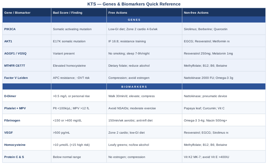
Moving from a general understanding of KTS to a targeted monitoring strategy starts with knowing which numbers to watch and why.
6 Biomarkers to Track in Klippel-Trenaunay Syndrome
KTS is not purely a skin or limb condition. At its core, it involves abnormal blood vessel development and chronically disrupted blood flow patterns. That makes certain blood markers far more informative for people with KTS than they would be in the general population. The six markers below were chosen because they reflect the mechanisms that actually drive KTS complications: coagulation activation, vascular inflammation, platelet dysfunction, and abnormal growth signaling. Each tells a different part of the story.
1. D-Dimer: Your Real-Time Clotting Activity Signal
Why it matters: People with KTS have a significantly elevated risk of deep vein thrombosis (DVT) and pulmonary embolism (PE) — not just from the structural abnormality of their veins, but from the chronic low-grade coagulation activation that accompanies venous stasis and turbulent flow within malformations. D-dimer is a fibrin degradation product: it rises whenever clot formation and breakdown are occurring simultaneously in the body. A persistently elevated D-dimer in someone with KTS may indicate that this process is ongoing even when outward symptoms appear stable.
Research by Mazoyer and colleagues published in Thrombosis and Haemostasis demonstrated that a substantial proportion of KTS patients show abnormal coagulation parameters — including elevated D-dimer and reduced fibrinogen — consistent with chronic localized intravascular coagulation within malformations. This state can remain clinically silent for extended periods before producing an acute event.
How to measure it: Standard D-dimer is available at any clinical laboratory through a simple blood draw. Cost ranges from $30–$80 depending on setting. High-sensitivity D-dimer assays are preferred. For KTS monitoring, establish a personal baseline and repeat every 6–12 months or whenever symptoms change — leg heaviness increasing, new skin changes, or unusual fatigue.
If the score is bad — the plan without supplements: Sustained elevation should prompt medical evaluation for active DVT, typically with Doppler ultrasound of the affected limb. Beyond clinical review, strict compression garment adherence is the most evidence-supported non-pharmacological approach. Daily walking (30+ minutes) activates the calf muscle pump and reduces venous stasis. Avoid prolonged immobility — particularly during travel — and elevate the affected limb during rest whenever possible. These are not optional lifestyle suggestions; they are functional anticoagulant mechanisms.
If the score is bad — the plan with supplements or equipment: Nattokinase (2,000–4,000 FU/day) has fibrinolytic properties and has been studied for mild antiplatelet and thrombolytic activity in cardiovascular contexts. Cycle 12 weeks on, 4 weeks off. Do not combine with prescribed anticoagulants without physician clearance. Serrapeptase (10–60mg/day, enteric-coated) may reduce fibrin accumulation; evidence remains weaker and largely from smaller trials. Sequential pneumatic compression devices — used at home for 30–60 minutes daily — dramatically improve venous return in KTS-affected limbs and are particularly valuable during illness or post-surgical immobility periods.
2. Platelet Count and Mean Platelet Volume (MPV): Monitoring Thrombocytopenia Risk
Why it matters: Large vascular malformations can trap and consume platelets — a process called Kasabach-Merritt phenomenon in its severe expression — leading to dangerously low counts and bleeding risk. Even sub-clinical platelet consumption, reflected as a declining platelet count paired with elevated mean platelet volume (MPV, which indicates accelerated platelet production and activation), signals that vascular malformations may be absorbing clotting resources faster than the bone marrow replaces them.
MPV above 12 fL indicates platelet activation and high turnover. When platelet count falls below 100,000/μL in a KTS patient, urgent hematology review is warranted regardless of symptoms. The combination of declining count with rising MPV is the warning pattern most worth recognizing early.
How to measure it: Platelet count and MPV are part of a routine complete blood count (CBC) — one of the cheapest and most widely available tests in medicine, typically $10–$40. MPV is reported automatically on most modern analyzers. Reticulated platelet percentage (an early marker of accelerated consumption) requires a specialized order and costs $50–$120. Baseline once, then every 6–12 months or when symptoms shift.
If the score is bad — the plan without supplements: Platelet count below 100,000/μL warrants specialist review before decisions about compression or physical activity intensity. Avoid NSAIDs (ibuprofen, aspirin) — these impair the function of already-reduced platelet numbers. Maintain moderate physical activity rather than complete rest, since venous stasis worsens platelet consumption within malformations. Any new bruising pattern, prolonged bleeding after minor injuries, or petechiae should trigger same-week medical contact.
If the score is bad — the plan with supplements or equipment: Papaya leaf extract (standardized, approximately 1,000mg/day for 5 days during acute drops) has modest evidence for supporting platelet production — do not use during active thrombosis or alongside anticoagulants. Curcumin at lower doses (200–500mg/day as liposomal form) may reduce platelet over-activation through NF-κB and COX-2 pathways — useful when MPV is elevated, though doses above 1,000mg/day may paradoxically extend bleeding time. Cycle 8 weeks on, 2 weeks off. Vitamin C (500–1,000mg/day) supports platelet membrane integrity and is safe long-term.
3. Fibrinogen: The Clotting and Inflammation Bridge
Why it matters: Fibrinogen is simultaneously a clotting factor and an acute-phase inflammatory protein. In KTS, it can be low — consumed by localized coagulation within malformations — or high — reflecting systemic inflammation. Both extremes carry risk: low fibrinogen increases bleeding vulnerability, while high fibrinogen increases the risk of clot propagation beyond the malformation site. Tracking fibrinogen over time provides a moving picture of whether localized coagulation is accelerating or stabilizing.
Cohort data from KTS patients has shown that a meaningful proportion carry fibrinogen values significantly below normal, consistent with chronic consumption within malformations, rather than deficiency of synthesis. This distinction matters for interpretation.
How to measure it: Fibrinogen is measured as part of a standard coagulation panel (alongside PT/INR and aPTT). Cost: $40–$100 as a standalone addition or included in comprehensive coagulation workups. Test fasting in the morning. Repeat every 6–12 months or after any thrombotic event or surgery. Normal range is typically 200–400 mg/dL; values below 150 mg/dL in KTS warrant investigation.
If the score is bad — the plan without supplements: Low fibrinogen in KTS almost always reflects active consumption and requires specialist evaluation rather than lifestyle optimization alone. For elevated fibrinogen, regular aerobic exercise — 150 minutes per week of moderate intensity — consistently lowers fibrinogen across multiple cardiovascular studies. A Mediterranean-style anti-inflammatory diet, minimizing refined carbohydrates and industrial seed oils, reduces systemic fibrinogen-driving inflammation. Losing even 5–10% of excess body weight produces measurable fibrinogen reduction.
If the score is bad — the plan with supplements or equipment: For elevated fibrinogen: omega-3 fatty acids (3–4g/day of EPA+DHA combined, triglyceride-form fish oil) have robust meta-analytic support for reducing fibrinogen and general cardiovascular inflammation. Reassess with a lipid panel at 12 weeks. Niacin (flushing form, 500–1,000mg/day with food) also reduces fibrinogen, but requires liver monitoring at doses above 1,000mg/day. For low fibrinogen caused by active consumption, no supplement replaces medical management — fresh frozen plasma or cryoprecipitate are the clinical interventions when levels become critical.
4. VEGF (Vascular Endothelial Growth Factor): Tracking Malformation Activity
Why it matters: VEGF is the primary molecular signal driving blood vessel growth. In KTS, abnormal PIK3CA or AKT1 mutations constitutively activate pathways that stimulate VEGF expression — meaning the body is chronically signaling for new vessel formation, even when that growth is structurally abnormal and harmful. Elevated circulating VEGF correlates with more extensive or progressively active malformations in vascular anomaly research, and several small studies have found elevated VEGF in KTS patients compared to controls.
While serum VEGF testing is not yet standard in KTS clinical protocols, it offers something conventional imaging alone cannot: a functional read on the activity of the underlying pathology, not just its anatomical extent. Tracking it may help identify periods of accelerated disease activity.
How to measure it: Serum VEGF-A is measured by ELISA-based assays. Cost: $80–$200 depending on the laboratory; it requires a specific physician order and is not part of standard metabolic panels. Values consistently above 500 pg/mL in adults are generally considered elevated in most laboratory reference ranges. Test in the morning under stable conditions — acute exercise transiently raises VEGF and should be avoided in the 24 hours before sampling.
If the score is bad — the plan without supplements: Sustained aerobic exercise at moderate intensity paradoxically helps normalize VEGF signaling — while VEGF spikes acutely during exertion, chronic exercise training induces receptor downregulation and improved endothelial function at rest. Zone 2 cardio (30–45 minutes, 4–5 days per week on low-impact equipment) is the most effective non-pharmacological approach to improving vascular health and stabilizing VEGF regulation. A low-glycemic, anti-inflammatory dietary pattern also reduces the constitutive VEGF expression associated with insulin resistance and chronic inflammation.
If the score is bad — the plan with supplements or equipment: Resveratrol (250–500mg/day of trans-resveratrol) modulates VEGF signaling downstream by influencing SIRT1 and PI3K pathways. Cycle 12 weeks on, 4 weeks off; mild GI discomfort is the most common side effect. Green tea extract standardized to EGCG (400–600mg EGCG/day with food) inhibits VEGF receptor phosphorylation and has demonstrated anti-angiogenic effects in cell and animal studies — human evidence remains limited but the safety profile is favorable. In more advanced presentations, sirolimus (rapamycin, an mTOR inhibitor) has been used off-label in PIK3CA-driven vascular anomalies with published case series showing partial regression of malformations. This requires management by a vascular anomaly specialist or relevant oncology center.
5. Homocysteine: The Vascular Damage Amplifier
Why it matters: Homocysteine is an intermediate amino acid that, when elevated, directly damages vascular endothelium through oxidative stress and impairs the function of natural anticoagulant proteins. In a condition already defined by structurally compromised blood vessels, elevated homocysteine can accelerate endothelial injury, increase thrombosis risk, and worsen the fragility of vessel walls that KTS has already altered. Homocysteine elevation is common in the general population — particularly in people carrying MTHFR variants, covered in the genetics section — and its impact is amplified against the background of existing vascular disease.
Thomas Dayspring, one of the most respected lipidologists in clinical practice, has consistently argued that homocysteine deserves standard inclusion in cardiovascular risk panels, not occasional ad hoc testing. For KTS patients, it deserves even higher priority given the dual vulnerability.
How to measure it: Fasting plasma homocysteine is measured by standard immunoassay at most clinical labs. Cost: $30–$80. Optimal range is below 10 μmol/L; values above 15 μmol/L represent a meaningful concern. Always test fasting. If elevated, retest at 3 months after intervention. Ongoing monitoring every 6–12 months is reasonable if MTHFR variants are also present.
If the score is bad — the plan without supplements: Moderate dietary methionine reduction (reducing very high red meat intake, which provides methionine as homocysteine's direct precursor) combined with consistently high green leafy vegetable intake (natural folate) addresses the primary metabolic drivers. Regular moderate exercise reduces homocysteine independently through multiple mechanisms. Avoiding excessive alcohol, which impairs B-vitamin metabolism and methylation capacity, is also important and often overlooked.
If the score is bad — the plan with supplements or equipment: This is one of the most reliably supplement-responsive biomarkers in medicine. The methylation stack — methylfolate (5-MTHF, 400–800mcg/day rather than folic acid), methylcobalamin B12 (500–1,000mcg/day), and pyridoxal-5-phosphate B6 (25–50mg/day) — lowers homocysteine in the majority of people within 8–12 weeks. Use the active methylated forms, particularly if MTHFR C677T is present. Trimethylglycine (betaine, 1,000–3,000mg/day) provides an alternative methylation pathway and can be added if the core stack is insufficient. No cycling required for these nutritional supports. Recheck at 3 months and adjust dose accordingly.
6. Protein C and Protein S: Natural Anticoagulation Capacity
Why it matters: Protein C and Protein S are natural anticoagulants — proteins that limit clot propagation once it begins. Deficiency of either significantly amplifies DVT and PE risk. In KTS, where venous flow is structurally abnormal and coagulation activation is ongoing, a coexisting Protein C or S deficiency transforms elevated risk into near-certain serious thrombotic complications at some point in life. Several published KTS cohort studies have noted that coexisting thrombophilia — including Protein C/S deficiency and Factor V Leiden — is disproportionately represented among KTS patients who present with acute thromboembolism.
This is arguably the single most actionable piece of thrombosis risk information you can obtain alongside D-dimer. Knowing this allows you and your specialist to make informed decisions about prophylactic anticoagulation long before an acute event forces the decision.
How to measure it: Functional Protein C and Protein S assays require a standard blood draw but must be performed when the patient is not currently anticoagulated and not during an acute inflammatory episode or active thrombosis (both can falsely lower levels). Cost: $80–$200 per test. Best ordered by a hematologist who can interpret the result in clinical context. Repeat testing is rarely needed unless initial results are borderline or clinical status changes significantly.
If the score is bad — the plan without supplements: Confirmed Protein C or S deficiency in KTS warrants specialist hematology consultation and an explicit discussion about prophylactic anticoagulation. Lifestyle modifications focus on eliminating additional thrombosis triggers: maintaining healthy body weight, rigorous compression adherence, consistent physical activity, aggressive hydration, and — most critically — complete avoidance of estrogen-containing contraceptives and hormone replacement therapy. Estrogen dramatically amplifies DVT risk in Protein S deficiency and should be treated as contraindicated until a thrombophilia specialist weighs in.
If the score is bad — the plan with supplements or equipment: No supplement restores Protein C or S levels in the setting of genetic deficiency — this is fundamentally a clinical management issue. That said, vitamin K2 (MK-7 form, 100–200mcg/day) supports the carboxylation reactions that activate Protein C and S, potentially optimizing their function at whatever level they are produced. Avoid high-dose vitamin E supplementation above 400 IU/day, which can further impair Protein C activity. For mechanical prevention, pneumatic compression devices remain the most evidence-supported non-pharmacological tool for reducing thrombosis risk in the affected limb.
---
With the blood markers addressed, it is worth looking one layer deeper — at the genetic architecture that shapes why these biomarkers behave the way they do in KTS.
Genes and Epigenetics: What the Research Reveals
Klippel-Trenaunay syndrome has historically been considered a sporadic, non-inherited condition. That understanding has been dramatically revised in the past decade. KTS is now recognized as part of the PIK3CA-Related Overgrowth Spectrum (PROS) — a family of conditions caused by somatic (acquired after conception, not inherited from parents) activating mutations in specific genes of the PI3K/AKT/mTOR signaling pathway. Because the mutations are somatic and mosaic — present in only a subset of cells — they cannot be detected on standard germline genetic tests, and they explain the asymmetric, patchy nature of the condition.
Understanding your genetic landscape, even through inference and available germline testing for modifier genes, helps anticipate which complications are most likely and which targeted interventions — including some now in clinical trial — may eventually apply. Ali Torkamani's work on variant interpretation and Gary Brecka's clinical application of genetic functional testing both highlight the value of moving from genotype to actionable biochemistry. That is the goal here.
Gene 1: PIK3CA — The Primary Molecular Driver
What it does: PIK3CA encodes the catalytic subunit (p110α) of Phosphoinositide 3-kinase — a pivotal enzyme in cell growth, survival, and proliferation. Activating mutations in PIK3CA lock the PI3K → AKT → mTOR signaling cascade in the "on" position in cells that carry the mutation. In KTS, these mutations are found within the abnormal vascular tissue itself. The result is constitutive signaling for cell growth and vessel formation, creating the overgrowth and vascular malformation phenotype. Keppler-Noreuil and colleagues formally described PROS in 2015, establishing PIK3CA as the central genetic driver across this spectrum.
Because PIK3CA mutations in KTS are somatic and mosaic, they are not detectable through consumer genetic testing (23andMe, etc.) — identifying them requires tissue biopsy and next-generation sequencing from the affected tissue, typically performed in specialized centers.
If the gene variant is active — the plan without supplements: While the somatic mutation itself cannot be modified by lifestyle, you can significantly reduce the signaling environment that amplifies its effects. A low-glycemic, calorie-sufficient diet reduces circulating insulin and IGF-1 — both of which stimulate PI3K independently, adding to the constitutive signal from the mutation. Chronic caloric excess and hyperinsulinemia feed the same pathway that PIK3CA mutations activate. Sustained Zone 2 aerobic exercise (4–5 sessions weekly, 30–45 minutes) improves insulin sensitivity and reduces PI3K pathway overdrive in peripheral tissues. Avoiding exogenous anabolic steroids or growth hormone is essential — these directly amplify PI3K/AKT/mTOR.
If the gene variant is active — the plan with supplements or equipment: Sirolimus (rapamycin, an mTOR inhibitor) is the most evidence-based pharmacological approach currently available. Multiple case series and small trials have shown partial regression of vascular malformations in PROS patients on low-dose sirolimus — this requires specialist prescribing, trough blood level monitoring, and awareness of infection risk. Alpelisib, a selective PI3Kα inhibitor, is approved for PIK3CA-mutant cancers and is being evaluated in PROS conditions including KTS. Nutraceutically: berberine (500mg, 2–3 times/day with meals) activates AMPK, which naturally suppresses mTOR signaling downstream. Cycle 12 weeks on, 4 weeks off; GI side effects common at higher doses. Quercetin (500–1,000mg/day) has demonstrated PI3K-inhibitory properties in cell studies with a favorable safety profile.
Gene 2: AKT1 — The Downstream Amplifier
What it does: AKT1 is the principal kinase immediately downstream of PIK3CA. Somatic AKT1 mutations — most commonly the E17K hotspot — cause Proteus syndrome, a condition that overlaps with KTS in its vascular and tissue overgrowth features. In atypical or borderline KTS presentations, AKT1 mutations should be considered during diagnostic workup. AKT1 activation promotes cell survival, growth factor independence, and suppression of normal programmed cell death — all of which contribute to uncontrolled vascular tissue proliferation.
If the gene variant is active — the plan without supplements: Time-restricted eating (16:8 intermittent fasting, eating within an 8-hour window) reduces PI3K/AKT activation during fasting windows by suppressing insulin and activating counter-regulatory pathways — AMPK increases, FOXO transcription factors are activated, mTOR is suppressed. This is one of the cleanest dietary levers for the PI3K/AKT axis. Regular resistance training, contrary to some concerns, does not meaningfully increase PI3K/AKT in vascular tissue when performed without anabolic agents.
If the gene variant is active — the plan with supplements or equipment: AKT inhibitors (capivasertib, ipatasertib) are in active pharmaceutical development but not available outside clinical trials for KTS currently. Nutraceutically: EGCG from green tea (400–600mg/day standardized extract) and resveratrol (250–500mg/day) both inhibit AKT phosphorylation in human cell studies. Metformin — available by prescription for type 2 diabetes — activates AMPK and suppresses AKT/mTOR; it has been discussed as a potential adjunct in PIK3CA/AKT1-driven overgrowth conditions. Clinical evidence in KTS is absent but the mechanistic rationale is substantive enough that specialist discussion is warranted.
Gene 3: AGGF1 (VG5Q) — The Angiogenesis Regulator
What it does: AGGF1 (Angiogenic Factor with G patch and FHA domains 1), also known as VG5Q, was the first gene specifically linked to KTS in a landmark 2004 Cell publication by Tian and colleagues. AGGF1 normally regulates angiogenesis — the formation of new blood vessels — and its overexpression or functional variants may contribute to the aberrant vessel proliferation seen in KTS. Its role is less clearly established than PIK3CA, and not all KTS patients carry identifiable AGGF1 variants. The mechanism appears to involve disruption of Wnt signaling and VEGF-independent angiogenesis pathways.
If the gene variant is present — the plan without supplements: Reducing chronic inflammatory load reduces baseline angiogenic signaling broadly. Smoking cessation is critical for anyone carrying AGGF1 variants — nicotine is a direct VEGF and angiogenesis stimulant that amplifies existing dysregulated vessel growth signals. Sleep optimization (7–9 hours consistent, timed sleep) supports physiological regulation of growth factors and dampens nocturnal inflammatory activity.
If the gene variant is present — the plan with supplements or equipment: Resveratrol (250–500mg/day) modulates Wnt/β-catenin and angiogenic signaling in multiple tissue contexts with a reasonable evidence base. Cycle 12 weeks on, 4 weeks off. Melatonin (0.5–3mg at bedtime) has demonstrated anti-angiogenic and anti-inflammatory properties in several human trials, and may be particularly relevant for KTS patients given common sleep disruption and the known nocturnal amplification of inflammatory cascades. No cycling typically needed at low doses; most benefit appears at consistent nightly use.
Gene 4: MTHFR — The Methylation Modifier
What it does: MTHFR (Methylenetetrahydrofolate reductase) is not a KTS-specific gene — it is one of the most common genetic variants in the general population, with C677T homozygosity present in approximately 10–15% of many populations. However, in KTS patients, MTHFR C677T homozygosity significantly amplifies the already-elevated thrombosis risk by impairing folate metabolism and raising homocysteine levels. This is the direct mechanistic bridge between Biomarker #5 above and your genetic profile. People with both KTS and MTHFR C677T homozygosity carry a compounded vascular burden that neither condition alone predicts.
Unlike PIK3CA, MTHFR variants are germline and fully detectable through consumer genetic testing (23andMe, AncestryDNA) or clinical SNP panels. Gary Brecka has brought significant public attention to MTHFR as an underappreciated driver of vascular and psychiatric pathology — his framework for practical methylation cycle support aligns closely with the clinical evidence, even where some extrapolations exceed what published trials strictly support.
If the gene is bad — the plan without supplements: Prioritize dietary folate from dark leafy greens (spinach, asparagus, kale, lentils) over synthetic folic acid, which requires conversion by the impaired MTHFR enzyme. Increase choline intake (eggs, liver) to support alternative methylation through the PEMT pathway. Moderate alcohol consumption, which impairs folate metabolism and methylation capacity, is particularly important to control when MTHFR variants are present.
If the gene is bad — the plan with supplements or equipment: The methylated B-vitamin stack is the evidence-supported intervention: methylfolate (5-MTHF, 400–800mcg/day), methylcobalamin B12 (500–1,000mcg/day), and pyridoxal-5-phosphate B6 (25–50mg/day). For MTHFR C677T homozygous individuals with KTS, this is arguably the highest-priority supplementation consideration given the dual thrombosis risk. Trimethylglycine (betaine, 1,000–2,000mg/day) provides the alternative methylation pathway and is additive when homocysteine remains elevated on the B-vitamin stack alone. No cycling required. Recheck homocysteine at 3 months.
Gene 5: Factor V Leiden (F5) — The Inherited Thrombophilia
What it does: The Factor V Leiden mutation (F5 R506Q) causes resistance to activated Protein C — the natural anticoagulant described in Biomarker #6. Heterozygous carriers face approximately 3–8× elevated lifetime DVT risk; homozygous carriers face 25–50× elevated risk compared to non-carriers. In KTS, a coexisting Factor V Leiden mutation essentially multiplies an already-significant thrombosis risk. Multiple published case series have noted Factor V Leiden in KTS patients who presented with DVT or pulmonary embolism as their first severe complication — a scenario that becomes more predictable when the genotype is known in advance.
Factor V Leiden is a germline variant and detectable through standard clinical genetic testing or consumer panels.
If the gene is bad — the plan without supplements: Factor V Leiden combined with KTS almost always warrants hematology consultation and explicit discussion of prophylactic anticoagulation, particularly around high-risk events (surgery, pregnancy, long-distance travel, immobility). Lifestyle focuses on eliminating all modifiable thrombosis triggers: maintaining healthy BMI, rigorous compression garment use, consistent daily movement, aggressive hydration, and absolute avoidance of estrogen-containing contraceptives and hormonal therapy. For air travel exceeding 4 hours, medical-grade compression stockings and scheduled in-flight calf exercises every 60 minutes are not optional precautions.
If the gene is bad — the plan with supplements or equipment: Nattokinase (2,000 FU/day) provides mild fibrinolytic adjunct activity — not as a replacement for prescribed anticoagulation, but as a supportive measure in consultation with your physician. Omega-3 fatty acids (3g EPA+DHA/day) reduce platelet aggregation and fibrinogen, addressing two of the three Virchow's triad components that Factor V Leiden amplifies. The combination of Factor V Leiden and KTS-related venous stasis is a recognized high-risk constellation. Portable Doppler ultrasound devices for home monitoring of the affected leg are increasingly available for consumer use and provide meaningful early warning for DVT development.
---
The biomarker and genetics frameworks above reflect the most current mechanistic understanding of KTS. The following section translates related principles from one of the most comprehensive practical resources on vascular monitoring currently available.
What Peter Attia's Outlive Teaches About Vascular Monitoring in KTS
Outlive: The Science and Art of Longevity by Peter Attia, MD, is not a book about rare vascular disorders. It is about the science and practice of living longer and healthier — but its most relevant chapters for anyone with KTS are those covering cardiovascular disease, coagulation biology, and what Attia calls "Medicine 3.0": aggressive early biomarker monitoring and intervention, years before disease becomes clinically obvious. For someone with KTS, this framework is not preventive luxury — it is operational necessity. The ten most impactful insights follow.
1. Biomarkers Are Meaningless Without a Personal Baseline
Attia argues throughout Outlive that reference ranges are population averages, not personal targets. Your D-dimer at 0.8 mg/L might fall within the "normal" laboratory range while representing a tripling from your stable personal baseline of 0.25 mg/L. In KTS, establishing and tracking your own trend over time is far more informative than any single value compared to a population reference. The goal is to know your normal so you can recognize your abnormal.
2. Lp(a) — The Hidden Vascular Risk Almost Nobody Tests
Attia considers lipoprotein(a) — Lp(a) — the most underappreciated vascular risk marker in standard medical practice. While not KTS-specific, elevated Lp(a) (above 30 mg/dL or 75 nmol/L) significantly increases both thrombosis and atherosclerotic plaque vulnerability. KTS patients with elevated Lp(a) carry a compounded vascular burden. Test it once — it is largely genetically determined and changes minimally with lifestyle — to understand your baseline risk. Cost: $30–$80. If elevated, it informs risk-threshold decisions with your vascular specialist.
3. ApoB — Better Than LDL for Assessing Vascular Particle Burden
Attia consistently advocates replacing LDL-C with ApoB (apolipoprotein B) as the superior measure of atherogenic particle burden. Every atherogenic lipoprotein carries exactly one ApoB molecule, making it the most direct count of particles that can embed in vascular walls. In KTS, atherosclerotic change in already-abnormal vessels can accelerate faster than in structurally normal vasculature. Keeping ApoB below 70 mg/dL is the threshold Attia recommends for individuals with elevated vascular risk. Cost: $30–$60.
4. Zone 2 Cardio as Foundational Vascular Medicine
Zone 2 training — low-intensity sustained aerobic effort at a pace where conversation is possible without gasping — is the cornerstone of Attia's exercise prescription for cardiovascular health. For KTS patients, it provides venous calf-pump benefit, improves insulin sensitivity (directly reducing PI3K pathway amplification), and supports endothelial function — all without the impact loading that aggravates affected limbs. Cycling, swimming, and elliptical work are the ideal KTS-compatible Zone 2 modalities. Four to five sessions weekly of 30–60 minutes.
5. Sleep Is a Vascular Intervention
Attia draws on extensive sleep research to demonstrate that insufficient or fragmented sleep raises inflammatory markers, elevates fibrinogen, increases platelet aggregation, and impairs vascular endothelial repair. For KTS — where each of these pathways runs chronically elevated — poor sleep is not a quality-of-life nuisance; it is a physiological risk multiplier. Seven to nine hours of consistent, properly timed sleep, with overnight pulse oximetry screening for sleep apnea (home devices available under $50), is a rational component of KTS risk management.
6. hs-CRP as the Inflammation Thermostat
High-sensitivity C-reactive protein (hs-CRP) is Attia's preferred general inflammation marker. In KTS, hs-CRP tracks systemic inflammatory load and can indicate when malformations are generating broader inflammatory activation beyond the affected limb. Optimal target: below 1 mg/L. Cost: $20–$60. Elevated hs-CRP in KTS should prompt assessment of sleep quality, dietary pattern, omega-3 status, and dental health — chronic low-grade gingival infection is a frequently overlooked hs-CRP driver.
7. Insulin and HOMA-IR — Metabolic Health Is Vascular Health
Attia emphasizes that insulin resistance is a foundational upstream driver of vascular dysfunction. In PIK3CA-driven conditions like KTS, where the PI3K pathway is constitutively active in affected cells, systemic insulin resistance potentially amplifies the signaling environment across all tissue. HOMA-IR (calculated from fasting insulin and fasting glucose) below 1.0 is Attia's optimal target. Improving HOMA-IR through time-restricted eating, resistance training, and low-glycemic dietary patterns directly reduces the biochemical context that PIK3CA mutations exploit. Cost for fasting insulin: $30–$60, alongside routine fasting glucose.
8. Fibrinogen as the Underrated Coagulation-Inflammation Bridge
While not always the headline of Attia's standard panel, fibrinogen appears throughout Outlive as a marker that uniquely bridges inflammation and clotting risk — the two primary pathological drivers in KTS. Including fibrinogen in regular KTS monitoring reflects exactly the kind of multi-axis thinking Attia advocates for high-risk vascular individuals. Paired with D-dimer, it provides complementary information that neither offers alone.
9. The Centenarian Reverse-Engineering Framework Applied to KTS
One of Attia's most compelling ideas is working backward from the physical capacities required for a good life at age 85–90 and training specifically for those benchmarks today. For someone with KTS, this means identifying which functional capacities matter most — cardiovascular endurance, single-leg stability, grip strength — and building toward them specifically, adapting methods to avoid loading affected anatomy while preserving real-world independence. Muscle mass also serves as an active venous pump, making resistance training an indirect vascular medicine.
10. The 5-Year (and 15-Year) Lead Time Rule
Attia's most challenging idea for conventional medicine is that every major cardiovascular event is preceded by a decade or more of detectable biomarker drift. The time to intervene is during that silent drift period, not at the clinical event. For KTS, where thrombotic complications can appear to arrive suddenly but typically develop from a background of progressive coagulation activation, this framework is directly applicable. The time to start tracking D-dimer, Protein C/S, homocysteine, and ApoB is not after a DVT. It is now.
---
Beyond biomarkers and genetics, several evidence-based physical and mind-body approaches have meaningful data for managing specific KTS-related complications.
Complementary Approaches Worth Considering
Low-Level Laser Therapy (Photobiomodulation)
Low-level laser therapy (LLLT) — also called photobiomodulation — uses red and near-infrared light (630–850nm) to stimulate cellular energy production, reduce localized inflammation, and promote tissue repair through mitochondrial activation. In KTS, where chronic wound healing difficulties, persistent edema, and recurrent soft tissue complications are common, LLLT offers a non-invasive adjunct to standard wound care. Photobiomodulation promotes ATP production in mitochondria, stimulates collagen synthesis, and has demonstrated anti-inflammatory effects on vascular endothelial cells in human tissue studies.
A controlled trial by Carati and colleagues (2003, Cancer) examined LLLT for lymphedema in cancer patients and found significant reductions in limb volume. While KTS-related lymphatic dysfunction differs mechanistically, the overlapping pathology — impaired lymphatic channels, chronic tissue edema — supports potential relevance. Smaller case reports using LLLT for chronic venous ulcers in vascular malformation contexts have reported faster healing rates compared to standard wound care alone.
In practice: use a 630–850nm device applying 3–5 J/cm² over affected skin areas excluding large malformations without prior specialist anatomical review. Sessions of 10–20 minutes, 3–5 times per week. Avoid direct application over actively infected wounds. Commercial home panels (Joovv, Mito Red, BioMax) are available in the $300–$600 range and provide sufficient irradiance for superficial tissue applications.
Massage Therapy — Manual Lymphatic Drainage
Manual lymphatic drainage (MLD) is a highly specialized form of massage that uses extremely gentle, skin-surface strokes to stimulate lymphatic vessel movement and reduce soft tissue edema. It is categorically different from standard deep tissue massage, which is contraindicated directly over vascular malformations. KTS frequently involves lymphatic channel impairment alongside venous anomalies, making the lymphedema component a distinct, often undertreated aspect of the condition for which MLD has the most relevant evidence base.
A systematic review by Lasinski and colleagues (2012, PM&R) examining complex decongestive therapy — which includes MLD — for limb lymphedema found consistent evidence for volume reduction and quality-of-life improvement. In KTS specifically, CDT incorporating MLD is increasingly included in specialist management protocols, though formal randomized controlled trial data specific to KTS-related edema remains limited.
In practice: MLD must be performed by a certified lymphedema therapist who is specifically informed of the KTS anatomy, particularly the location and extent of vascular malformations. Self-administered MLD following trained instruction from a certified therapist is a viable daily supplement to professional sessions. Combine with compression garment application immediately following MLD for maximal sustained effect. Avoid deep pressure over port-wine stain areas showing active skin changes.
Mindfulness Meditation and MBSR
Klippel-Trenaunay syndrome carries a significant chronic pain burden — neuropathic pain, musculoskeletal pain from limb asymmetry and gait compensation, and vascular congestion pain. This is compounded by the psychological weight of diagnostic uncertainty, the rarity of the condition, and the ongoing unpredictability of symptom progression. Mindfulness-Based Stress Reduction (MBSR) — an 8-week structured program originally developed by Jon Kabat-Zinn at UMass — has robust evidence for reducing chronic pain intensity, anxiety, and inflammatory markers including CRP and IL-6.
A meta-analysis by Hilton and colleagues (2017, Annals of Internal Medicine) examining mindfulness meditation for chronic pain found moderate evidence for pain reduction and meaningful improvements in quality of life across multiple conditions, with minimal adverse effects. In vascular contexts specifically, MBSR's documented reduction in cortisol and sympathetic nervous system activation is relevant — chronic sympathetic tone increases peripheral vascular resistance and platelet reactivity, both of which matter directly in KTS.
In practice: the full MBSR program is available through hospital wellness centers, university programs, and online platforms including Kabat-Zinn's original recordings. Apps such as Headspace and Waking Up provide accessible entry points. A commitment of 30–45 minutes daily for 8 weeks produces the most studied outcomes. The body scan practice is particularly relevant for KTS, as it builds nuanced bodily awareness and the capacity to observe sensations without amplification — a genuinely useful clinical skill when ongoing self-monitoring is a daily reality.
Breathing-Based Therapies
Controlled breathing practices — specifically slow-paced breathing at 4–6 cycles per minute with extended exhalation — activate the parasympathetic nervous system through vagal stimulation, reduce heart rate variability in the therapeutic direction, lower blood pressure, and decrease pro-inflammatory cytokine expression. These are measurable physiological effects with consistent replication across human studies, not theoretical proposals. For KTS patients, where elevated fibrinogen, D-dimer, and platelet activity can be partially amplified by chronic stress-inflammatory coupling, controlled breathing is a free, daily-use tool that addresses a genuine pathway.
A systematic review by Zaccaro and colleagues (2018, Frontiers in Human Neuroscience) provided a comprehensive overview of slow breathing's physiological effects, demonstrating consistent reductions in sympathetic activation and inflammatory markers across multiple conditions. Cardiac rehabilitation programs routinely incorporate slow breathing for its vascular and autonomic benefits, providing a parallel evidence base in structurally relevant populations.
Protocol: 5 minutes of slow breathing (4-count inhale through the nose, 6-count exhale through the mouth) 2–3 times daily — upon waking, midday, and before sleep. Box breathing (4-4-4-4) is an alternative used in cardiac rehabilitation. For acute KTS pain episodes or pre-procedure anxiety, 10 minutes of slow breathing has demonstrated analgesic and anxiolytic effects in several human trials. No equipment required, though guided apps (Breathwrk, Othership) support consistency in establishing the habit.
---
Conclusion
Klippel-Trenaunay syndrome sits in an uncomfortable space in modern medicine — rare enough that many clinicians have limited hands-on experience with it, complex enough that standard vascular guidance misses its most specific risks. The framework in this article does not replace specialist care. It gives you the tools to make that care more precise, more proactive, and better calibrated to your actual biological situation.
The most actionable immediate step is to start tracking. A D-dimer, complete blood count, fibrinogen, homocysteine, and Protein C/S assay are available at any clinical laboratory at modest cost and can be ordered by most general practitioners. If you have access to consumer genetic testing, identifying your MTHFR and Factor V Leiden status can meaningfully clarify your underlying thrombosis risk profile. If your D-dimer or homocysteine is elevated, you now have a starting framework for addressing it — through lifestyle modifications first, targeted supplementation second, and clinical escalation when markers demand it.
Bring this biomarker list to your next appointment with a vascular specialist or hematologist. Ask which are already being monitored and which are not. That conversation — grounded in specific, measurable data — may open a clinical dialogue that generic KTS management never quite reached.
Cardiovascular: Blood Vessel Conditions Vascular Conditions
Autoimmune: Inflammatory Conditions Connective Tissue Conditions