This article contains AI generated content.
· UpdatedKnee Ankylosis — 7 Biomarkers And 6 Genes To Track
Introduction
If your knee has lost significant range of motion — whether after surgery, infection, prolonged immobilization, or progressive inflammatory disease — you are likely familiar with how inadequate most general advice feels. "Stay active," "try physical therapy," "reduce inflammation" are all reasonable suggestions, but they rarely explain why your joint keeps stiffening, why your recovery stalls where others progress, or what is actually happening at the tissue level. Knee ankylosis, the partial or complete loss of joint motion due to fibrous adhesions or bony fusion, is a condition where individual biology matters enormously — and where surface-level recommendations often fall short.
The processes that drive ankylosis — chronic inflammation, excessive fibrosis, abnormal bone remodeling, cartilage breakdown — are measurable. They leave traces in your blood, your urine, and increasingly in your genetic profile. Ignoring these signals and simply hoping that movement and time will reverse the process means treating everyone the same, even though the underlying drivers can be completely different from one person to the next. One person's ankylosis is fueled by an overactive immune system. Another's is primarily a fibrosis problem. A third may have a nutritional deficiency that quietly amplifies joint damage at every step.
This is where precision tracking makes a real difference. Knowing which biomarkers are elevated — and understanding which genetic variants might make you more susceptible to fibrosis, aggressive inflammation, or poor vitamin D utilization — gives you a targeted starting point instead of a list of generic interventions. It allows you to work with a physician who can prioritize the right interventions for your specific biology, rather than cycling through the standard protocol without understanding why it is or is not working.
This article is built around that more precise approach. The primary section covers seven biomarkers that are most informative for anyone dealing with knee ankylosis, including what each one reveals, how to measure it, and concrete plans for when a result comes back unfavorable. A shorter genetics section follows, covering six key genes linked to ankylosis susceptibility. Beyond that, you will find a summary of one of the most relevant frameworks on inflammation and longevity, along with evidence-backed complementary approaches. Better information does not guarantee recovery, but it consistently leads to better decisions — and that is where meaningful change begins.
Summary
This article reveals 7 blood and urine biomarkers that track the core processes driving knee ankylosis — including inflammation, cartilage breakdown, fibrosis risk, and bone remodeling — with exact measurement methods, target ranges, and specific action plans for when results are poor. It then covers 6 genes that influence how aggressively your joint degrades, how much fibrosis you tend to produce, and how well your body regulates the inflammatory response — with protocols for each unfavorable variant, with and without supplements. After that, the article distills 10 key insights from Peter Attia's science-backed framework that directly challenge conventional thinking about joint inflammation and recovery. Finally, four complementary modalities with actual clinical evidence for joint conditions are reviewed — including the Autoimmune Protocol from Sarah Ballantyne, low-level laser therapy, tai chi, and mindfulness-based stress reduction — each with a realistic application protocol.
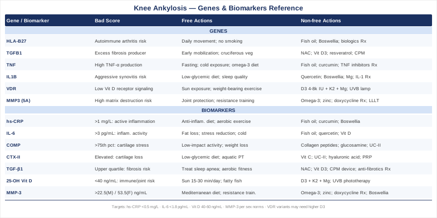
7 Biomarkers That Reveal What Is Driving Your Knee Ankylosis
Most clinicians managing knee ankylosis focus on structural findings — X-rays, MRI, physical examination. These are essential, but they describe the consequence, not the process. Biomarkers describe the process, which means they can reveal whether inflammation is still active, whether fibrosis is progressing, whether bone remodeling is dysregulated, and whether nutritional gaps are silently making everything worse. The seven markers below were selected for their specificity to the biological pathways most relevant to ankylosis, their clinical availability, and their direct connection to actionable interventions.
Biomarker 1 — hs-CRP (High-Sensitivity C-Reactive Protein)
Why it matters. C-reactive protein is produced by the liver in response to IL-6 and other pro-inflammatory cytokines. In the context of knee ankylosis, sustained elevation of hs-CRP signals that chronic systemic inflammation is still active — the same inflammatory environment that keeps the joint locked in a destructive, fibrotic cycle. Elevated hs-CRP is also independently associated with progression of joint disease in both rheumatoid arthritis and post-traumatic arthritis, two of the most common antecedents to ankylosis. Peter Attia, in his work on longevity biomarkers, considers hs-CRP one of the core panels any patient with chronic disease should be tracking routinely.
How to measure it. Standard blood draw processed at any clinical laboratory. Cost: $20–50 in the United States, often included in panels. Request the high-sensitivity version (hs-CRP), not standard CRP, as it detects lower levels of inflammation. Optimal target: below 0.5 mg/L (Attia's recommendation for low cardiovascular and inflammatory risk). Borderline concern: 1–3 mg/L. Active inflammatory concern: above 3 mg/L.
If the score is bad — the plan without supplements. The lifestyle factors most strongly linked to reduced hs-CRP are: adopting an anti-inflammatory dietary pattern (minimizing refined carbohydrates, ultra-processed foods, and seed oils; increasing vegetables, legumes, and fatty fish), improving sleep quality and duration (sleep deprivation raises CRP independently of other factors), engaging in moderate aerobic exercise three to five times per week (which acutely raises then chronically lowers CRP), and eliminating or significantly reducing alcohol. Maintain this approach for at least 12 weeks before reassessing. If you smoke, this is one of the strongest independent drivers of elevated CRP and addressing it is non-negotiable.
If the score is bad — the plan with supplements or equipment. Fish oil at 3–4 g combined EPA+DHA per day has consistent evidence for reducing hs-CRP (cycle 12 weeks on, reassess). Curcumin with piperine (500–1000 mg curcumin, 20 mg piperine daily) reduces CRP in multiple RCTs — take with a fat-containing meal for absorption. Boswellia serrata (300–400 mg AKBA-standardized extract, twice daily) has anti-inflammatory properties relevant to joint conditions. Side effects to monitor: fish oil can increase bleeding time at high doses; curcumin may interact with anticoagulants. Use cold thermotherapy (cold immersion or cold packs) on the knee joint to help locally reduce inflammatory signaling.
Biomarker 2 — IL-6 (Interleukin-6)
Why it matters. Interleukin-6 sits upstream of CRP in the inflammatory cascade. It is produced locally in the synovial tissue and systemically in response to infection, autoimmune activity, metabolic stress, and tissue injury. In knee ankylosis, IL-6 drives both the inflammatory phase that initiates joint damage and the fibrotic signaling that cements structural changes. It also stimulates osteoclast activity and bone remodeling in ways that may contribute to abnormal bone formation at the joint margins. Importantly, IL-6 can be elevated even when CRP is borderline, making it a more sensitive upstream marker.
How to measure it. Serum IL-6 is available through specialty laboratories and increasingly through standard labs. Cost: $80–150. Optimal range: below 1.8 pg/mL. Values above 3 pg/mL in a resting state suggest clinically meaningful inflammatory activity. Note that IL-6 fluctuates with exercise (it spikes acutely after intense training), so draw blood in a rested state, at least 48 hours after intense exercise.
If the score is bad — the plan without supplements. Visceral fat is a major independent source of IL-6 production. If body composition is a factor, targeted fat loss through caloric management and resistance training has a measurable impact on serum IL-6 within 8–16 weeks. Chronic stress and poor sleep also independently sustain elevated IL-6; structured stress reduction (daily diaphragmatic breathing, consistent sleep schedule, reduction of psychosocial stressors) can meaningfully lower IL-6 within weeks. Brief cold water exposure (cold showers or cold immersion) has been shown in controlled studies to transiently suppress IL-6.
If the score is bad — the plan with supplements or equipment. EPA and DHA (from fish oil) reduce IL-6 at doses of 2–4 g per day, with effects accumulating over 8–12 weeks. Quercetin (500–1000 mg/day) inhibits IL-6 production in macrophages and has shown effect in human inflammatory trials — cycle 8 weeks on, 4 weeks off to avoid receptor desensitization. Vitamin D supplementation has consistently shown IL-6-lowering effects in deficient individuals (see Biomarker 6). If IL-6 remains persistently elevated despite lifestyle and supplementation, this warrants discussion with a rheumatologist, as targeted biologics (IL-6 inhibitors such as tocilizumab) are available by prescription for specific inflammatory conditions.
Biomarker 3 — COMP (Cartilage Oligomeric Matrix Protein)
Why it matters. COMP is a glycoprotein found abundantly in cartilage, tendon, and ligament. When cartilage is mechanically stressed or biochemically degraded, COMP is released into the circulation. Elevated serum COMP is a specific and relatively early indicator of cartilage breakdown — it can rise before structural changes are visible on imaging. In the context of knee ankylosis, COMP tracks whether the cartilage within or surrounding the joint is actively degrading, which informs whether protective interventions are urgently needed or whether the process has stabilized.
How to measure it. Serum COMP is available through specialty laboratories (e.g., Hospital for Special Surgery affiliated labs, specialty rheumatology panels). Cost: $100–250. Reference ranges vary by laboratory and are age- and sex-adjusted, but generally values above the 75th percentile for your demographic warrant attention. Request trending over time rather than a single value, as trajectory is more informative than a snapshot.
If the score is bad — the plan without supplements. Reduce impact loading on the joint immediately — transition to low-impact activities (water exercise, cycling, elliptical) while elevated COMP normalizes. Excess body weight dramatically increases mechanical stress on the knee; even a 5–10% reduction in body weight measurably lowers COMP in individuals with knee joint disease. Physical therapy focused on quadriceps strengthening reduces joint loading through improved muscular support, which lowers ongoing cartilage damage signals. Avoid prolonged static loading (long periods of standing or kneeling) during the acute phase.
If the score is bad — the plan with supplements or equipment. Collagen peptides (specifically type I and II hydrolyzed collagen, 10–15 g/day, with vitamin C) have shown in RCTs to reduce COMP and improve joint symptoms — take at least 30 minutes before exercise on training days. Glucosamine sulfate (1500 mg/day) and chondroitin sulfate (1200 mg/day) have mixed evidence overall but show the strongest signal specifically in patients with elevated COMP and early joint space narrowing. Undenatured type II collagen (UC-II, 40 mg/day) works via oral tolerance mechanisms and may be more targeted. Side effect note: glucosamine should be used with caution in individuals with shellfish allergies or insulin resistance. Cycle supplements over 3-month periods and reassess COMP levels.
Biomarker 4 — CTX-II (C-Terminal Telopeptide of Type II Collagen)
Why it matters. CTX-II is a urinary marker of type II collagen degradation — the specific collagen that makes up hyaline cartilage. While COMP tracks cartilage matrix stress broadly, CTX-II is more directly linked to proteolytic cartilage breakdown by enzymes like MMPs and ADAMTS. Elevated CTX-II predicts progression of joint space narrowing in knee osteoarthritis and is considered one of the most sensitive biochemical markers for active cartilage loss. In a joint prone to ankylosis, tracking CTX-II reveals whether the cartilage component is still being actively destroyed or has reached a stable state, which changes the priority of interventions significantly.
How to measure it. First or second-morning urine sample, analyzed at specialty laboratories. Cost: $150–300. Values are corrected for urinary creatinine. Standard reference ranges are age- and sex-specific. For practical purposes, aim for the lower half of the reference range. Several European research laboratories and US specialty labs (e.g., Nordic Bioscience) offer validated CTX-II assays.
If the score is bad — the plan without supplements. Weight management is the single most impactful lifestyle intervention for reducing CTX-II in overweight individuals. A low-glycemic dietary pattern independently lowers cartilage degradation markers, likely through reduction of advanced glycation end-products (AGEs) that cross-link collagen and accelerate its breakdown. Avoid high-impact repetitive loading until CTX-II normalizes. Aquatic physical therapy is particularly appropriate here — it maintains joint mobility and muscle function without adding compressive loading to the cartilage.
If the score is bad — the plan with supplements or equipment. Vitamin C (500–1000 mg/day, from whole food or supplement) is essential for collagen synthesis and cross-linking — deficiency accelerates cartilage matrix breakdown. Type II collagen supplementation (UC-II, 40 mg/day) has the most direct mechanism for lowering CTX-II. Hyaluronic acid supplements (oral, 80–200 mg/day) have some evidence for reducing cartilage breakdown markers, though intra-articular injection remains more studied than oral administration. For severely elevated CTX-II, discuss with your orthopedic specialist whether intra-articular platelet-rich plasma (PRP) injections may be appropriate — PRP has shown measurable reductions in cartilage degradation markers in controlled studies.
Biomarker 5 — TGF-β1 (Transforming Growth Factor Beta-1)
Why it matters. TGF-β1 is arguably the most underappreciated biomarker in ankle ankylosis. It is the master regulator of fibrosis — the process by which normal tissue is replaced with scar tissue. In the knee, elevated TGF-β1 drives the formation of fibrous adhesions that physically restrict joint motion, the hallmark of fibrous ankylosis. TGF-β1 also promotes the differentiation of fibroblasts into myofibroblasts, cells that actively contract and shorten connective tissue. If your ankylosis is primarily fibrous (rather than bony), TGF-β1 is almost certainly a central player — and it is a marker that most standard joint panels miss entirely.
How to measure it. Serum or plasma TGF-β1 via ELISA, available through specialty and research-affiliated laboratories. Cost: $150–350. Values must be interpreted carefully as TGF-β1 in platelets can contaminate samples if not processed correctly — request platelet-poor plasma when possible. Reference ranges: serum values in healthy adults typically fall between 5–25 ng/mL, but research consistently shows that values in the upper quartile correlate with significantly increased fibrotic activity.
If the score is bad — the plan without supplements. Chronic hypoxia (from obesity-hypoventilation, untreated sleep apnea, or sedentary deconditioning) is a major independent driver of TGF-β1 upregulation. Addressing sleep apnea with CPAP, improving aerobic conditioning, and optimizing breathing mechanics (including through diaphragmatic breathing practice) can reduce TGF-β1 meaningfully. Eliminating excess alcohol reduces hepatic and systemic TGF-β1 production. A dietary pattern low in refined carbohydrates and high in sulforaphane-rich cruciferous vegetables (broccoli, cauliflower) supports detoxification pathways that regulate TGF-β1 activity.
If the score is bad — the plan with supplements or equipment. N-acetylcysteine (NAC, 600–1200 mg/day) inhibits TGF-β1-induced fibroblast activation and has been studied in fibrotic lung conditions with promising results. Vitamin D at optimal levels (see Biomarker 6) actively suppresses TGF-β1 signaling. Pirfenidone and nintedanib are prescription anti-fibrotic agents used primarily in pulmonary fibrosis — discussion with a specialist about off-label use in severe joint fibrosis may be warranted if all other approaches fail. For local application, continuous passive motion (CPM) devices, when used under physiotherapy supervision, apply mechanical stimulus that counteracts fibrotic contracture by applying shear stress to the adhesions. Side effects of NAC: occasional nausea at high doses; take with food. Cycle 8 weeks on, 4 weeks off.
Biomarker 6 — 25-OH Vitamin D
Why it matters. Vitamin D is far more than a bone mineral — it is a potent immunomodulator with receptors expressed on nearly every immune cell type. In the context of knee ankylosis, vitamin D deficiency is associated with increased synovial inflammation, impaired resolution of inflammatory responses, elevated TGF-β1, and reduced muscle strength (which accelerates mechanical joint stress). Multiple studies in rheumatoid arthritis, ankylosing spondylitis, and post-traumatic joint disease have found that low vitamin D correlates with worse disease activity and more rapid structural progression. Peter Attia lists 25-OH vitamin D as one of the essential tests in any comprehensive health panel.
How to measure it. Standard blood draw, widely available. Cost: $30–80, often covered by insurance for joint conditions. Test: serum 25-hydroxyvitamin D (25-OH D3). Optimal range for immune and musculoskeletal function: 40–60 ng/mL (100–150 nmol/L), per Attia's recommendations. Insufficiency: 20–40 ng/mL. Deficiency: below 20 ng/mL. Retest every 3–6 months when correcting levels.
If the score is bad — the plan without supplements. Midday sun exposure on arms, legs, and torso for 15–30 minutes (depending on skin tone and geographic latitude) can generate 10,000–20,000 IU of vitamin D per session. For individuals in northern latitudes, this approach has significant seasonal limitations. Increase dietary sources: wild salmon (600–1000 IU per serving), sardines, egg yolks, and UV-exposed mushrooms. Combined, dietary and sun exposure strategies can raise levels 5–10 ng/mL over 2–3 months, which may be insufficient if baseline is very low.
If the score is bad — the plan with supplements or equipment. Vitamin D3 supplementation (not D2) is effective and inexpensive. For deficiency (below 20 ng/mL): 5000 IU/day with reassessment at 3 months. For insufficiency (20–40 ng/mL): 2000–3000 IU/day. Always co-supplement with vitamin K2 (MK-7, 100–200 mcg/day) and magnesium glycinate (200–400 mg/day) — both are required for proper vitamin D metabolism and direct calcium appropriately to bone rather than soft tissue. Side effects: toxicity is rare below 10,000 IU/day but possible; always retest before increasing dose. A UVB phototherapy lamp ($100–300, medical-grade) can substitute for sun exposure in deficient northern-latitude individuals.
Biomarker 7 — MMP-3 (Matrix Metalloproteinase-3 / Stromelysin-1)
Why it matters. MMP-3 is a zinc-dependent enzyme produced by synovial fibroblasts and chondrocytes in response to inflammatory stimuli. It degrades collagen, proteoglycans, and other extracellular matrix components — driving both cartilage destruction and the reorganization of fibrous tissue within the joint. Elevated MMP-3 is found in rheumatoid arthritis, psoriatic arthritis, osteoarthritis, and post-traumatic joint disease. It predicts progression of joint damage in RA patients and is used in some European clinical protocols as a disease activity marker. In a joint developing ankylosis, MMP-3 tracks the activity of tissue-destructive enzymatic processes and can guide the intensity of anti-inflammatory interventions.
How to measure it. Serum MMP-3 via immunoassay, available at specialty labs and some hospital-based labs. Cost: $100–200. Sex-specific reference ranges: males, below 22.5 ng/mL; females, below 53.5 ng/mL (women have physiologically higher baseline MMP-3). Values significantly above these thresholds indicate active matrix-destructive activity.
If the score is bad — the plan without supplements. Anti-inflammatory dietary patterns (Mediterranean diet) consistently reduce MMP production in joint tissues. Eliminating or minimizing refined sugar reduces the AGE load that independently stimulates MMP production in chondrocytes. Resistance training — even at moderate intensity — reduces synovial inflammation and downstream MMP signaling over time (6–12 weeks of consistent training). Maintaining a healthy weight reduces the mechanical and metabolic drivers of MMP-3 overexpression in the knee joint.
If the score is bad — the plan with supplements or equipment. Omega-3 fatty acids (EPA+DHA, 3–4 g/day) inhibit MMP-3 transcription through NF-κB suppression — one of the most direct nutritional interventions for MMP reduction. Zinc (15–30 mg/day, as zinc picolinate) provides the cofactor for proper MMP regulation; both deficiency and excess zinc are problematic, so test serum zinc before supplementing. Doxycycline at sub-antimicrobial doses (20 mg twice daily, prescription only) is an MMP inhibitor that has been studied specifically for joint preservation; discuss with your prescribing physician. Boswellia serrata (400 mg AKBA-standardized extract, twice daily) inhibits MMP-3 through 5-LOX pathway suppression. Cycle omega-3 and boswellia together for 12 weeks, then reassess serum MMP-3.
6 Genes That Shape Your Ankle Ankylosis Risk and Recovery
Genetic testing through whole-genome sequencing or targeted SNP panels (23andMe, AncestryDNA, or clinical whole-genome options through companies like Invitae or GeneDx) can reveal meaningful predispositions that explain why your joint responds differently to standard interventions. The six genes below are the most clinically relevant to the pathways driving knee ankylosis. For each, understanding the implications allows you to pre-empt the downstream effects — not eliminate risk, but manage it more precisely.
Gene 1 — HLA-B27
What it does. HLA-B27 is not a single gene but an allele of the HLA-B gene on chromosome 6 that encodes a cell-surface protein involved in immune antigen presentation. Carrying HLA-B27 significantly raises the risk of seronegative spondyloarthropathies — including ankylosing spondylitis, reactive arthritis, and psoriatic arthritis — all of which can drive progressive joint ankylosis, including at the knee. Approximately 8% of the general population carries HLA-B27, but the prevalence among ankylosing spondylitis patients is 90–95%.
If the gene is bad — the plan without supplements. HLA-B27 carriers benefit strongly from maintaining spinal and joint mobility through daily structured movement. Low-impact exercise (swimming, cycling) performed consistently is more protective than occasional intense training. Avoid prolonged immobilization of the knee at all costs — even brief periods of disuse accelerate ankylosis in genetically susceptible individuals. A strict anti-inflammatory diet (eliminating refined starches and grains, which some researchers associate with triggering bacterial molecular mimicry in HLA-B27 carriers) is worth a 12-week trial. Smoking cessation is mandatory — smoking dramatically worsens outcomes in HLA-B27-positive inflammatory arthritis. Maintain good sleep posture using a firm, flat mattress.
If the score is bad — the plan with supplements or equipment. Fish oil (3–4 g EPA+DHA daily, ongoing) is the most well-supported anti-inflammatory supplement for HLA-B27-related inflammation. Boswellia serrata (400 mg twice daily) reduces 5-LOX-driven inflammation that is particularly active in spondyloarthropathies. For HLA-B27-positive patients with confirmed inflammatory arthritis and ankylosis progression, a rheumatologist should be consulted about disease-modifying therapies (DMARDs) or biologic agents (TNF inhibitors, IL-17 inhibitors). These are prescription therapies but represent the most powerful available intervention for halting ankylosis progression in HLA-B27-driven disease. Continuous passive motion (CPM) equipment can be used at home under physiotherapy guidance to maintain mechanical joint mobility.
Gene 2 — TGFB1 (TGF-β1 Gene)
What it does. Specific SNPs in the TGFB1 gene — particularly the rs1800470 (codon 10 Leu/Pro) and rs1800471 (codon 25 Arg/Pro) variants — influence baseline TGF-β1 production. Individuals with high-producer variants tend to mount more aggressive fibrotic responses to tissue injury, inflammation, or surgical intervention. In the context of knee ankylosis, this matters significantly: a post-surgical or post-infectious joint that would produce modest fibrous adhesions in most people may develop dense, joint-obliterating scar tissue in someone with high-TGFB1 variants.
If the gene is bad — the plan without supplements. Prioritize early mobilization after any knee injury or surgery — every additional day of immobilization is more fibrosis-promoting in high-TGFB1 individuals. Treat sleep apnea aggressively if present (hypoxia is a key TGF-β1 upregulator). Limit alcohol intake (promotes TGF-β1 hepatically). Adopt a dietary pattern rich in sulforaphane (from broccoli sprouts, cruciferous vegetables) — sulforaphane activates Nrf2 pathways that counteract TGF-β1-driven fibrosis. Manage chronic stress (elevated cortisol promotes TGF-β1 signaling in connective tissue).
If the gene is bad — the plan with supplements or equipment. NAC (600–900 mg/day) is the most accessible intervention for high-TGFB1 individuals — cycle 8 weeks on, 4 off. Vitamin D3 at optimal levels (40–60 ng/mL) actively suppresses TGF-β1-mediated fibroblast activation. Resveratrol (250–500 mg/day, with a fat-containing meal) has shown TGFB1-suppressive effects in cell and animal studies — human evidence is early but the safety profile is good. CPM devices used post-surgery or post-injury (under physiotherapy supervision) apply the mechanical signaling needed to counteract fibrotic contracture in high-risk individuals.
Gene 3 — TNF (TNF-α Gene)
What it does. The TNF gene encodes tumor necrosis factor-alpha, one of the most potent pro-inflammatory cytokines in the body. The rs1800629 SNP (G-308A promoter variant) increases TNF-α transcription, resulting in higher baseline and stimulus-induced TNF-α levels. This translates to more aggressive synovial inflammation, more rapid cartilage destruction, and a higher risk of progressive joint damage in inflammatory conditions. TNF-α also promotes osteoclast activity, which can contribute to bone erosion and abnormal remodeling at the joint — a contributor to bony ankylosis.
If the gene is bad — the plan without supplements. Intermittent fasting (16:8 or 5:2 protocols) has been shown to reduce TNF-α production independent of weight loss — a useful baseline lifestyle intervention for high-TNF individuals. Time-restricted eating reduces the metabolic endotoxemia that drives chronic TNF-α signaling. Cold exposure (brief cold showers or cold water immersion 3–4 times per week, 2–3 minutes at under 15°C) transiently suppresses TNF-α via norepinephrine release. An anti-inflammatory diet that eliminates seed oils (high omega-6) and prioritizes omega-3 sources shifts the eicosanoid balance away from TNF-promoting pathways.
If the gene is bad — the plan with supplements or equipment. Fish oil (4 g EPA+DHA/day) reduces TNF-α transcription; this is the best-supported nutritional intervention. Curcumin (500–1000 mg with piperine, twice daily) inhibits NF-κB, the transcription factor that drives TNF-α gene expression — cycle 12 weeks. Resveratrol (250–500 mg/day) also suppresses NF-κB. For confirmed inflammatory arthritis in high-TNF genotype carriers with ankylosis progression, TNF inhibitor biologics (etanercept, adalimumab, infliximab — prescription only) are the most powerful intervention and should be discussed with a rheumatologist.
Gene 4 — IL1B (Interleukin-1 Beta Gene)
What it does. IL-1β is one of the earliest and most potent drivers of synovial inflammation. The IL1B gene variants (particularly rs16944, the -511 C/T promoter SNP) influence how much IL-1β is produced in response to inflammatory triggers. High-producer IL1B variants are associated with more severe joint destruction in rheumatoid arthritis, faster progression of knee osteoarthritis, and greater pain sensitivity through central sensitization mechanisms. IL-1β also promotes pannus formation — the invasive synovial tissue overgrowth that contributes to joint destruction and subsequent fibrous ankylosis.
If the gene is bad — the plan without supplements. A low-glycemic, low-refined-carbohydrate diet reduces the repeated IL-1β spikes driven by postprandial glucose and insulin surges. Prioritize sleep quality (even one night of poor sleep measurably elevates IL-1β the following day). Manage psychosocial stress through structured daily practice — chronic stress maintains a low-grade IL-1β elevation that cumulatively damages joint tissue. Probiotic-rich foods (fermented vegetables, kefir, yogurt) support gut barrier integrity and reduce the bacterial translocation that triggers IL-1β in systemic circulation.
If the gene is bad — the plan with supplements or equipment. Quercetin (500–1000 mg/day) is the most accessible IL-1β inhibitor available as a supplement — it blocks NLRP3 inflammasome activation, the pathway that produces active IL-1β. Boswellia serrata reduces IL-1β-driven prostaglandin production. Magnesium (400 mg/day as glycinate or malate) reduces NLRP3 inflammasome activation in deficient individuals. For severe disease in high-IL1B carriers, prescription IL-1 inhibitors (anakinra, canakinumab) are the most targeted intervention — discuss with a specialist.
Gene 5 — VDR (Vitamin D Receptor Gene)
What it does. Even adequate serum vitamin D does not guarantee adequate vitamin D function — the VDR gene encodes the receptor through which vitamin D exerts its effects in immune cells, bone, and connective tissue. Key VDR SNPs — including FokI (rs2228570), BsmI (rs1544410), and TaqI (rs731236) — influence receptor efficiency. Individuals with low-efficiency VDR variants may show normal serum 25-OH vitamin D but inadequate downstream vitamin D signaling, resulting in continued immune dysregulation, poor bone quality, and impaired resolution of inflammation even with standard supplementation doses.
If the gene is bad — the plan without supplements. Maximize direct sun exposure (UVB light at the skin level produces vitamin D that enters the liver and kidneys through a pathway slightly different from supplemental vitamin D, potentially with some advantages for VDR signaling). Emphasize dietary sources of vitamin K2 and magnesium (essential cofactors for vitamin D receptor activation). Weight-bearing exercise promotes VDR expression in bone and muscle cells independently of circulating vitamin D levels.
If the gene is bad — the plan with supplements or equipment. VDR low-efficiency carriers often need higher supplementation targets to achieve the same functional outcome — targeting serum 25-OH vitamin D at the higher end (55–65 ng/mL) rather than the standard 40 ng/mL minimum. This may require 4000–8000 IU of D3 per day (under physician supervision with regular testing). Always co-administer vitamin K2 (MK-7, 200 mcg/day) and magnesium (400 mg/day). Some practitioners also use vitamin D analogs under prescription for VDR resistance states. A medical-grade UVB phototherapy lamp ($150–400) provides an additional source of vitamin D activation for VDR-variant individuals.
Gene 6 — MMP3 (Matrix Metalloproteinase-3 Gene)
What it does. The MMP3 gene contains a well-studied promoter polymorphism at position -1171 (rs3025058), involving a run of adenosines: the 5A allele is associated with higher MMP-3 transcription compared to the 6A allele. Individuals carrying the 5A/5A or 5A/6A genotype produce more MMP-3 in synovial fibroblasts in response to inflammatory stimuli, translating to more aggressive enzymatic degradation of cartilage and extracellular matrix. In RA patients, 5A carriers show accelerated joint space narrowing and higher rates of structural damage. For knee ankylosis, elevated MMP-3 in genetically susceptible individuals means the joint's extracellular matrix is more vulnerable to progressive remodeling.
If the gene is bad — the plan without supplements. Joint protection strategies are critically important for 5A carriers: avoid high-impact repetitive loading, use appropriate footwear, and consider knee bracing during higher-load activities. Weight management reduces mechanical MMP-3 stimulation. An anti-inflammatory dietary pattern (Mediterranean) reduces the cytokine signals that trigger MMP-3 transcription at the synovial level. Resistance training to strengthen the quadriceps and hamstrings reduces knee joint stress and therefore lowers mechanical MMP-3 induction.
If the gene is bad — the plan with supplements or equipment. Omega-3 fatty acids (3–4 g EPA+DHA/day) remain the strongest nutritional inhibitor of MMP-3 transcription. Zinc (15–25 mg/day, as picolinate — test serum levels first) regulates MMP activity. Doxycycline at sub-antimicrobial doses (20 mg twice daily, prescription) is a direct MMP-3 inhibitor studied in clinical trials for joint disease — discuss with a prescribing physician. LLLT/photobiomodulation applied locally to the knee (see Strategy 4) has also shown MMP-3-reducing effects in joint tissue in human studies.
What Peter Attia's Framework Reveals About Joint Inflammation You Won't Hear at a Routine Appointment
Peter Attia's Outlive: The Science and Art of Longevity (2023) is primarily a book about extending healthy lifespan, but its detailed framework on chronic inflammation, metabolic health, and biomarker-driven decision-making is directly applicable to anyone managing a joint condition like knee ankylosis. Attia challenges the passive, symptom-management approach that dominates conventional care and replaces it with a proactive, data-driven model. Here are the ten most impactful ideas from that framework as they apply to ankle ankylosis.
1 — Chronic inflammation is the silent accelerant behind most joint destruction
Attia argues that most of what we call "degenerative" joint disease is actually inflammatory joint disease playing out over years. The implication for ankylosis: treating the structural problem without addressing the inflammatory driver that created it is like mopping up a flood without turning off the tap. Tracking hs-CRP and IL-6 routinely is step one.
2 — hs-CRP below 0.5 mg/L should be the target, not the "normal range"
Most labs flag CRP as normal up to 3 mg/L or even 10 mg/L. Attia considers anything above 0.5 mg/L a marker of meaningful inflammatory activity that deserves investigation. Applied to knee ankylosis, being "within the normal range" does not mean the inflammatory environment is joint-safe. Push toward 0.5 or below.
3 — Zone 2 cardio is one of the most powerful anti-inflammatory interventions available without a prescription
Attia dedicates substantial attention to aerobic base training at low intensity (Zone 2: conversational pace, nose breathing). Consistent Zone 2 training (at least 3 hours per week, spread across 4–5 sessions) lowers IL-6, reduces visceral fat, improves mitochondrial function, and has a cumulative anti-inflammatory effect that compounds over months. For knee ankylosis, this means cycling, swimming, or elliptical — not running — at sustained low intensity, consistently.
4 — Muscle mass is not vanity — it is the primary insulin sink and an anti-inflammatory organ
Attia emphasizes that skeletal muscle is metabolically active tissue that absorbs glucose from the bloodstream (reducing inflammatory glycation) and produces myokines — anti-inflammatory signaling molecules released during contraction. Building and maintaining muscle mass around the knee is not just about mechanical support; it directly reduces the systemic inflammatory load that drives joint destruction.
5 — Sleep is the most underused recovery and anti-inflammation tool
Poor sleep (under 7 hours, fragmented sleep, or undiagnosed sleep apnea) independently elevates CRP, IL-6, TNF-α, and TGF-β1 — essentially all the markers that drive knee ankylosis progression. Attia treats sleep as a non-negotiable clinical intervention, not a lifestyle preference. If sleep is poor, no supplement stack will compensate.
6 — Vitamin D is a hormone, not a vitamin — and most people are taking inadequate doses
Attia's clinical target for serum 25-OH vitamin D is 40–60 ng/mL, which for most deficient adults requires 2000–5000 IU/day of D3 along with K2 and magnesium. He treats vitamin D deficiency as clinically relevant to musculoskeletal health, immune regulation, and inflammation control — three systems directly relevant to ankylosis.
7 — Continuous glucose monitoring reveals hidden inflammatory triggers in your diet
Even without diabetes, Attia recommends periodic use of continuous glucose monitors (CGMs) to identify foods that create outsized blood sugar spikes in your specific metabolism. Each spike triggers an inflammatory cascade that stresses the joint environment. Identifying and eliminating your personal glycemic triggers — not just "eating healthy" in general — can meaningfully reduce daily inflammatory burden.
8 — Omega-3 fatty acids require higher doses and longer timelines than most people use
Attia's clinical protocol uses 4 g combined EPA+DHA per day as a meaningful anti-inflammatory dose, sustained over months. Most people who "try fish oil" use 1 g/day for a few weeks and conclude it doesn't work. The REDUCE-IT trial (which Attia references) showed dramatic effects at 4 g/day of EPA — doses that require pharmaceutical-grade fish oil rather than a random drugstore supplement.
9 — Emotional stress is not separable from physical inflammation — it must be treated as a medical input
Attia draws directly on research showing that psychological stress elevates CRP, IL-6, and TNF-α to clinically meaningful degrees — comparable to dietary or exercise influences. For someone managing knee ankylosis, chronically elevated stress hormones maintain a low-grade inflammatory state that defeats even optimal pharmaceutical and nutritional interventions. Structured daily stress management (breathwork, NSDR, mindfulness) is not optional.
10 — Prevention is always easier than reversal — early biomarker tracking changes outcomes
Attia's central thesis is that waiting for symptoms or structural findings to make decisions is always too late. Tracking inflammatory and metabolic biomarkers early — before irreversible changes occur — is the defining difference between maintaining function and managing decline. For anyone with early signs of joint stiffening, beginning this biomarker tracking protocol now rather than in two years may be the single most impactful decision available.
Complementary Approaches With Clinical Evidence for Knee Ankylosis
The following modalities each have meaningful human clinical evidence relevant to knee joint conditions and inflammatory joint disease. They are most effective as additions to — not replacements for — the primary biomarker-guided and genetic approaches above.
Low-Level Laser Therapy (LLLT) / Photobiomodulation
Low-level laser therapy uses specific wavelengths of light (typically 630–1000 nm) at non-thermal intensities to stimulate mitochondrial cytochrome c oxidase, promoting cellular energy production, reducing oxidative stress, and modulating inflammatory mediators in joint tissue. For knee conditions specifically, LLLT has been shown to reduce MMP-3 expression in synovial tissue, lower levels of IL-1β and TNF-α locally, and improve microcirculation in tissues with adhesive changes. The mechanism is directly relevant to ankylosis: promoting cellular repair, reducing inflammatory enzyme activity, and improving tissue oxygenation in regions of fibrotic change.
A 2016 systematic review published in Photomedicine and Laser Surgery evaluated LLLT for knee osteoarthritis across multiple RCTs and found significant reductions in pain and improved range of motion compared to sham treatment, with the strongest effects using 904 nm infrared wavelengths at 1–3 J/cm² applied directly over the joint. The World Association of Laser Therapy (WALT) has issued dosing guidelines based on accumulated clinical trial evidence, recommending 1–4 J per point over the joint for anti-inflammatory effect and 4–8 J per point for tissue regeneration targets.
For home application, medical-grade LLLT panels or handheld devices (e.g., Joovv, Mito Red, or Thor Photobiomodulation — clinical-grade devices) can be applied to the knee for 10–20 minutes per session, 5 days per week. Consumer-grade devices cost $200–800; clinical devices $1000–5000. Realistic expectation: 6–12 weeks of consistent application before meaningful change in joint mobility or inflammatory markers. No significant side effects at therapeutic doses; avoid use if there is any suspicion of active joint infection or malignancy.
Tai Chi
Tai chi is a slow, continuous, low-impact movement practice derived from Chinese martial traditions. Its relevance to knee ankylosis lies in its unique combination of proprioceptive training, low-load progressive joint movement, dynamic muscular support training, and stress reduction — all without the compressive loading that would exacerbate damaged joint tissue. The gentle arc movements characteristic of tai chi continuously move the knee through its available range of motion without impact, providing the kind of sustained, low-grade mechanical stimulus that opposes fibrous adhesion formation.
A landmark randomized controlled trial published in Arthritis & Rheumatology (Wang et al.) compared tai chi to physical therapy in patients with knee osteoarthritis and found equivalent functional improvements, with tai chi additionally producing greater reductions in depression and greater patient adherence. Meta-analyses covering joint disease broadly consistently find significant improvements in pain scores, functional range of motion, and quality of life with 12+ weeks of regular tai chi practice compared to inactive controls.
For practical application, begin with a beginner tai chi program (video-guided is sufficient for starting) three times per week for 30–45 minutes. Tai chi for arthritis — a specific program developed by Dr. Paul Lam — has the most direct evidence for joint disease and provides a structured protocol appropriate for individuals with limited mobility. Within 4–6 weeks, most practitioners begin to notice improved balance, reduced stiffness, and gradually increasing range of motion. It is a safe practice with no meaningful adverse events reported in joint disease populations, though rapid progression should be avoided in the early weeks.
The Autoimmune Protocol (AIP) from Sarah Ballantyne
The Autoimmune Protocol, developed by Dr. Sarah Ballantyne (author of The Paleo Approach), is a dietary and lifestyle elimination-reintroduction framework specifically designed for conditions with autoimmune drivers. Given that a major proportion of knee ankylosis cases arise from or are worsened by autoimmune inflammatory arthritis — including rheumatoid arthritis, ankylosing spondylitis, and psoriatic arthritis — the AIP is directly relevant as a disease-management adjunct. The protocol removes foods hypothesized to increase intestinal permeability and immune activation (gluten, dairy, legumes, nightshades, eggs, nuts, seeds, alcohol, refined foods) and simultaneously emphasizes gut-healing nutrient density.
The evidence base for AIP is growing. A 2017 clinical trial in Inflammatory Bowel Diseases (Konijeti et al.) demonstrated significant reductions in inflammatory markers and symptom scores in Crohn's disease patients following AIP — establishing that the protocol does produce measurable anti-inflammatory effects in autoimmune conditions in humans. An expanding body of research on the gut-joint axis (the bidirectional relationship between intestinal microbiome, gut barrier integrity, and joint inflammation) supports the mechanistic rationale: many ankylosing spondylitis and RA patients show altered gut microbiomes and subclinical intestinal permeability, which the AIP directly addresses.
For practical application, commit to the full AIP elimination phase for 30–60 days — partial adherence significantly reduces efficacy. After the elimination phase, foods are reintroduced one at a time, every 5–7 days, while tracking joint symptoms, stiffness, and inflammatory biomarkers (hs-CRP, IL-6) to identify personal triggers. This diagnostic-reintroduction step is what makes AIP a personalized tool rather than a generic diet. Ballantyne's book and website provide detailed protocols including nutrient density tracking to avoid deficiencies during elimination. Consult a registered dietitian familiar with AIP if nutritional adequacy is a concern during the elimination phase.
Mindfulness-Based Stress Reduction (MBSR)
MBSR is an 8-week structured program developed by Dr. Jon Kabat-Zinn combining mindfulness meditation, body scan, and yoga-based movement with formal didactic instruction on stress physiology. Its relevance to knee ankylosis is both direct and indirect: directly, MBSR reduces perceived pain intensity and pain-related disability in chronic musculoskeletal conditions; indirectly, it reduces cortisol, IL-6, CRP, and TNF-α — the same inflammatory markers driving joint destruction. Importantly, MBSR addresses the central sensitization component of joint pain, which often develops alongside structural ankylosis and amplifies the experience of stiffness and immobility independently of tissue damage.
A 2015 systematic review published in JAMA Internal Medicine covering 47 randomized controlled trials (3515 participants) found that mindfulness meditation programs produced significant reductions in pain, depression, and psychological distress, with effects that persisted at follow-up. A specific RCT in rheumatoid arthritis patients showed that MBSR reduced psychological distress and improved subjective joint function measures compared to controls — relevant to the inflammatory joint disease population most likely to develop ankylosis.
For application, the gold standard is an in-person 8-week MBSR program (widely available through hospitals, universities, and wellness centers; cost $300–600 for the full program). Equivalent results have been demonstrated with rigorous online programs. Daily practice is key: formal sitting meditation, 20–45 minutes per day, plus informal mindfulness application throughout daily activities. Specific body scan practices that focus attention directly on the knee joint can help reduce central sensitization of knee pain signals. Realistic timeline: symptom benefits typically emerge at weeks 4–6 of consistent practice, with continued improvement over months.
Conclusion
Knee ankylosis is not a single problem with a single solution. It is the downstream consequence of specific, measurable biological processes — inflammation, fibrosis, cartilage breakdown, bone remodeling — that vary considerably in their intensity, their genetic susceptibility, and their responsiveness to different interventions. The seven biomarkers covered in this article give you a laboratory-grounded map of which processes are most active in your specific situation. The six genetic factors help explain why your joint may behave differently from someone else's under the same conditions. Together, they replace guesswork with targeted direction.
The most actionable next step is to start tracking. Request hs-CRP, IL-6, vitamin D, and MMP-3 from your physician as an initial panel — these are the most accessible and most immediately actionable markers. If you have access to genetic testing, HLA-B27 and the TGFB1 and TNF SNP profiles are the highest-yield additions. From there, work with a rheumatologist or sports medicine physician who is comfortable interpreting these results in the context of your joint condition. Complementary approaches like LLLT, tai chi, and dietary protocols can be layered in without waiting for test results.
Better data leads to better decisions. That is where recovery, or at least meaningful preservation of function, begins.
Musculoskeletal: Bone Conditions Joint Conditions
Autoimmune: Inflammatory Conditions