This article contains AI generated content.
Knee Cavernous Hemangioma: 4 Genes And 6 Biomarkers To Track
If you've spent time searching for answers about a vascular growth in your knee, you've probably noticed the information is scattered and inconsistent. Some pages describe a "hemangioma" the way they'd describe a birthmark. Others jump straight into orthopedic surgery terms without explaining why the joint keeps swelling on its own, seemingly without a fall or injury to blame. That gap between the plain-language description and the actual biology is where most people get stuck.
Generic advice tends to fail here because a knee vascular malformation isn't one uniform thing. It behaves differently depending on how it interacts with local blood flow, clotting, and inflammation, and depending on the underlying genetic mutation driving its growth. Telling someone to "rest and monitor it" or "see a specialist" is true but not actionable. It doesn't tell you what to actually ask for at your next blood draw, what an MRI report is really measuring, or why your surgeon might mention a drug like sirolimus instead of a supplement.
This article takes a more specific approach. It walks through the laboratory and imaging markers that clinicians actually use to characterize these lesions, the genes known to drive them, and where real supportive measures — with realistic limits — might fit into your routine. None of this replaces a hematologist, vascular anomalies specialist, or orthopedic surgeon, but it gives you a sharper vocabulary for that conversation.
None of this is about finding a miracle fix, because for a structural vascular lesion, there usually isn't one. But understanding which biomarkers are worth tracking, which genes are actually involved, and which supportive strategies have real evidence behind them can change how confidently you manage the condition day to day. That's the more grounded kind of hope this article is built around.
Summary
Before going deeper, it helps to know what you're actually working with. What's commonly called a "cavernous hemangioma" of the knee is, in the vast majority of cases, a slow-flow venous malformation of the joint lining (synovium) or surrounding soft tissue — not a true hemangioma in the strict pathological sense, and not the same disease as a brain "cavernous malformation" despite the overlapping name. That distinction matters because it changes which lab tests are useful and which genes are actually relevant.
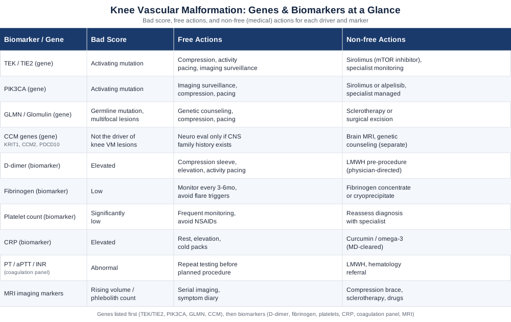
The most clinically useful biomarkers aren't exotic. They're markers of a phenomenon called localized intravascular coagulopathy — a low-grade, contained clotting process that happens inside the malformation itself and can explain pain flares, phlebolith formation, and bleeding risk during any future procedure. D-dimer, fibrinogen, platelets, coagulation panels, inflammatory markers, and MRI characteristics each tell a different part of that story, and each has a fairly clear "if this is abnormal, here's what tends to help" answer.
On the genetic side, two genes — TEK (also called TIE2) and PIK3CA — account for the overwhelming majority of sporadic venous malformations, including many in the knee. A rarer gene, GLMN, causes a related but distinct condition. And the CCM genes associated with cerebral cavernous malformations are, despite the shared naming convention, a different disease in a different tissue — worth knowing about mainly so you don't chase the wrong test. The sections below unpack all of this, plus a set of realistic supportive strategies for the days between medical appointments.
The Six Biomarkers Worth Tracking In A Knee Vascular Malformation
Before looking at genes, it's worth understanding what's actually measurable in your blood and on imaging right now, because these are the numbers that guide day-to-day decisions — including how safe a future surgery or sclerotherapy session will be. Most of what matters here traces back to one phenomenon: localized intravascular coagulopathy (LIC), a slow, contained clotting-and-breakdown cycle that happens inside the abnormal, sluggish-flowing vessels of the malformation itself. It's distinct from the more severe, platelet-depleting Kasabach-Merritt phenomenon seen with a different type of vascular tumor, and recognizing that distinction is itself clinically important. Research from Dompmartin and colleagues established that this coagulopathy is present in roughly 40% of people with venous malformations, and is strongly linked to lesion size and the presence of phleboliths — the small calcified clots that show up on imaging (Dompmartin et al., Archives of Dermatology).
D-dimer
Why it matters
D-dimer is a breakdown product of clot formation. In someone with a knee vascular malformation, an elevated D-dimer is one of the clearest signals that LIC is active inside the lesion. It correlates with lesion surface area and phlebolith burden, and clinicians use it to help distinguish a venous malformation from other vascular anomalies that don't cause this coagulation signature (Dompmartin et al.).How to measure it
A standard blood draw, processed as part of a coagulation panel. Cost is typically in the $20 to $60 range in the United States when billed outside of a bundled panel, and it's often already covered as part of pre-surgical workups.If the level is elevated: the plan without supplements
The evidence-based non-supplement options are compression garments over the affected knee to reduce venous pooling, activity modification during flares (avoiding prolonged standing or high-impact loading), and elevation. None of these change the underlying mutation, but they reduce the mechanical trigger for clot turnover. Frequency: daily wear of a fitted compression sleeve during waking hours, reassessed with your specialist every 3 to 6 months alongside repeat D-dimer testing.If the level is elevated: the plan with supplements or equipment
When D-dimer is significantly elevated, especially before a planned procedure, the actual medical intervention is low molecular weight heparin (LMWH) — a prescription anticoagulant, not a supplement — used short-term to reduce clot burden and surgical bleeding risk. This must be physician-directed. Outside of that context, there is no supplement with solid evidence for correcting LIC, and blood-thinning supplements such as high-dose fish oil or nattokinase are generally discouraged without direct supervision, since they can theoretically increase bleeding risk in a lesion that already bleeds and clots abnormally. If your specialist does approve adjunct omega-3 intake for general cardiovascular reasons, a typical dose is 1 to 2 grams daily, cycled with a pause if any unusual bruising appears, and stopped at least a week before any procedure.Fibrinogen
Why it matters
Fibrinogen tends to run low in active LIC because it's being consumed faster than the liver replaces it. A low fibrinogen alongside a high D-dimer is one of the more specific patterns for a clinically significant coagulopathy, and it's a key number surgeons check before any invasive procedure on the lesion (Venous Malformations and Blood Coagulation in Children).How to measure it
Included in a standard coagulation panel drawn with D-dimer; cost is usually $15 to $40 as an add-on, or bundled into a pre-operative panel.If the level is low: the plan without supplements
There isn't a lifestyle fix for consumptive fibrinogen depletion — it reflects what's happening mechanically inside the lesion. The realistic non-supplement approach is monitoring frequency (every 3 to 6 months, or before any planned procedure) and avoiding activities that visibly provoke swelling, since flares tend to track with lower fibrinogen readings.If the level is low: the plan with supplements or equipment
Severely low fibrinogen before surgery is corrected with fibrinogen concentrate or cryoprecipitate in a hospital setting — again a medical, not supplement-based, intervention. There's no supplement shown to safely raise fibrinogen in this context, and attempting to do so outside medical supervision isn't advisable given the bleeding risk already present.Platelet count
Why it matters
In LIC, platelets are usually normal or only mildly reduced. This is actually a useful negative: a significantly low platelet count points away from simple LIC and toward a different, more aggressive vascular tumor (like kaposiform hemangioendothelioma with Kasabach-Merritt phenomenon), which needs different management entirely. Platelet count is therefore as much a differential-diagnosis tool as a monitoring one.How to measure it
Part of a routine complete blood count (CBC), typically $10 to $30, often already included in annual bloodwork.If the count is abnormal: the plan without supplements
If platelets are mildly low, the standard non-supplement approach is simply more frequent monitoring and avoiding NSAIDs, which impair platelet function further. If they're substantially low, this should prompt reassessment of the diagnosis itself with your specialist rather than symptom management.If the count is abnormal: the plan with supplements or equipment
There's no supplement protocol for correcting platelet counts tied to a vascular lesion. Vitamin K, iron, or folate supplementation is only relevant if a separate, unrelated deficiency is identified on bloodwork — it will not change platelet behavior driven by the malformation itself.C-reactive protein (CRP)
Why it matters
CRP is a general inflammation marker. It tends to rise modestly during painful flares, thrombosis episodes within the lesion, or after a procedure, and can help separate an inflammatory flare from a mechanical or infectious cause of new knee swelling — an important distinction since infection in a joint is a medical emergency.How to measure it
A simple blood test, usually $15 to $50 depending on whether it's the standard or high-sensitivity assay.If the level is elevated: the plan without supplements
Rest, elevation, and short-term activity reduction during a flare are the mainstays. Applying cold packs for 15 to 20 minutes a few times a day during an acute flare is a reasonable, low-risk adjunct.If the level is elevated: the plan with supplements or equipment
Omega-3 fatty acids and curcumin have general anti-inflammatory evidence in musculoskeletal contexts, though not specifically studied in venous malformations. If your physician clears it, a typical curcumin dose is 500 to 1000 mg daily with piperine for absorption, taken with food, cycled for 8 to 12 weeks with a break to reassess necessity — and stopped before any procedure, since curcumin has mild antiplatelet effects that compound the lesion's own coagulation abnormalities. Mild GI upset is the most common side effect.Coagulation panel: PT, aPTT, and INR
Why it matters
Prothrombin time (PT), activated partial thromboplastin time (aPTT), and international normalized ratio (INR) give a broader picture of clotting function beyond D-dimer and fibrinogen alone. Imaging studies characterizing LIC severity in venous malformations use this fuller panel, not D-dimer in isolation, to grade risk before intervention (MRI phenotypes of localized intravascular coagulopathy in venous malformations).How to measure it
Standard venous blood draw, typically $20 to $70 as a bundled panel, almost always ordered together with D-dimer and fibrinogen before any procedure.If results are abnormal: the plan without supplements
This is a case where the "non-supplement plan" is really a scheduling plan: repeat testing closer to any planned procedure date, and looping in a hematologist if results are trending worse, since abnormal values here directly affect surgical or sclerotherapy safety.If results are abnormal: the plan with supplements or equipment
Correction, when needed, is done medically (LMWH pre-treatment, as discussed above, sometimes for several weeks before a planned procedure). There is genuinely no supplement equivalent here, and this is one of the clearer places in this article where the honest answer is "this needs a physician, not a supplement aisle."MRI-based imaging markers
Why it matters
MRI is the primary diagnostic and monitoring tool for knee vascular malformations, and it isn't just a yes/no test — specific features function as biomarkers in their own right. T2-weighted signal intensity, lesion volume, and phlebolith count all correlate with symptom severity and coagulopathy risk, and tracking them over time shows whether a lesion is stable or progressing (MRI phenotypes of LIC in venous malformations; case series in pediatric synovial hemangioma of the knee).How to measure it
An MRI of the knee, with or without contrast. In the United States this typically runs $500 to $3,000 depending on facility and insurance status; it's the single most expensive item on this list but also the most diagnostically central one.If findings are concerning: the plan without supplements
Serial imaging on a schedule set by your specialist (commonly every 6 to 12 months for a stable lesion, sooner if symptoms change), combined with a symptom diary tracking swelling, pain, and any bleeding episodes, gives you and your care team an early signal if the lesion is changing faster than expected.If findings are concerning: the plan with supplements or equipment
This is where equipment matters more than supplements: a properly fitted knee compression sleeve, and in some cases a hinged knee brace during higher-load activity, can reduce mechanical strain on a growing lesion. Image-guided treatments — sclerotherapy or, for mutation-confirmed cases, targeted drug therapy — are the actual interventions for a progressing lesion, and are discussed with your vascular anomalies specialist rather than self-directed.Taken together, these six markers describe a coherent picture: how much clotting activity is happening locally, whether that activity is stable or escalating, and how safe any future procedure would be. That picture becomes even clearer once you understand what's driving the lesion at the genetic level — which is where the mutations discussed next come in.
What Genetic Research Reveals About Why These Malformations Form
Genetic research on vascular malformations has moved fast over the last fifteen years, and it's worth understanding both what's solid and what's still preliminary. Unlike a metabolic gene variant that a supplement or lifestyle change might meaningfully offset, these are structural mutations driving abnormal vessel growth — so the honest framing here is less "how do I fix this gene" and more "what does this gene tell me about my options."
TEK (TIE2)
What it affects
TEK encodes the TIE2 receptor, which normally regulates how endothelial cells (the cells lining blood vessels) respond to growth signals. Somatic mutations in TEK — meaning they occur only in the lesion's cells, not in blood or other tissue — were identified as the cause of about half of sporadic venous malformations in a landmark study, with a rare inherited form caused by a germline version of the same mutation (Limaye et al., Nature Genetics; overview in GeneReviews, TEK-Related Venous Malformations). The mutation causes the receptor to signal constantly, even without its normal trigger, which drives the abnormal vessel channels seen on imaging.If the gene is implicated: the plan without supplements
Since this is a local, structural mutation, the realistic non-drug plan is the same monitoring and mechanical support described in the biomarker section — compression, activity pacing, and imaging surveillance — plus genetic counseling if there's a family history suggesting the inherited form, since that changes screening recommendations for relatives.If the gene is implicated: the plan with supplements or equipment
There is no supplement that addresses a TIE2 gain-of-function mutation. The actual targeted approach under active study is sirolimus, an mTOR-pathway inhibitor, which has shown measurable improvement in the coagulopathy associated with slow-flow vascular malformations in clinical study (Effect of sirolimus on coagulopathy of slow-flow vascular malformations). This is a prescription medication requiring specialist oversight and regular lab monitoring, not a self-directed option, and carries real side effects (mouth sores, elevated cholesterol, increased infection risk) that require monitoring during use.PIK3CA
What it affects
PIK3CA encodes a subunit of an enzyme (PI3-kinase) central to cell growth signaling. Somatic activating mutations in PIK3CA account for a substantial share of venous malformations that don't carry a TEK mutation, and sit within a broader group of conditions called PIK3CA-related overgrowth spectrum disorders (Somatic PIK3CA mutations as a driver of sporadic venous malformations).If the gene is implicated: the plan without supplements
Identical monitoring logic to TEK: imaging surveillance, mechanical support, and symptom tracking, since a PIK3CA mutation doesn't change the day-to-day non-pharmacological management much.If the gene is implicated: the plan with supplements or equipment
The mutation directly activates the same PI3K-AKT-mTOR pathway that sirolimus targets, which is why some PIK3CA-driven lesions respond to the same drug, and newer PI3K-specific inhibitors like alpelisib are being studied for more severe cases. Again, these are prescription interventions with defined side-effect profiles (elevated blood glucose, GI upset, rash) requiring specialist management — there is no over-the-counter equivalent, and framing it otherwise would be misleading.GLMN (Glomulin)
What it affects
GLMN is a different situation: it's typically a germline (inherited) mutation, with a "second hit" somatic loss occurring in the lesion itself. It causes glomuvenous malformations — a related but distinct entity from typical venous malformations, often described as bluish, cobblestone-like, and more tender to direct pressure. Four common founder mutations account for roughly two-thirds of familial cases (Four common glomulin mutations cause two thirds of glomuvenous malformations; genotype-phenotype data in Genotypes and phenotypes of 162 families with a glomulin mutation).If the gene is implicated: the plan without supplements
Because these lesions are often multifocal and hereditary, the useful non-drug step is a genetics consultation for the patient and first-degree relatives, plus the same compression and activity-pacing approach for symptomatic lesions.If the gene is implicated: the plan with supplements or equipment
No supplement alters glomulin function. Management is procedural — sclerotherapy or surgical excision for troublesome lesions — decided case by case with a dermatologist or vascular anomalies specialist familiar with glomuvenous malformation, per current clinical descriptions (StatPearls, Glomangioma (Glomuvenous Malformation)).The CCM genes: KRIT1, CCM2, and PDCD10
What it affects
This one needs a clear caveat. KRIT1 (CCM1), CCM2, and PDCD10 (CCM3) cause cerebral cavernous malformations — clusters of abnormal, low-flow vessels in the brain and spinal cord. Because the term "cavernous hemangioma" is loosely used across very different tissues, people researching a knee lesion sometimes come across these genes and assume they're relevant. For a soft-tissue or intra-articular knee lesion, they generally are not the driver; the biology, risk profile, and management are specific to the central nervous system. If a family history of cerebral cavernous malformations exists independently of a knee lesion, that's worth raising with a physician as its own, separate matter.If there's a family history: the plan without supplements
No supplement or lifestyle change is protective here. The evidence-based path is neurological evaluation and, if indicated, targeted MRI brain imaging and genetic counseling — unrelated to knee lesion management.If there's a family history: the plan with supplements or equipment
There is no supplement or device-based intervention with meaningful evidence for CCM gene carriers. This is squarely a specialist-managed condition, and any supplement claims targeting these genes specifically should be treated with skepticism.Genetic testing on lesion tissue (rather than blood) is increasingly used to confirm which of these mutations is present, mainly because it can determine eligibility for targeted drug therapy like sirolimus or alpelisib. That's a conversation worth having with a vascular anomalies center if standard monitoring and mechanical support aren't keeping symptoms manageable.
Ten Ideas From Outlive That Apply Directly To Vascular And Joint Health
Peter Attia's book Outlive: The Science and Art of Longevity isn't written about vascular malformations specifically, but its core argument — that most people wait too long to measure the things that actually predict future problems — maps unusually well onto a condition like this one, where coagulation markers and imaging are exactly the kind of "measure before it becomes a crisis" data Attia argues for. Here are the ideas from it most worth applying if you're managing a knee vascular malformation.
1. Medicine 3.0 means acting before the crisis, not after
Attia's central distinction is between reactive medicine, which waits for a diagnosis or an emergency, and proactive medicine, which tracks trends over years. For a slow-flow malformation, that means regular D-dimer and imaging checks even when you feel fine, not just after a painful flare.2. Lp(a) is worth testing once, because it's genetically fixed
Lipoprotein(a) is roughly 70 to 90% genetically determined and stable across most of adulthood, which is why major lipid guidelines now recommend testing it once in a lifetime rather than repeatedly (Lipoprotein(a): An important piece of the ASCVD risk factor puzzle). This is a useful mental model to borrow: some numbers, once known, don't need constant rechecking — which is exactly how genetic testing on your lesion tissue can work too.3. Cardiorespiratory fitness is one of the strongest predictors of long-term survival
A large cohort study following over 120,000 adults found no upper limit to the mortality benefit of higher fitness, with the highest fitness group showing roughly an 80% lower mortality risk compared to the least fit (Mandsager et al., JAMA Network Open). For someone managing a knee condition, this argues strongly for finding low-impact ways to maintain aerobic fitness (cycling, swimming, incline walking) rather than giving up on cardiovascular exercise altogether.4. Zone 2 training builds a metabolic base without joint strain
Attia repeatedly emphasizes low-intensity, sustainable aerobic training (roughly 70-80% of max heart rate) as a foundation most people neglect. This style of training happens to be lower-impact than sprinting or heavy plyometrics, making it a reasonable fit for a knee that needs to avoid sudden mechanical loading.5. Stability and strength training prevent the injuries that force bad decisions
A recurring theme is that most people don't die of frailty directly — they die of the fall or fracture frailty causes. Applied to a knee malformation, this reframes strength training around the surrounding hip and ankle musculature as protective: better stability means fewer awkward movements that provoke swelling or trauma to the lesion.6. ApoB is a more accurate risk marker than standard cholesterol
Attia argues that particle count (ApoB) predicts cardiovascular risk more precisely than LDL cholesterol alone. This isn't specific to vascular malformations, but anyone already dealing with one coagulation-related condition has a good reason to get a fuller, more accurate cardiovascular risk picture rather than relying on a basic lipid panel.7. Continuous glucose data reveals problems standard labs miss
Attia is an early advocate of continuous glucose monitoring in non-diabetics to catch metabolic dysfunction years before it shows up on a fasting glucose test. Chronic inflammation and metabolic dysfunction can compound baseline inflammatory markers like CRP, so this is a reasonable complementary data point if your CRP is running persistently high.8. The "Centenarian Decathlon" reframes goals around function, not numbers
Rather than chasing an abstract fitness score, Attia suggests defining the specific physical tasks you want to be able to do decades from now (carrying groceries, getting up off the floor, hiking) and training backward from those. For someone protecting a vulnerable knee, this is a more motivating and realistic framework than generic exercise advice.9. Emotional health is a fourth, often-ignored pillar
Attia devotes real space to the idea that emotional and relational health directly affects how well people adhere to every other health strategy. Managing a chronic, sometimes painful joint condition is genuinely stressful, and this is a legitimate reason to take that stress seriously rather than treating it as separate from the physical problem.10. Small, consistent inputs beat occasional intense interventions
The book's overall argument is that longevity is built from unglamorous daily consistency — sleep, movement, nutrition — rather than dramatic one-time fixes. That's a fitting note to end on for a condition where the actual leverage points are things like wearing a compression sleeve daily and keeping up with scheduled labs, not searching for a single breakthrough treatment.Complementary Approaches That Can Support Comfort And Recovery
None of the approaches below change the underlying malformation. They're realistic, evidence-informed additions for the anxiety, pain, and recovery periods that come with living alongside this condition — particularly around procedures like sclerotherapy or surgery.
Guided imagery
Guided imagery is a structured relaxation technique where a person is walked through vivid, calming mental scenes, often paired with slow breathing, to reduce the physiological stress response. It's relevant here specifically because people with knee vascular malformations frequently face recurring procedures — imaging, blood draws, sclerotherapy sessions — and procedural anxiety is a real, recurring burden rather than a one-time issue.A randomized clinical trial found that guided imagery relaxation therapy meaningfully reduced preoperative anxiety compared with standard care (Guided imagery relaxation therapy on preoperative anxiety: a randomized clinical trial). Similar approaches have also been studied for reducing procedural pain and distress during medical procedures in younger patients, which is directly relevant given how often this condition is diagnosed in children and young adults.
To apply it practically, a 10 to 15 minute recorded guided imagery session in the day or two before an imaging appointment or procedure, and again in the waiting room, is a low-cost, no-risk addition to standard care. It doesn't need equipment beyond headphones and a quiet space, and there are no meaningful side effects — it simply shouldn't be mistaken for a substitute for anxiety or pain medication when those are genuinely needed.
Progressive muscle relaxation
Progressive muscle relaxation (PMR) involves systematically tensing and releasing muscle groups to reduce overall physical tension and, secondarily, pain perception. It's a reasonable fit for a chronic joint condition because pain and muscle guarding around an affected knee often reinforce each other, and PMR directly targets that feedback loop.While large condition-specific trials in vascular malformation patients don't exist, PMR has a long track record in general chronic pain and preoperative anxiety literature, and its low cost and lack of side effects make it a sensible adjunct rather than a primary treatment. The technique is simple enough to self-administer once learned from a physical therapist or a reputable guided recording.
A realistic protocol is a 15 to 20 minute session, 3 to 4 times a week, ideally in the evening or before a stressful appointment, done seated or lying down to avoid unnecessary knee loading. There's no cycling requirement and no known side effects, though it should be paused if it triggers unusual dizziness (uncommon, but reported with deep relaxation techniques in a small subset of people).
Low-level laser therapy and photobiomodulation
This category needs a careful distinction. Higher-powered medical lasers (notably Nd:YAG) are an established, direct treatment for venous malformations themselves, with reported success rates above 90% for reducing lesion size and symptoms in appropriate candidates (Laser treatment of venous malformations). That is a specialist-administered medical procedure, not a wellness therapy, and it's mentioned here mainly so you recognize the term if a vascular anomalies specialist brings it up.Separately, lower-powered photobiomodulation (true "low-level laser therapy" in the wellness sense) has more general evidence for reducing pain and inflammation in various musculoskeletal contexts, though it has not been specifically studied in knee vascular malformations, and the evidence for chronic pain overall is mixed — some reviews show benefit only in the short term. This distinction matters: don't assume a consumer-grade photobiomodulation device will shrink the lesion the way a medical laser might.
If you and your physician decide a home-use, low-powered device is reasonable for general joint discomfort (not as lesion treatment), typical protocols use short sessions of 5 to 10 minutes over the painful area, 3 to 5 times weekly, for several weeks before reassessing benefit. Side effects are minimal at low power, but any device use over or near the malformation itself should be cleared first, since heat and vasodilation are generally handled with more caution in vascular lesions than in ordinary muscle soreness.
Conclusion
A knee vascular malformation is manageable largely through good information: knowing that D-dimer, fibrinogen, platelets, CRP, a full coagulation panel, and serial MRI findings together describe how active the lesion is, and knowing that TEK, PIK3CA, and occasionally GLMN mutations explain why it formed in the first place — while the unrelated CCM genes shouldn't be confused with this condition despite the shared naming. None of this replaces monitoring by a vascular anomalies specialist, hematologist, or orthopedic surgeon, but it should make those conversations sharper and less passive.
The practical next step is straightforward: if you haven't had a baseline coagulation panel and MRI, ask for one; if you have older results, compare them against the markers described here to see what's actually trending; and if genetic testing on lesion tissue hasn't been discussed, ask whether it would change your treatment options. Better information doesn't guarantee an easy path, but it consistently leads to better, less reactive decisions — which is the most realistic form of progress available for a condition like this one.