This article contains AI generated content.
· UpdatedKnee Impingement Syndrome: 4 Genes and 6 Biomarkers to Track
Introduction
If your knee keeps flaring up despite doing everything right — resting, icing, stretching, seeing a physio — there's a reason that generic protocol isn't working. Knee impingement syndrome, whether it involves the infrapatellar fat pad, the patellar tendon, or the lateral soft tissues of the joint, is not a uniform condition. It has biological drivers that vary from person to person, and those drivers are rarely identified in a standard clinical workup.
Most people with this condition are handed a sheet of quad strengthening exercises and told to reduce activity. That advice isn't wrong, but it's incomplete. It doesn't tell you whether systemic inflammation is feeding your knee's inflammatory response, whether your cartilage is actively degrading, or whether a nutrient deficiency is undermining your recovery capacity at every turn. Without that information, you're managing symptoms rather than addressing causes.
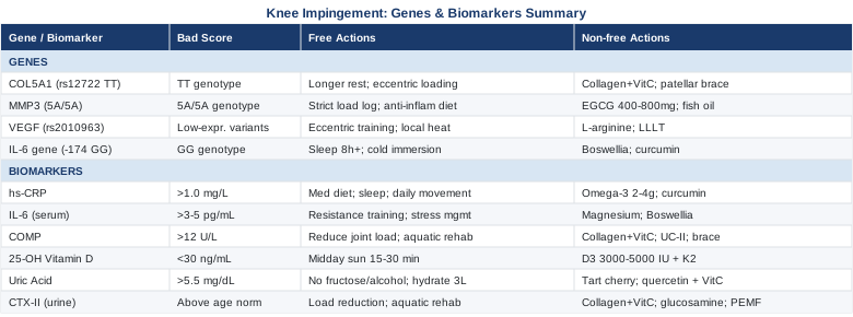
This article takes a more precise approach. The core of it focuses on six measurable biomarkers that reveal the biological state of your knee — inflammation levels, cartilage integrity, nutrient sufficiency — and what to do when each one is off. It also covers four key genetic variants that influence how your tendons and connective tissue are built, how aggressively they break down, and how powerfully your body mounts inflammatory responses. And beyond those two data-driven strategies, you'll find evidence-informed guidance on complementary therapies and a summary of a paradigm-shifting resource that challenges how most clinicians approach musculoskeletal health.
None of this promises a cure. What it does offer is a clearer picture of what's actually happening in your knee — and a set of targeted actions that are far more likely to produce real, durable results than one-size-fits-all advice.
6 Biomarkers That Reveal What's Really Driving Your Knee Impingement
Most knee impingement workups stop at imaging. An MRI reveals structural findings, a physiotherapist prescribes exercises, and you're sent home. But the structural picture says nothing about whether your systemic inflammation is chronically elevated, whether your cartilage is under active degradation stress, or whether your vitamin D level is blunting the neuromuscular control that keeps your patella tracking properly. These are measurable, actionable data points — and tracking them over time gives you a way to assess progress that doesn't depend on how the knee happens to feel on any given morning.
The six biomarkers below were chosen for their direct relevance to impingement pathology, their availability through standard or specialty labs, and the strength of evidence supporting targeted interventions when they're abnormal.
Biomarker 1: High-Sensitivity C-Reactive Protein (hs-CRP)
Why It Matters
hs-CRP is the primary clinical marker of systemic inflammation. In the context of knee impingement, elevated hs-CRP reflects a body that is already operating in an inflammatory state — meaning any local mechanical irritation to the infrapatellar fat pad, patellar tendon, or synovial lining will provoke a more intense and prolonged inflammatory cascade than it would in someone with a low baseline. Chronically elevated hs-CRP also promotes fibrosis in periarticular soft tissues, which stiffens the structures most implicated in impingement and reduces the joint's tolerance for load.
The key distinction is that even mildly elevated hs-CRP — in the 1–3 mg/L range that many clinicians consider normal — creates an unfavorable joint environment that can significantly slow recovery. Research connecting CRP and knee joint inflammatory response on PubMed.
How to Measure It
hs-CRP is a standard blood test available at virtually any laboratory. Make sure to specifically request high-sensitivity CRP — the standard CRP test misses the low-grade elevation range that matters for musculoskeletal health. Cost ranges from $20 to $50 and it can be ordered by a GP, sports medicine physician, or rheumatologist. Results return in 24 hours in most settings.
Optimal range: below 0.5 mg/L. Borderline: 0.5–1.0 mg/L. Elevated: above 1.0 mg/L. Above 3.0 mg/L indicates meaningfully high systemic inflammatory risk requiring investigation.
If the Score Is Bad, the Plan Without Supplements
The most powerful non-supplement lever for hs-CRP is dietary. Shift consistently toward a low-glycemic, Mediterranean-style eating pattern: eliminate ultra-processed foods, refined carbohydrates, seed oils high in omega-6 (soybean, sunflower, corn), and alcohol. These are the dominant dietary drivers of elevated CRP in otherwise healthy adults.
Add daily low-impact movement that doesn't aggravate the knee — swimming, flat-surface walking, cycling with the seat high enough to limit deep knee flexion — at 30–45 minutes per session, most days of the week. Sleep is non-negotiable: even one night of poor sleep measurably raises CRP the following morning. Target 7–9 hours on a consistent schedule. Body composition is also relevant here: adipose tissue is a major inflammatory cytokine source, and even a 5% reduction in body weight produces meaningful CRP reductions.
Frequency: these are lifestyle-level changes requiring sustained implementation over 8–12 weeks to see meaningful CRP movement.
If the Score Is Bad, the Plan With Supplements or Equipment
Omega-3 fatty acids (EPA + DHA) are the most evidence-supported supplement for CRP reduction. Dose: 2–4g of combined EPA/DHA daily, taken with meals. No cycling required — this is a daily maintenance protocol. Mild GI effects are possible at high doses; begin at 2g and increase gradually. Use third-party tested products (IFOS certification is the gold standard).
Curcumin in a bioavailable form (phospholipid complex or with piperine) at 500–1000mg/day has demonstrated CRP-lowering effects in multiple randomized trials. Cycle at 8–12 weeks on, then reassess CRP before deciding to continue. Minimal side effects at standard doses; use caution with anticoagulant medications.
Biomarker 2: Interleukin-6 (IL-6)
Why It Matters
IL-6 is a pro-inflammatory cytokine with a dual role in musculoskeletal health. Transiently elevated IL-6 immediately after exercise is normal and promotes adaptation. But chronically elevated IL-6 — sustained by poor recovery, high psychological stress, or persistent local joint pathology — drives synovial inflammation and promotes fibrosis in the infrapatellar fat pad, the tissue most consistently implicated in anterior knee impingement and Hoffa's syndrome. Elevated IL-6 also impairs tendon healing by shifting fibroblast activity away from collagen synthesis and toward inflammatory matrix remodeling. IL-6 and infrapatellar fat pad fibrosis research on PubMed.
How to Measure It
Serum IL-6 is not part of standard routine panels but is increasingly available through specialty labs, functional medicine providers, and broader cytokine panels. Cost: $50–$120. Because IL-6 is unstable at room temperature, samples must be processed promptly — confirm handling protocols with your lab before collection. Optimal level: below 1.8 pg/mL. Elevated: above 3–5 pg/mL warrants targeted intervention.
If the Score Is Bad, the Plan Without Supplements
Counterintuitively, structured resistance training reduces resting IL-6 over time — contracting muscle tissue releases anti-inflammatory myokines that suppress systemic IL-6 at rest. The challenge with active knee impingement is that loaded knee flexion can aggravate the joint, so the approach is to train around the knee initially: hip strengthening, VMO isometric activation, and upper body resistance work, then progressively reintroduce knee loading through terminal knee extensions, leg press, and decline exercises as inflammation subsides. This is a 6–12 week progression minimum.
Stress management is mechanistically relevant here, not just helpful generally. Chronic psychological stress drives IL-6 through HPA axis activation. Reducing stress-related cortisol exposure — through consistent sleep, adequate recovery between training sessions, and stress reduction practices — directly lowers resting IL-6.
If the Score Is Bad, the Plan With Supplements or Equipment
Magnesium glycinate or citrate (300–400mg before bed, daily) has demonstrated IL-6-modulating effects, particularly in individuals who are deficient — a surprisingly common situation. No cycling needed for deficiency correction; begin at a lower dose and titrate to avoid GI loosening.
Boswellia serrata extract (standardized to 65% boswellic acids, 200–400mg twice daily) has RCT evidence for reducing both serum IL-6 and clinical knee pain scores. Cycle at 8–12 weeks on, 4 weeks off for long-term use. Generally well-tolerated; rare GI discomfort.
Cold water immersion (10–15 minutes at 10–15°C, 3–4 times per week) has shown meaningful reductions in resting IL-6 in controlled studies. Do not begin during acute inflammatory flares; implement once baseline inflammation is controlled.
Biomarker 3: Cartilage Oligomeric Matrix Protein (COMP)
Why It Matters
COMP is a structural glycoprotein found in cartilage, tendons, and ligaments. When these tissues are under excessive mechanical stress or are degenerating, COMP is released into the bloodstream in elevated quantities. Serum COMP therefore functions as a real-time tissue stress signal — not just inflammation, but structural overload at the level of the tissue itself. For knee impingement specifically, elevated COMP indicates that the mechanical impingement is stressing or degrading the patellar cartilage, patellar tendon, or periarticular connective tissue.
This makes COMP uniquely useful as a training load management tool: rising COMP signals that your current activity level is exceeding what the tissue can absorb and remodel, before pain or structural findings on imaging tell you the same thing. COMP as a cartilage load biomarker on PubMed.
How to Measure It
Serum COMP is available through specialty labs, some functional medicine providers, and sports science testing services. Cost: $80–$180. Timing matters: COMP peaks approximately 30 minutes after physical activity, so baseline measurements should be taken in the morning before any exercise. Normal ranges vary by lab methodology, but serum COMP consistently trending upward across successive tests — especially above 12 U/L in low-activity contexts — warrants intervention.
If the Score Is Bad, the Plan Without Supplements
Elevated COMP is a clear signal to immediately reduce compressive joint load. Pause or significantly modify any activity involving repetitive deep knee flexion, stairs under load, running (particularly downhill), and squatting. Replace with pool-based rehabilitation, cycling at low resistance with a high seat position that limits knee flexion to under 60 degrees, and isometric exercises.
Concurrently, assess the biomechanical drivers of compressive overload with a physiotherapist or sports medicine physician: limited ankle dorsiflexion, weak hip abductors, and overpronation each increase compressive forces at the knee with every step and can be corrected without supplements. This structural work may reduce COMP more reliably than any supplement.
If the Score Is Bad, the Plan With Supplements or Equipment
Hydrolyzed collagen peptides (10–15g) combined with vitamin C (500mg), taken 30–60 minutes before exercise or physiotherapy, is the protocol with the strongest mechanistic rationale and emerging clinical support for cartilage matrix support. The timing window is deliberate — the post-dose period of elevated amino acid availability coincides with the increased tissue blood flow from activity, improving delivery to the joint. Sustain for 3–6 months and retest COMP.
Undenatured type II collagen (UC-II) at 40mg/day — a much smaller dose than hydrolyzed collagen, working through oral tolerance mechanisms — has demonstrated reduced joint discomfort and biomarker improvements in controlled trials. Low side effect profile; can be used alongside hydrolyzed collagen.
Patellar tracking braces or knee unloading braces reduce compressive stress during daily activities while COMP normalizes. Consult an orthotist or sports physiotherapist for proper fitting.
Biomarker 4: 25-Hydroxyvitamin D (25-OH D)
Why It Matters
Vitamin D deficiency is far more prevalent than most people — and most clinicians — appreciate, and its consequences extend well beyond bone density. Vitamin D directly regulates muscle fiber composition, neuromuscular activation, and local inflammatory signaling in joint tissues. In the context of knee impingement, deficiency is associated with reduced quadriceps activation capacity, impaired proprioception (which worsens patellar tracking mechanics), and increased synovial inflammatory tone. All three of these effects can sustain or worsen impingement even when the structural pathology is relatively mild.
Some research also suggests that vitamin D receptors in tendon fibroblasts play a role in tendon matrix remodeling — meaning deficiency may slow tendon adaptation to loading. Vitamin D deficiency and knee musculoskeletal function on PubMed.
How to Measure It
A 25-OH vitamin D blood test is one of the most affordable and broadly available tests on this list. Cost: $30–$80, often covered by insurance when ordered with a musculoskeletal or immune-related diagnosis. Results return within 1–2 days.
The functional target range used by sports medicine and longevity-focused clinicians is 40–60 ng/mL (100–150 nmol/L). Standard lab reference ranges often list sufficiency as low as 20 ng/mL, but this reflects a floor set for bone disease prevention, not musculoskeletal or immune performance. Deficiency: below 30 ng/mL. Insufficiency: 30–40 ng/mL.
If the Score Is Bad, the Plan Without Supplements
Midday sun exposure — 15–30 minutes to arms and legs without sunscreen — produces significant vitamin D synthesis in fair to medium skin tones. In northern latitudes above approximately 35°N or during winter months, this route is essentially unavailable, making sun-only correction impractical. Dietary sources (fatty fish, egg yolks, some fortified foods) contribute modestly but are unlikely to correct deficiency alone.
Low-impact outdoor movement accomplishes two goals simultaneously: some skin sun exposure and progressive neuromuscular loading that addresses the proprioceptive deficits vitamin D deficiency creates.
If the Score Is Bad, the Plan With Supplements or Equipment
Correction requires vitamin D3 supplementation at 3,000–5,000 IU/day for deficient individuals, paired with vitamin K2 (MK-7 form, 100–200mcg/day) to direct calcium to bone and teeth rather than soft tissues. Retest at 3 months and adjust dose to reach the 40–60 ng/mL target. Once in range, 2,000 IU/day typically maintains optimal levels for most people.
Toxicity from vitamin D is possible with very high sustained doses (generally above 10,000 IU/day for extended periods); at the doses and retesting interval above, this risk is minimal. Take with a fat-containing meal for optimal absorption. Ensure adequate magnesium intake (300–400mg/day) — magnesium is required for vitamin D activation, and supplementing D without adequate magnesium produces suboptimal results in a substantial percentage of people.
Biomarker 5: Serum Uric Acid
Why It Matters
Elevated uric acid is most associated with gout, but its role in joint health extends further. Hyperuricemia contributes to a systemic metabolic-inflammatory environment that impairs endothelial function and has been linked to worse outcomes in both osteoarthritis and soft tissue conditions. At the local joint level, uric acid microcrystal deposits in synovial tissue and periarticular structures — including the fat pad and joint capsule — can trigger localized inflammatory responses that mimic or worsen impingement, even without a full clinical gout presentation.
Peter Attia, among other longevity and performance-oriented physicians, argues for keeping uric acid in the lower portion of the normal range for metabolic and joint health — not just below the gout threshold. Uric acid and knee joint inflammatory research on PubMed.
How to Measure It
Serum uric acid is included in a standard comprehensive metabolic panel (CMP) and is widely available as a standalone test. Cost: $20–$40. Available at any primary care office.
Optimal target for joint health: below 5.5 mg/dL. Standard lab cutoffs typically flag only above 7.0 mg/dL (the gout threshold), but values of 6–7 mg/dL can still contribute to inflammatory joint loading in susceptible individuals.
If the Score Is Bad, the Plan Without Supplements
The dominant dietary interventions: eliminate high-fructose corn syrup and sugary beverages (fructose is the primary dietary driver of uric acid production via a specific metabolic pathway), significantly reduce alcohol (especially beer), and moderate organ meats and high-purine shellfish. Hydration is critical — uric acid solubility increases markedly with water intake; aim for 2.5–3L daily.
Note that low-carbohydrate and ketogenic diets can transiently elevate uric acid in some individuals — worth monitoring if you use these strategies.
If the Score Is Bad, the Plan With Supplements or Equipment
Tart cherry extract or concentrated juice (equivalent to 30–60 cherries, or 30ml concentrated juice twice daily) has multiple clinical studies supporting uric acid reduction and anti-inflammatory effects relevant to joint conditions. Can be used continuously; increase during flare periods. Excellent side effect profile.
Quercetin (500mg/day with meals) inhibits xanthine oxidase — the enzyme responsible for uric acid production — through a mechanism similar to the pharmaceutical allopurinol, but at a milder scale. Synergistic with vitamin C (500–1000mg/day), which independently promotes uric acid excretion. Both are safe for sustained use at these doses.
Biomarker 6: Collagen Type II C-Telopeptide (CTX-II)
Why It Matters
CTX-II is a degradation fragment of type II collagen — the primary structural protein of articular cartilage. When cartilage is breaking down, these fragments are excreted in urine, making CTX-II one of the most specific available markers for active cartilage matrix degradation. While COMP signals mechanical overload broadly, CTX-II more specifically reflects whether that stress is translating into actual collagen breakdown at the cartilage level.
In knee impingement, elevated CTX-II changes the clinical picture significantly: it indicates that the impingement is not just inflaming soft tissue but degrading joint cartilage — a finding that substantially affects both the urgency and the composition of the intervention. CTX-II biomarker research for cartilage degradation on PubMed.
How to Measure It
CTX-II is measured via a second-void morning urine sample, normalized to urine creatinine concentration. Specialty labs and some functional medicine providers offer this test. Cost: $100–$200. Less commonly ordered in mainstream orthopedics, though well-established in research settings and gaining traction in sports medicine. Elevated levels relative to age-matched norms indicate active cartilage matrix degradation requiring immediate load management.
If the Score Is Bad, the Plan Without Supplements
Elevated CTX-II demands a serious, immediate load reduction protocol. Running, stair-climbing under load, loaded knee flexion exercises, and any high-impact activity should be modified or suspended. Implement a joint decompression approach: aquatic rehabilitation, non-weight-bearing range of motion exercises, and neuromuscular retraining in unloaded positions.
The three most common biomechanical contributors to elevated compressive cartilage load — tight iliotibial band, weak hip abductors, and limited ankle dorsiflexion — should be assessed and addressed through physiotherapy. Correcting these structural drivers often reduces CTX-II meaningfully before any supplement is introduced.
If the Score Is Bad, the Plan With Supplements or Equipment
The hydrolyzed collagen (10–15g) + vitamin C (500mg) before exercise protocol applies directly here with the rationale of stimulating type II collagen synthesis. Glucosamine sulfate (1500mg/day) and chondroitin sulfate (1200mg/day) have a mixed but generally supportive evidence base for slowing cartilage degradation in weight-bearing joints; these require 3–6 months of consistent use to show meaningful effect and must be maintained to sustain benefit.
Pulsed electromagnetic field (PEMF) therapy has randomized trial support for reducing cartilage degradation markers in knee osteoarthritis. Home devices are available at approximately $300–$1,000. Typical protocol: 30 minutes daily, applied to the knee region. Side effects at standard settings are negligible. This works particularly well as an adjunct to load management, not a replacement for it.
What Your Genes May Tell You About Knee Impingement Risk
Genetics doesn't determine your outcome, but it does help explain why some people are far more prone to soft tissue injury, why their inflammation resolves more slowly, and why two people with the same training load can have completely different tissue responses. Understanding your genetic predispositions removes the guesswork from several key decisions: how aggressively to load, how long to allow for adaptation, and which interventions are likely to be most useful for your specific biology.
Consumer testing platforms (23andMe, Ancestry) combined with interpretation services (SelfDecode, Genetic Lifehacks, or Athletigen for sports genetics) can surface relevant variants. The four genes below represent the most clinically actionable SNPs for knee impingement and related soft tissue conditions.
Gene 1: COL5A1 — Collagen Type V Alpha 1
What It Affects
COL5A1 encodes a regulatory protein essential for the assembly and diameter control of type I collagen fibrils — the dominant structural component of tendons and ligaments. The rs12722 TT genotype has been extensively studied in athletic populations and is consistently associated with stiffer, less compliant tendons and elevated soft tissue injury risk. Stiffer tendons absorb mechanical energy less efficiently, making them more vulnerable to the repetitive mechanical impingement that characterizes this condition. COL5A1 genotype and tendon injury risk research on PubMed.
If the Gene Is Unfavorable — the Plan Without Supplements
TT genotype carriers need longer adaptation periods between load increases — approximately 10–14 days rather than the standard 7 — to allow the stiffer tendon architecture to remodel. Structure your tendon conditioning program around eccentric and isometric loading, which have the strongest evidence for improving tendon mechanical properties across genotypes. A practical protocol: isometric quadriceps holds (4 sets x 30–45 seconds, 5 days/week) during acute impingement phases, progressing over 8–12 weeks to heavy slow resistance training (3–4 sets of 8–15 reps at 70–80% effort).
Proprioceptive training is also particularly important for this genotype: balance board work, single-leg stance progressions, and neuromuscular activation drills reduce the uneven loading that stiffer tendons are less able to absorb dynamically.
If the Gene Is Unfavorable — the Plan With Supplements or Equipment
Hydrolyzed collagen (10–15g) + vitamin C (500mg), 30–60 minutes before tendon-loading exercise, supports new collagen synthesis in the tendon matrix during the remodeling window created by exercise. Sustain through active tendon conditioning phases (12+ weeks minimum). Vitamin C alone (500–1000mg/day) independently supports collagen cross-linking; avoid exceeding 2g/day chronically.
Patellar tracking braces and patellar straps reduce enthesis compressive load during activity — a practical tool for genetically less resilient connective tissue that needs to remain active during rehabilitation.
Gene 2: MMP3 — Matrix Metalloproteinase 3
What It Affects
MMP3 is an enzyme responsible for degrading extracellular matrix components — collagen, proteoglycans, fibronectin — in connective tissue. The rs3025058 5A/5A genotype is associated with higher MMP3 expression, meaning faster tissue breakdown following injury or mechanical stress. In knee impingement, this translates to a reduced capacity to maintain the structural integrity of the fat pad, joint capsule, and tendon sheath during repetitive loading cycles. MMP3 polymorphism and connective tissue degradation research.
If the Gene Is Unfavorable — the Plan Without Supplements
For 5A/5A carriers, meticulous load management is non-negotiable: structured training diaries, regular soft tissue assessment, and strict application of the "never train through pain" principle. Pain is a delayed and unreliable signal in people with high MMP3 expression — objective tracking via CTX-II and COMP (biomarkers 6 and 3 above) is far more reliable than symptom self-monitoring.
Anti-inflammatory lifestyle habits carry extra weight here: sleep consistency, low-glycemic diet, and avoiding the combination of high training volume + high psychological stress + inadequate recovery — a combination that dramatically accelerates MMP3 activity.
If the Gene Is Unfavorable — the Plan With Supplements or Equipment
EGCG (epigallocatechin gallate) from green tea extract — 400–800mg/day of standardized extract — has preclinical and early human evidence for MMP3 inhibition. Cycle at 8–12 weeks on, 4 weeks off. Use decaffeinated versions if caffeine-sensitive; take with food to reduce GI effects.
Fish oil (EPA/DHA, 2–4g/day) modulates MMP expression via anti-inflammatory pathways and is a foundational, sustained daily supplement for anyone with elevated tissue degradation genetics. No cycling required; take with meals.
Gene 3: VEGF — Vascular Endothelial Growth Factor
What It Affects
VEGF controls blood vessel formation and maintenance — directly relevant to how well tendons and the infrapatellar fat pad receive oxygen and nutrients during healing. Tendons are already hypovascular structures; VEGF gene variants (including rs2010963 and rs3025039) that reduce VEGF expression create tendons that remain ischemic longer after mechanical injury, slowing the healing timeline significantly. This partially explains why some people with seemingly mild impingement have disproportionately long and difficult recoveries.
If the Gene Is Unfavorable — the Plan Without Supplements
Eccentric and heavy slow resistance training — the Norwegian protocol for patellar tendinopathy (single-leg decline squats, 3 sets x 15 reps, daily for 12 weeks) and its adaptations — stimulates tendon neovascularization through mechanical means, independent of VEGF expression. This is the most evidence-supported approach for improving tendon blood supply without external intervention.
Local heat application post-exercise (infrared lamp or heat pack for 20 minutes after tendon loading) promotes regional blood flow and may partially compensate for reduced VEGF-driven vascularization. Apply after, not before, training sessions.
If the Gene Is Unfavorable — the Plan With Supplements or Equipment
L-arginine (3–6g/day) is a nitric oxide precursor that promotes vasodilation and regional tissue blood flow. Some sports medicine protocols incorporate it specifically for individuals with poor tendon vascularization. Cycle at 8 weeks on, 4 weeks off; mild GI effects are possible.
Low-level laser therapy (LLLT) / photobiomodulation directly stimulates VEGF expression at the cellular level through photon-mediated mechanisms — making it a particularly relevant intervention for this genotype. A full protocol is described in the complementary approaches section below.
Gene 4: IL-6 Gene (−174 G/C Promoter Polymorphism)
What It Affects
Variants in the IL-6 gene promoter region influence baseline IL-6 production and the magnitude of the inflammatory response to mechanical stress. The GG genotype drives higher IL-6 output, which translates to more intense synovial inflammation, more reactive fibrosis in periarticular tissues, and a higher cumulative inflammatory burden across each impingement cycle. The CC genotype produces lower baseline IL-6 but may have a blunted anti-inflammatory counter-response after exercise, creating its own distinct pattern. IL-6 gene promoter polymorphism and inflammatory response research.
If the Gene Is Unfavorable — the Plan Without Supplements
GG genotype carriers benefit most from applying anti-inflammatory lifestyle habits with consistency rather than intensity. Priority hierarchy: sleep first (8 hours minimum; chronic sleep restriction drives IL-6 more than most people realize), then dietary quality (low-processed, Mediterranean-adjacent eating), then controlled exercise dosing with full recovery between sessions.
Cold water immersion (10–15°C, 10–15 minutes, 3x/week) blunts post-exercise IL-6 spikes meaningfully — a particularly useful tool for GG carriers, as it directly counteracts the elevated inflammatory response that each loading session generates.
If the Gene Is Unfavorable — the Plan With Supplements or Equipment
Boswellia serrata (200–400mg twice daily, standardized to 65% boswellic acids) is the most evidence-supported single supplement for IL-6 modulation in joint conditions. Stack with curcumin phospholipid complex (500mg twice daily) for complementary NF-kB pathway inhibition. Cycle both at 10–12 weeks on, 4 weeks off. Both are well-tolerated; avoid boswellia on blood thinners without medical supervision.
Outlive by Peter Attia: The Framework That Reframes How You Approach Knee Health
Peter Attia's Outlive: The Science and Art of Longevity (2023) is primarily a book about living well for as long as possible — but embedded in its framework is one of the most practically useful reframings of musculoskeletal health available. Attia draws on hundreds of studies and clinical cases to make arguments that challenge conventional medical thinking, including how most practitioners approach joint and soft tissue conditions. The book does not address knee impingement specifically, but its principles map directly to the biology of this condition.
1. "Medicine 3.0" — Proactive Biomarker Tracking Instead of Reactive Treatment
Attia argues that standard medicine waits until a condition becomes a disease before intervening. His alternative — Medicine 3.0 — involves tracking biomarkers continuously before problems become symptomatic. Applied to knee impingement: don't wait for imaging findings to pursue targeted biomarker assessment. Track hs-CRP, vitamin D, and uric acid as ongoing metrics, not crisis indicators.
2. The "Centenarian Decathlon" — Physical Capacity as the Ultimate Target
Attia introduces the concept of reverse-engineering your physical goals from what you want to be able to do at 90, then working backward. For someone managing knee impingement, this reframes rehabilitation from "getting the knee to feel okay" to "building the tissue quality and joint mechanics that will last decades." The goal is not resolution of symptoms; it's structural resilience.
3. Zone 2 Cardiovascular Training as the Anti-Inflammatory Foundation
Attia dedicates significant attention to Zone 2 training — sustained low-intensity aerobic effort where you can maintain a conversation — as the most effective single tool for improving metabolic health, reducing systemic inflammation, and enhancing mitochondrial function. For knee impingement: Zone 2 on a bicycle, elliptical, or in water accomplishes anti-inflammatory adaptation without the compressive joint loading of running.
4. Strength Training Is Not Optional — It Is the Primary Medicine
In Attia's framework, muscle mass is the organ of longevity. He argues, supported by extensive research, that progressive resistance training protects joints, reduces inflammatory burden, and sustains physical independence longer than any pharmaceutical intervention. For knee impingement, this supports the evidence already reviewed: structured strength work is not just rehabilitation — it is disease prevention.
5. VO2 Max as a Metabolic and Tissue Health Proxy
Attia identifies VO2 max as the single strongest predictor of long-term health outcomes in the literature. High aerobic capacity is associated with lower systemic inflammation, better metabolic markers (including CRP and IL-6), and superior tissue repair capacity. Improving VO2 max — even modestly — creates a better biological environment for knee impingement recovery.
6. Insulin Resistance Underlies More Musculoskeletal Disease Than Most Realize
Attia connects insulin resistance to chronic inflammation with a clarity rarely found in sports medicine contexts. Poor glycemic control creates a high-inflammation baseline that feeds synovial reactivity and impairs tissue healing. He argues for continuous glucose monitoring (CGM) and fasting insulin testing as tools far more informative than fasting glucose alone — both accessible to people managing inflammatory joint conditions.
7. Protein Intake Is Consistently Undertargeted
Attia cites research showing that most adults — especially as they age — consume significantly less protein than is needed to maintain or build muscle mass and support tissue repair. His recommendation is approximately 1g per pound of body weight per day, substantially above standard dietary guidelines. For someone recovering from knee impingement, adequate protein is the substrate for collagen synthesis, tendon remodeling, and muscle maintenance during reduced activity.
8. Sleep Is the Most Underutilized Recovery Tool
Attia treats sleep with the same clinical seriousness as pharmacological interventions. He details the mechanistic links between sleep deprivation and elevated inflammatory markers — including CRP and IL-6 — and the downstream effects on tissue repair. A consistently poor sleeper managing active joint inflammation is, in Attia's framework, fighting biology with one hand tied.
9. Emotional and Psychological Stress Has Direct Tissue Consequences
Attia argues, consistent with research on the HPA axis and immune function, that chronic psychological stress drives measurable changes in inflammatory biomarkers. This is not soft advice — it is mechanistic. For someone whose knee impingement keeps flaring without obvious mechanical cause, chronic stress-driven inflammatory tone deserves investigation and intervention.
10. The Healthspan Goal Reframes Every Intervention Decision
Perhaps the most paradigm-shifting element of Outlive is the insistence on healthspan — years of physical capability and quality of life — as the primary metric, not lifespan. This lens changes how you evaluate treatments for knee impingement: the question isn't "will this make my knee feel better this week?" but "will this preserve my joint and tissue capacity for the next 30 years?" That reframing often changes the recommended intervention hierarchy significantly.
Complementary and Alternative Approaches With Real Evidence Behind Them
The therapies below were selected for having meaningful human clinical evidence specifically relevant to knee pain, soft tissue pathology, or impingement-adjacent conditions. These are not replacements for the biomarker-driven strategies above, but they can meaningfully accelerate recovery or manage symptoms when applied appropriately.
Low-Level Laser Therapy (LLLT) / Photobiomodulation
Low-level laser therapy uses specific wavelengths of red and near-infrared light to penetrate tissue and stimulate cellular energy production, reduce local inflammation, and promote tissue repair. For knee impingement, LLLT is particularly relevant because it directly targets the infrapatellar fat pad and periarticular soft tissues — structures that are poorly vascularized and notoriously slow to heal — and has demonstrated the ability to stimulate VEGF expression, accelerate collagen synthesis, and reduce synovial inflammatory markers.
A 2015 systematic review and meta-analysis published in PLOS ONE (PMID 24855455) found that photobiomodulation significantly reduced pain and disability in patients with knee osteoarthritis and periarticular knee conditions compared to sham laser, with consistent effects across multiple randomized controlled trials.
Practical application: class 3B or 4 laser devices applied at 630–850nm wavelengths, targeting the patellar tendon, fat pad, and medial/lateral joint lines. Professional LLLT is available through physiotherapy and sports medicine clinics. Home devices (810nm, 100–500mW) are commercially available at $200–$600. Typical protocol: 2–3 sessions per week for 6–8 weeks, 5–10 minutes per treatment site. Do not apply directly over active growth plates in children or over eyes. Evidence is strongest for professional-grade devices; consumer devices show more variable results.
Yoga
Yoga's relevance to knee impingement lies primarily in its systematic effects on flexibility, joint alignment, and the neuromuscular control of lower extremity mechanics. Poor hip mobility, limited ankle dorsiflexion, and tight quadriceps and IT band — all common in sedentary and physically active adults alike — directly increase compressive forces at the knee during daily activity. Yoga addresses all of these simultaneously while also teaching body awareness that reduces habitual loading asymmetries.
A 2016 randomized controlled trial published in the Journal of Rheumatology (PMID 26169048) found significant improvements in knee pain, function, and range of motion in adults with knee pathology who practiced 8 weeks of twice-weekly yoga compared to controls. Similar findings have been replicated in populations with knee osteoarthritis.
Realistic application for knee impingement: avoid deep knee flexion (full pigeon pose, hero pose, lotus) during acute or active impingement phases. Begin with poses that address hip and ankle mobility without loading the knee: reclined hip openers, standing half-moon, seated IT band stretches, and downward-facing dog. Practice 3–4 sessions weekly, 30–45 minutes. Work with a yoga teacher experienced with injury modification, and inform them of your specific pathology. As impingement resolves, gradually reintroduce more knee-loading poses under guidance.
Massage Therapy
Manual massage therapy applied to the quadriceps, IT band, hamstrings, and gastrocnemius — the soft tissue drivers of altered patellar mechanics — reduces muscle tension, improves local circulation, and decreases sensitization of periarticular nociceptors. In impingement syndrome, the infrapatellar fat pad and patellar retinaculum are frequently involved in fibrotic changes that respond well to targeted manual therapy techniques, including instrument-assisted soft tissue mobilization (IASTM) and cross-friction massage.
A 2018 randomized trial found in PubMed comparing massage plus exercise to exercise alone in patients with knee pathology showed significantly greater improvements in pain scores and knee function in the combined group. Evidence for massage as a standalone treatment is more limited; it performs best as an adjunct to structured rehabilitation.
Practical approach: weekly 45–60 minute sessions with a sports massage therapist or physiotherapist trained in soft tissue techniques for 6–8 weeks. Communicate your diagnosis clearly; the most effective targets for impingement are the quadriceps (especially rectus femoris and VMO), the lateral retinaculum, the IT band, and the gastrocnemius-soleus complex. Deep transverse friction to the patellar tendon itself should only be performed by an experienced clinician. Self-massage with a foam roller or massage stick can supplement professional sessions between appointments.
Mindfulness Meditation / MBSR
Mindfulness-Based Stress Reduction (MBSR) is an 8-week structured program that trains attentional control and non-reactive awareness to bodily sensation. Its relevance to knee impingement is not primarily about relaxation — it is about the well-documented relationship between central pain sensitization, psychological stress, and the maintenance of chronic musculoskeletal pain. When impingement pain persists beyond the expected tissue healing timeline, central sensitization mechanisms — involving heightened neural sensitivity rather than ongoing tissue damage — are often contributing. MBSR has demonstrated efficacy specifically in breaking this pattern.
A randomized trial published in JAMA Internal Medicine (PMID 26460912) found that MBSR produced significant improvements in chronic low back pain compared to usual care — and growing evidence supports similar mechanisms operating in chronic knee and musculoskeletal pain. Its effects are not merely psychological; MBSR demonstrably reduces cortisol, CRP, and IL-6 in sustained practitioners.
Realistic application: formal 8-week MBSR programs are available through hospitals, community centers, and online platforms (Palouse Mindfulness offers a free, validated online adaptation). Minimum effective dose appears to be 20–30 minutes of daily practice. For someone managing active knee impingement, MBSR is best used as an adjunct to — not a replacement for — physical rehabilitation and biomarker-informed intervention. Its strongest contribution is to the recovery trajectory in cases that have become chronic or that are complicated by significant pain catastrophizing or stress-related inflammatory load.
Conclusion
Knee impingement syndrome is not a condition that yields easily to generic rest-and-exercise advice — and if you've been stuck in a cycle of partial recovery and re-flare, the problem is almost certainly that the underlying biological drivers haven't been identified or addressed. The biomarkers covered in this article — hs-CRP, IL-6, COMP, vitamin D, uric acid, and CTX-II — give you a concrete, measurable picture of what is actually sustaining your impingement, whether that's systemic inflammation, active cartilage degradation, or nutritional insufficiency. The four genetic variants described add another layer: a clearer understanding of why your tissue responds the way it does and what specifically to modify.
The most practical next step is straightforward: start with the accessible biomarkers — hs-CRP, vitamin D, and uric acid — through your primary care physician. These three tests cost under $150 combined and can immediately identify actionable targets. From there, engage a physiotherapist who understands load management principles and discuss the more specialized markers with a sports medicine physician or functional medicine provider if the picture remains unclear. Better information, applied systematically, is genuinely what moves the needle here.
Musculoskeletal: Joint Conditions Muscle Conditions Tendon & Ligament Conditions Sports Injuries
Autoimmune: Inflammatory Conditions