This article contains AI generated content.
Knee Malunion: 5 Genes And 7 Biomarkers To Track
If you've been told your knee healed "in a slightly wrong position" after a fracture near the joint, or a follow-up scan shows a few degrees of angulation that never fully corrected, you've probably already heard the standard reassurance: some wear and tear is expected, keep up with physical therapy, maybe a brace, wait and see. That advice isn't wrong. It's just built around an average patient with an average healing rate — and averages don't explain why some knees remodel cleanly after a fracture while others stall into a fixed malalignment that eventually needs correcting.
What actually determines whether a fracture around the knee heals straight, heals crooked, or drifts toward a corrective osteotomy down the line is a mix of mechanics and biology. The mechanical part — how well the bone was reduced and fixed — is largely in your surgeon's hands. The biological part is different: mineral status, hormone signaling, inflammation, collagen quality, and how efficiently your bone-building cells respond to the injury. That side is measurable, and in many cases modifiable, well before or after a malunion is diagnosed.
Generic orthopedic advice rarely goes there, mostly because a 15-minute follow-up appointment isn't built for it. This article is.
The goal here isn't to replace your surgeon's judgment or promise that a blood panel will straighten a bone that has already healed crooked — that's a mechanical problem, usually solved surgically. The goal is narrower and more useful: to show you which internal signals are worth tracking, what a bad number actually means, and what a realistic response looks like, with and without supplements. Better information here doesn't guarantee a different outcome, but it does mean fewer decisions made blind.
Summary
Knee malunion is rarely just a surgical outcome — it's also a readout of how well your underlying bone-building machinery is working. Below you'll find the seven blood markers most consistently tied to fracture consolidation and remodeling, including what a "bad" result actually looks like and how to respond to it; the five genes most often linked to slower or faster bone healing, with an honest read on how strong that evidence really is; a breakdown of what a widely discussed longevity book gets right about bone and joint resilience; and five complementary approaches with genuine human evidence for recovery around the knee. If you've ever wondered whether a slow-healing fracture was bad luck, imperfect surgery, or something written into your own biology, the sections ahead should make that a lot less murky.
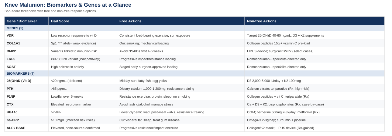
The 7 Biomarkers Behind Bone Healing Around The Knee
The logic behind biomarker tracking — measure the actual number, don't guess from population averages, and act on the number rather than a vague sense that something's off — is the same logic Peter Attia has pushed into mainstream longevity medicine, and the same rigor that lipidologists like Thomas Dayspring and Allan Sniderman have long applied to cardiovascular risk (treating apoB as the causal number rather than a proxy). Applied to bone healing, the same discipline matters: two people with an identical distal femur fracture and an identical fixation can have very different vitamin D, inflammatory, and glucose profiles — and those differences show up in healing time, callus quality, and ultimately in whether the bone settles straight or drifts into malunion.
The seven markers below are the ones with the clearest link to fracture healing biology, ordered roughly from most essential to most situational. Several are inexpensive enough to include in routine bloodwork; a few require a specialty draw.
Serum 25-hydroxyvitamin D (25-OH-D)
Vitamin D is technically a hormone precursor, and it's the single most-studied nutritional factor in fracture healing because it governs how efficiently calcium gets absorbed and deposited into new bone. Deficiency has been repeatedly linked to slower union and a higher rate of nonunion in trauma patients, and orthopedic literature increasingly treats it as a routine screen for anyone with a healing fracture, not just older patients with osteoporosis, as summarized in this narrative review of vitamin D in orthopaedic trauma.
How to measure it
A standard serum 25(OH)D blood draw, roughly $40–$100 out of pocket if not covered by insurance, available through any commercial lab or as part of a broader panel. Most orthopedic protocols target a level above 30 ng/mL (75 nmol/L); below 20 ng/mL is generally flagged as deficient.If the score is bad, the plan without supplements
Fifteen to thirty minutes of midday sun exposure on bare skin several times a week (duration depends on skin tone and latitude), more dietary fatty fish, egg yolks, and fortified dairy, and simply spending more time on outdoor weight-bearing activity once cleared by your surgeon. Retest in around three months rather than guessing.If the score is bad, the plan with supplements or equipment
Vitamin D3 (cholecalciferol), typically 2,000–5,000 IU daily, paired with vitamin K2 (MK-7, ~100 mcg) to help direct calcium toward bone rather than soft tissue. A randomized trial on bolus vitamin D3 dosing after distal radius fracture is one of the few controlled human looks at this exact question, using imaging to track healing directly — see the HR-pQCT randomized controlled trial. This is not a supplement you need to cycle — it's fat-soluble and taken daily — but retest every three to six months until stable, then annually. At sustained doses above roughly 10,000 IU/day, hypercalcemia becomes a real risk (nausea, kidney stones), so don't self-escalate past standard dosing without a repeat blood level.Parathyroid hormone (PTH)
PTH is the master regulator of calcium and phosphate balance. When it's chronically elevated — often secondary to low vitamin D or kidney issues — it drives net bone resorption, which is the opposite of what a healing fracture needs. Interestingly, PTH is also used therapeutically: intermittent teriparatide (synthetic PTH 1–34) is prescribed to accelerate healing in select high-risk fractures, as reviewed in this systematic review of PTH treatment in insufficiency fractures — a useful reminder that the same hormone can be either the problem or the treatment, depending on the pattern.
How to measure it
An intact PTH blood test, roughly $50–$90, ideally drawn fasting in the morning due to natural diurnal variation. Reference ranges vary by lab but generally sit around 10–65 pg/mL.If the score is bad, the plan without supplements
Correct the usual root cause first: adequate dietary calcium (roughly 1,000–1,200 mg/day from food), sun exposure and vitamin D status as above, moderating excess sodium and caffeine, and resistance training, which independently supports calcium retention in bone.If the score is bad, the plan with supplements or equipment
Calcium citrate or carbonate (500–600 mg elemental calcium per dose, once or twice daily with meals) only if dietary intake is genuinely insufficient — more isn't better here, and excess supplemental calcium without adequate vitamin D and K2 has been associated with vascular calcification concerns in some studies. Correcting vitamin D usually normalizes PTH on its own. In specific, physician-diagnosed high-risk nonunion cases, teriparatide (20 mcg subcutaneous injection daily) is a prescription-only option, generally capped at 24 months of lifetime use due to a bone tumor signal seen in rodent studies at high doses — this is a specialist-directed decision, never a self-directed one.P1NP (procollagen type I N-terminal propeptide)
P1NP is released when osteoblasts are actively laying down new collagen matrix, making it one of the more direct windows into bone formation activity. In a study of adults with tibial and femoral shaft fractures, P1NP (alongside CTX) rose predictably and elevated six-week levels tracked with better radiological healing at twelve weeks — see the bone turnover marker study in intramedullary-fixed fractures.
How to measure it
A specialized serum assay, roughly $60–$150, often a send-out test rather than a routine panel item — available through major labs or longevity-focused clinics. Best drawn fasting in the morning, since food and time of day both shift the result.If the score is bad, the plan without supplements
Progressive, surgeon-cleared resistance and load-bearing exercise is the strongest known non-pharmacological stimulus for osteoblast activity. Protein intake matters too (see below), as does quitting smoking — nicotine measurably suppresses bone formation markers — and prioritizing consistent sleep, since much of the anabolic signaling for tissue repair happens overnight.If the score is bad, the plan with supplements or equipment
Hydrolyzed collagen peptides (10–15 g/day) taken alongside vitamin C (roughly 500 mg) provide the raw materials and cofactor for collagen synthesis; evidence here is promising but still early for fracture-specific outcomes rather than general connective tissue turnover. A whole-body vibration platform is sometimes used as an adjunct load stimulus, though evidence for bone formation markers specifically is preliminary. For genuinely stalled healing, anabolic agents like teriparatide or abaloparatide are physician-prescribed, not self-directed.CTX (C-terminal telopeptide of type I collagen)
CTX is the mirror image of P1NP — a marker of osteoclast activity and collagen breakdown. Looking at the two together gives a formation-to-resorption ratio, which is more informative than either number alone; the same tibia/femur fracture study referenced above tracked both.
How to measure it
A serum test similar in cost to P1NP ($60–$150). CTX is notably sensitive to fasting status and time of day — a non-fasting afternoon draw can swing the result by 20–30%, so consistency in how it's measured matters more than for most other markers.If the score is bad, the plan without supplements
Avoid prolonged fasting or very low-calorie eating during active healing, since energy deficit pushes resorption up. Limit alcohol and smoking, both of which increase resorption markers, and manage chronic stress — sustained high cortisol has a well-documented resorptive effect on bone.If the score is bad, the plan with supplements or equipment
The same calcium, vitamin D3, and K2 combination described above is the first-line response. Bisphosphonates lower CTX dramatically, but they are prescription drugs generally reserved for osteoporosis rather than acute fracture care — in fact, some surgeons deliberately hold off on them during the first months of healing because blunting resorption can also blunt the remodeling phase. This is a conversation for your orthopedic team, not a self-directed choice.HbA1c
HbA1c reflects average blood glucose over roughly three months, and it's one of the most consistently reproduced risk factors for delayed union, nonunion, and post-surgical complications — including the kind of imperfect consolidation that becomes a malunion. Both type 1 and type 2 diabetes have been associated with roughly double the odds of fracture-healing complications, as summarized in this review of diabetes, diabetic complications, and fracture risk, with risk rising notably once HbA1c crosses the 7–8% range.
How to measure it
A standard blood draw, roughly $15–$40, no fasting required, included in most annual panels.If the score is bad, the plan without supplements
Lower the glycemic load of meals, add fiber, take a short walk after larger meals, build resistance training into the week (muscle is the largest glucose sink in the body), and keep sleep timing consistent — poor sleep independently worsens insulin sensitivity.If the score is bad, the plan with supplements or equipment
A continuous glucose monitor (roughly $60–$100/month) is genuinely useful here — not as a permanent fixture, but for two to four weeks at a time to identify which specific meals spike your glucose. Berberine (500 mg, two to three times daily with meals) has meta-analytic evidence for glucose-lowering effects comparable to some pharmaceutical options; cycle it in 8–12 week blocks with breaks, and expect possible GI side effects (bloating, loose stools) at higher doses. Prescription metformin is an option for diagnosed prediabetes or diabetes, but that's a decision for your physician, not a supplement aisle choice.High-sensitivity CRP (hs-CRP)
hs-CRP is a general inflammation marker. In the fracture setting it's used clinically mostly to rule out infection in a nonunion, as in this study on CRP and infection risk in long-bone non-unions, where the risk of infection rose from roughly 26% to 72% as CRP crossed the 10 mg/L threshold. Separately, chronically elevated baseline CRP (not the acute post-injury spike) has also been linked to lower bone strength and higher long-term fracture risk in the SWAN cohort study, suggesting a persistently inflamed system may be a less favorable environment for bone remodeling in general.
How to measure it
An hs-CRP blood test, roughly $15–$30, no fasting required, widely available.If the score is bad, the plan without supplements
Reduce visceral fat if present, keep up regular aerobic activity, prioritize sleep, address gum disease if it exists (a surprisingly common silent driver of baseline inflammation), and cut back on ultra-processed food and excess alcohol.If the score is bad, the plan with supplements or equipment
Omega-3 fish oil (2–3 g combined EPA/DHA daily) has modest, fairly consistent evidence for lowering CRP over months. Curcumin with piperine (500–1,000 mg daily, cycled 8–12 weeks on and roughly 4 weeks off) has some supporting evidence for inflammatory marker reduction, though study quality varies. Important caveat: if hs-CRP is elevated specifically in the weeks after a fracture or surgery and comes with fever, redness, or wound drainage, that's a signal for medical evaluation, not a supplement protocol.Alkaline phosphatase (ALP), ideally bone-specific (BSAP)
ALP is one of the oldest and cheapest bone formation markers, produced by active osteoblasts, and it's discussed in detail in this overview of biochemical markers of osteoporosis. Total ALP is nearly always already on your standard metabolic panel, which makes it a free first look — though it can also rise from liver issues, which is why an isolated elevation sometimes needs the bone-specific version to clarify the source.
How to measure it
Total ALP is essentially free — it's part of any basic or comprehensive metabolic panel most people already get annually. Bone-specific ALP is a separate send-out test, roughly $60–$100, worth ordering only if total ALP is elevated and the source is unclear.If the score is bad, the plan without supplements
The same osteogenic stimulus that helps P1NP applies here: progressive, cleared-for-loading resistance and impact exercise, adequate protein and calcium intake, and avoiding smoking.If the score is bad, the plan with supplements or equipment
The same collagen, vitamin C, and K2 stack described earlier applies. One equipment-based option worth knowing about: low-intensity pulsed ultrasound (LIPUS) devices, used at home under prescription guidance (roughly $1,500–$5,000 to purchase, sometimes available through rental programs), have clinical evidence supporting faster healing in delayed unions specifically — this is a conversation to have with your surgeon rather than a self-directed purchase.Biomarkers tell you how your bone biology is behaving right now, but they don't fully explain why two people respond so differently to the same nutrient status or the same healing stimulus. That's where genetics adds a layer — a smaller, earlier-stage body of evidence, but a useful one.
What Your Genes May Be Adding To The Picture
Genomics researchers like Ali Torkamani, who has spent years studying how genetic variation predicts real clinical outcomes, and health educators like Gary Brecka, who has popularized pairing genetic panels with functional blood work, both make a version of the same point: your genotype doesn't determine your outcome, but it can tell you where to look harder and where you might need to compensate more deliberately. Bone healing is a good example — a handful of genes have been repeatedly studied in relation to fracture nonunion and bone density, and knowing where you sit on them can inform how aggressively you manage the biomarkers above.
A note on evidence quality upfront: genetic association studies in bone healing are generally smaller and noisier than the biomarker literature. Some early single-study findings didn't hold up once larger, pooled analyses were done. Where that's the case below, it's stated plainly rather than oversold.
VDR (Vitamin D Receptor)
The VDR gene doesn't control how much vitamin D is in your blood — it controls how effectively your cells respond to the vitamin D that's there. Common variants (often labeled BsmI, ApaI, TaqI, and FokI) have been studied extensively for their relationship to bone mineral density and fracture risk, but a large meta-analysis found no consistent overall association between the most-studied variant (BsmI) and fracture susceptibility across genetic models, as reported in this updated meta-analysis of VDR polymorphisms and osteoporotic fracture risk. The honest read: this gene is biologically plausible and worth knowing, but the human evidence for it moving the needle on its own is mixed.
If the gene is bad, the plan without supplements
Since a less-sensitive receptor is (partly) offset by mechanical signaling that doesn't run through VDR at all, lean harder on consistent, surgeon-approved load-bearing activity and sun exposure rather than assuming diet alone will compensate.If the gene is bad, the plan with supplements or equipment
Aim for a higher-normal serum 25(OH)D target — closer to 40–60 ng/mL rather than just above 30 — on the logic that more circulating hormone can partially offset reduced receptor efficiency, alongside the same D3 plus K2 approach described in the biomarker section. Retest every three to six months rather than assuming the higher dose is working.COL1A1
COL1A1 codes for type I collagen, the structural scaffold of bone matrix. A well-studied variant at an Sp1 transcription factor binding site affects how much collagen protein gets produced, and early, smaller studies linked the less common "T" allele to lower bone density and higher fracture risk. However, the large GENOMOS study, pooling over 20,000 participants, found only a modest link between this variant and bone density and a weak association with vertebral fractures specifically — carriers did not show a clearly elevated overall fracture risk, as detailed in this GENOMOS study on COL1A1 polymorphism, bone density, and fracture risk. This is a case where a bigger, later study meaningfully tempered the earlier findings — worth remembering before treating any single-gene result as decisive.
If the gene is bad, the plan without supplements
Quit smoking if applicable — it directly interferes with collagen cross-linking and is one of the few modifiable factors known to independently worsen collagen quality. Controlled mechanical loading also stimulates matrix remodeling somewhat independently of baseline collagen genetics.If the gene is bad, the plan with supplements or equipment
Hydrolyzed collagen peptides (15 g) taken with vitamin C 30–60 minutes before loading is a reasonable, low-risk approach based on general connective tissue synthesis research, though there isn't yet a dedicated randomized trial pairing this specific protocol with COL1A1 genotype and bone outcomes — file this under promising but early.BMP2
Bone morphogenetic protein 2 is one of the key signaling molecules that converts the early cartilage-like callus at a fracture site into actual bone — a process reviewed in this study on chondrocyte BMP2 signaling in fracture healing. Certain BMP2 polymorphisms have shown up repeatedly across smaller studies of fracture nonunion, and a broader systematic review lists BMP2 among a cluster of genes (alongside ones like NOG and FGFR1) associated with nonunion risk — see this review of nonunion epidemiology, diagnosis, and clinical features.
If the gene is bad, the plan without supplements
There's no strong lifestyle lever that directly raises BMP2 expression, but avoiding NSAIDs during the first four to six weeks of active healing is one practical, evidence-aligned step — some research suggests NSAIDs can blunt BMP-driven callus formation during this early window (a discussion to have with your surgeon rather than a blanket rule, since pain control also matters).If the gene is bad, the plan with supplements or equipment
Recombinant BMP2 is an actual surgical product, applied directly at the fracture or fusion site by a surgeon in select nonunion cases — not something available or appropriate to self-administer. LIPUS devices, mentioned earlier, are thought to work partly by upregulating BMP-pathway activity and are the more accessible, physician-guided option here.LRP5
LRP5 is a co-receptor in the Wnt signaling pathway, one of the body's primary regulators of how much bone mass gets built and maintained. A common variant, rs3736228, has been associated with increased osteoporosis and fracture risk, and rare loss-of-function mutations cause a severe childhood bone fragility disorder, as described in this study on the LRP5 polymorphism and fracture risk.
If the gene is bad, the plan without supplements
Mechanical loading is one of the only known ways to activate this pathway independent of genotype — osteocytes physically sense strain and signal through Wnt/LRP5 in response. Once ccleared post-fracture, progressive impact and resistance loading is the most evidence-aligned response available without a prescription.If the gene is bad, the plan with supplements or equipment
No supplement targets LRP5 directly. Downstream of this same pathway, the prescription drug romosozumab (an anti-sclerostin antibody, discussed further below) is used for severe osteoporosis under specialist care — relevant context, not a self-directed option.SOST (sclerostin)
SOST codes for sclerostin, a protein that inhibits the LRP5/6 pathway above — essentially a brake on bone formation. Interestingly, the body naturally suppresses sclerostin at a fracture site as part of the normal healing response, and this same mechanism is now a drug target, as covered in this review of anti-sclerostin therapy applications.
If the gene is bad, the plan without supplements
Mechanical loading transiently lowers sclerostin production, which is part of the rationale behind carefully staged, surgeon-approved early loading protocols now being studied in fracture rehabilitation.If the gene is bad, the plan with supplements or equipment
There's no OTC supplement that meaningfully changes sclerostin activity. Romosozumab, the pharmaceutical option, is injectable, prescription-only, and carries a boxed warning around cardiovascular risk — it's approved for severe osteoporosis, not routine fracture healing, and included here for context rather than as a recommendation.Genes and biomarkers explain a lot about bone biology in isolation, but the bigger picture — how strength, stability, and metabolic health interact to determine injury resilience — is where a broader framework helps.
Ten Lessons From Outlive That Apply Directly To Knee Recovery
Peter Attia's book Outlive: The Science and Art of Longevity isn't written about fractures or malunion specifically, but its central argument — that most people wait for a crisis before addressing the physical capacities that would have prevented it — maps unusually well onto orthopedic injury and recovery. Below are ten of its most transferable ideas, each tied back to what they mean for a knee that's healing or at risk of healing badly.
1. "Medicine 3.0" means acting before the crisis, not after
Attia's core argument is that conventional medicine treats disease reactively, while a more useful model tracks risk markers early and intervenes before a bad outcome locks in. Applied here, that means checking vitamin D, HbA1c, and inflammatory markers before a fracture — or immediately after one — rather than only reacting once a malunion shows up on a follow-up X-ray.2. The "Centenarian Decathlon" forces specificity
Attia's exercise: name the physical tasks you want to still do in your final decade of life (carrying groceries, getting off the floor unassisted, hiking) and train backward from there. For someone recovering from a knee fracture, this reframes rehab away from "get back to normal" and toward specific, testable functional targets.3. Muscle strength predicts more than people expect
Grip strength, used in the book as a proxy for overall muscular capacity, has been shown to predict all-cause mortality more strongly than blood pressure in some large cohorts, per this study on handgrip strength and mortality. The knee-specific translation: quadriceps and hamstring strength after a fracture isn't cosmetic recovery, it's a marker of how well the rest of the body is holding up too.4. VO2 max is treated as the single most powerful lever
Attia repeatedly cites cardiorespiratory fitness as one of the strongest predictors of longevity in the literature, consistent with findings summarized in this review on VO2max as a predictor of longevity. For fracture recovery, maintaining aerobic capacity through non-impact modalities (cycling, swimming, rowing) during immobilization periods keeps the broader cardiovascular and metabolic system primed for tissue repair.5. Stability is the "fourth pillar" most people skip
Beyond strength, cardio, and nutrition, Attia argues stability training — balance, proprioception, controlled joint positioning — is systematically undertrained until an injury forces the issue. This is directly relevant to malunion: a knee healing at a slight angle changes load distribution, and proprioceptive/stability work is one of the few tools that can help the surrounding musculature adapt to compensate.6. Zone 2 training supports the metabolic engine behind repair
Sustained, low-intensity aerobic work is framed in the book as a way to build mitochondrial efficiency. Tissue repair, including bone remodeling, is metabolically demanding — a well-conditioned aerobic base is a plausible (if general) support for the energy requirements of healing.7. Protein intake recommendations are higher than most people assume
Attia argues the standard RDA (0.8 g/kg) is inadequate for anyone recovering from injury or trying to preserve muscle with age, aligning with research on sarcopenia showing that older adults benefit from intakes closer to 1.2 g/kg/day or higher — see this study on protein intake and muscle mass preservation. During active fracture healing, protein needs likely trend even higher, not lower.8. Resistance training is framed as a bone-health tool, not just a muscle-health tool
The book repeatedly ties strength training to skeletal density, not only muscle mass, consistent with clinical reviews such as this summary of resistance exercise effects on bone health. Once a surgeon clears loading, progressive resistance work is one of the most reliable levers for supporting the bone that's healing, not just the muscles around it.9. Sleep is treated as a recovery input, not a lifestyle nicety
Much of the hormonal signaling that supports tissue repair, including growth hormone release, is concentrated in deep sleep. Attia treats sleep quality as a measurable, trainable input rather than background noise — worth taking seriously during any healing window, not only for comfort.10. The book challenges the "rest and wait" default in orthopedics
Perhaps the most provocative idea for this specific topic: Attia's broader thesis is that passive waiting is rarely the optimal strategy once risk factors are known. Applied to a healing or malunited knee, that means proactively addressing modifiable biomarkers and building strength and stability around the joint, rather than treating "give it time" as a complete plan.These principles work best alongside, not instead of, hands-on approaches that have direct evidence in orthopedic recovery — which is where a few complementary modalities become genuinely useful.
Complementary Approaches Worth Considering
The following five approaches all have real human evidence in knee-specific or fracture-adjacent contexts. None of them replace surgical correction of an established malunion or the biomarker work above — they're additions, and the evidence quality varies from solid to modest.
Biofeedback
Biofeedback uses real-time visual, auditory, or electromyographic (EMG) signals to help a patient consciously activate a muscle that's become inhibited — a very common problem after knee surgery or prolonged immobilization, where the quadriceps in particular often "switch off" even when structurally intact. For anyone recovering from a knee fracture or post-malunion surgery, this addresses a specific, well-documented obstacle to regaining function.
A representative protocol comes from a randomized controlled trial in patients after total knee arthroplasty, where EMG biofeedback was paired with isometric quadriceps contractions (roughly 15-minute sessions of 10-second contract/relax cycles) and produced better functional recovery than the same exercises without biofeedback — see this randomized controlled trial on EMG biofeedback after total knee arthroplasty.
In practice, this means asking a physical therapist about EMG or visual biofeedback devices during early rehab, particularly if quadriceps activation feels weak or inconsistent weeks after surgery. Sessions are typically short (10–20 minutes), low-risk, and done a few times per week alongside standard physical therapy rather than as a standalone treatment.
Low-level laser therapy / photobiomodulation
Low-level laser therapy (LLLT), also called photobiomodulation, applies red or near-infrared light to tissue with the aim of stimulating cellular activity, including collagen production and circulation. It's relevant here because it's one of the few complementary modalities studied directly in human bone fracture healing, rather than only in soft tissue.
A controlled human study on closed fractures of the wrist and hand found measurable effects on healing outcomes, described in this study on LLLT for closed bone fractures, though it's worth noting that a broader systematic review of photobiomodulation across multiple fracture trials found benefits for pain that were statistically but not clearly clinically significant — evidence here is promising but not definitive.
Realistically, this is something to raise with a physical therapist or physiatrist who has access to a clinical-grade device rather than a consumer product bought online, and to treat as a possible adjunct to standard rehab rather than a primary healing strategy for a fracture near the knee.
Tai chi
Tai chi combines slow, controlled movement with weight shifting and balance challenges, which makes it relevant on two fronts for knee malunion: it has documented effects on bone mineral density, and its balance component is directly useful for anyone compensating for altered knee alignment.
A randomized controlled trial in middle-aged adults at risk of osteoporosis found meaningful improvements in bone mineral density with structured tai chi programs, detailed in this randomized controlled trial on tai chi and bone mineral density, and multiple meta-analyses have found similar effects, though most note that direct fracture-reduction data (as opposed to bone density) is still limited.
For someone with a knee malunion, tai chi is generally low-impact enough to start once basic weight-bearing is cleared, and the slow, deliberate movement patterns double as proprioceptive training for a joint that may be loading slightly asymmetrically. Two to three sessions a week of 20–40 minutes is a reasonable, low-risk starting frequency.
Yoga
Yoga's relevance here is mostly about bone density and controlled loading rather than the knee joint specifically, though certain poses also build the single-leg stability that's often compromised around a malunited knee.
The most cited human evidence is Dr. Loren Fishman's decade-long study of a twelve-pose daily yoga sequence, which found significant improvements in spine and femur bone density over ten years of follow-up, described in this study on a twelve-minute daily yoga regimen and bone density, with no serious yoga-related injuries reported among compliant participants.
Given that some yoga poses (deep lunges, certain twists) load the knee in ways that may not suit an altered alignment, this is best started with a physical therapist or an instructor experienced with post-fracture patients, avoiding deep knee flexion poses until cleared, and building up gradually rather than starting with a full advanced sequence.
Massage therapy
Massage is relevant mainly for the recovery period immediately around surgery or fracture treatment — reducing pain, supporting range of motion, and addressing the stiffness that often accompanies a knee that's been immobilized.
A systematic review and meta-analysis of massage after total knee arthroplasty found it improved pain and range of motion in early recovery, though it did not show a measurable effect on D-dimer levels (a clotting-related marker) specifically, as summarized in this systematic review of massage for rehabilitation after total knee arthroplasty. A separate dose-finding trial in knee osteoarthritis found a 60-minute weekly session produced significantly better outcomes than usual care.
In practice, this looks like scheduling massage sessions (30–60 minutes, once or twice weekly) during the early-to-mid recovery window, focused on the surrounding musculature rather than directly over surgical hardware or an incompletely healed fracture line, and always cleared with your surgeon first given the proximity to healing bone.
Conclusion
A knee that heals slightly out of alignment is rarely just bad luck or a surgical miss — it's usually the visible endpoint of a biological process that was already running slower, more inflamed, or less efficiently than it needed to be. The seven biomarkers above give you a concrete way to check that process directly, with clear thresholds and realistic responses if something's off. The five genes add context for why your biology might need to work harder in some areas than someone else's. And the broader strength, stability, and metabolic principles from Attia's framework, alongside the complementary approaches with real evidence behind them, round out a plan that's proactive rather than reactive.
None of this replaces a conversation with your orthopedic surgeon, especially if a malunion is already established and correction is being discussed. But it does give you something useful to bring into that conversation. A sensible next step: get the core panel drawn — 25(OH)D, HbA1c, hs-CRP, and total ALP are inexpensive and available almost anywhere — and use the results, not guesswork, to decide what to fix first.
Endocrine & Metabolic: Diabetes & Blood Sugar
Autoimmune: Connective Tissue Conditions