This article contains AI generated content.
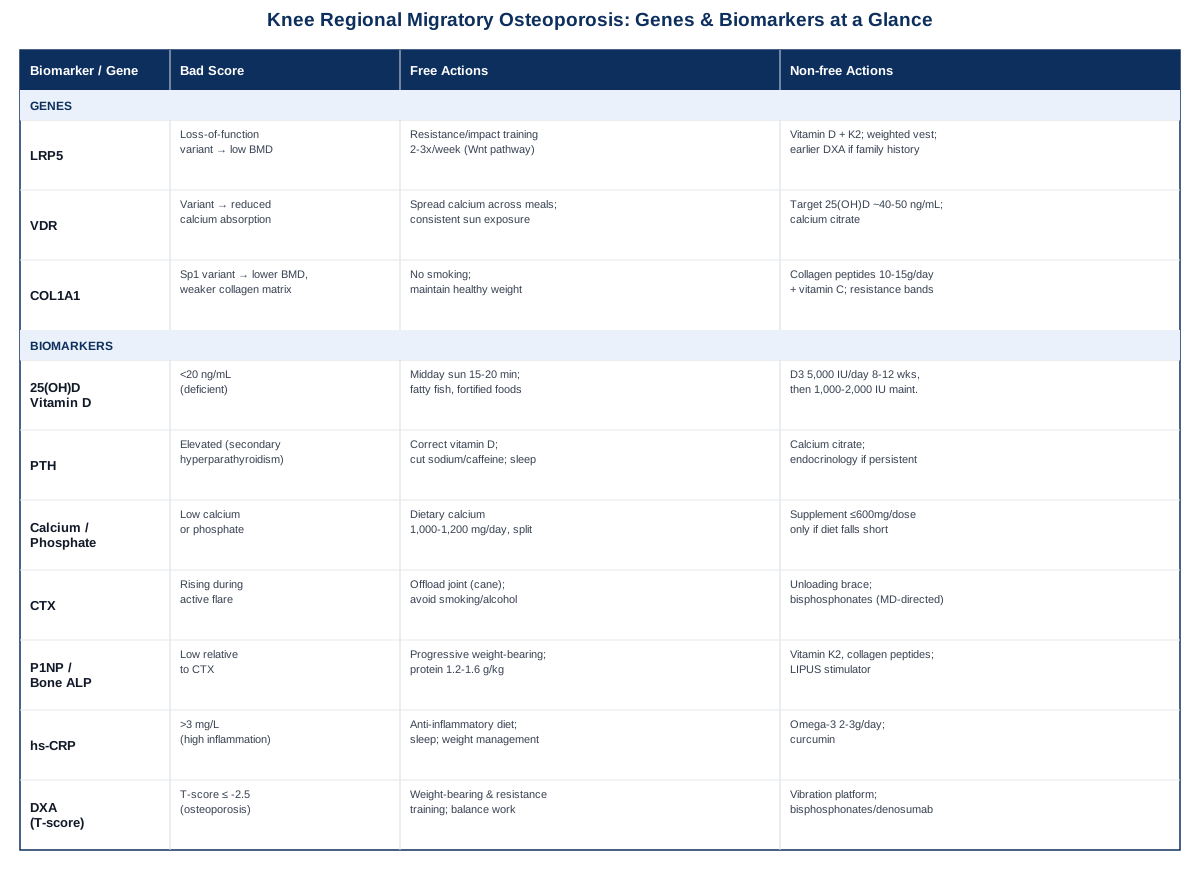
Knee Regional Migratory Osteoporosis: 3 Genes and 7 Biomarkers to Track
If your knee pain has been drifting — a hot, aching hip that faded only for the knee to flare next, or a foot that acted up months before the knee ever did — you have probably already noticed that most advice about knee pain does not fit your situation. Articles about arthritis, tendonitis, or overuse injuries assume a single joint, a single mechanical cause, and a single fix. Regional migratory osteoporosis (RMO) of the knee does not behave that way. It shows up as marrow edema and localized bone thinning that moves between weight-bearing joints, often without a clear trigger, and it tends to confuse clinicians who are not specifically looking for it.
Generic bone-health advice — "get more calcium," "do weight-bearing exercise," "take vitamin D" — is not wrong, but it is built for common postmenopausal osteoporosis, not for a rarer, migratory, marrow-edema-driven process that behaves more like a metabolic flare than a steady degenerative decline. Treating the two identically means missing the details that actually matter here: which lab values move during an active phase, which ones predict recovery, and which underlying genetic tendencies make a person more prone to this pattern in the first place.
This article takes the more specific route. Instead of repeating broad bone-health talking points, it walks through the biomarkers that clinicians and researchers actually use to characterize RMO and track its course, then looks at the genetic variants that shape baseline bone metabolism and may explain why some people are more susceptible. Both sections are written to be practical: what each marker or gene tells you, how to check it, and what a sensible response looks like, with and without supplements or equipment.
None of this replaces a diagnosis or a treatment plan from a clinician who has actually examined your imaging. But better information changes the questions you bring to that appointment. Knowing which biomarkers reflect active bone turnover, which genes influence your baseline bone metabolism, and which lifestyle-based approaches have real (if sometimes modest) evidence behind them gives you a more precise map — and a more grounded reason to expect that this migratory, unpredictable-feeling condition can actually be understood and managed.
Summary
Regional migratory osteoporosis of the knee is uncommon enough that most people never get a clear explanation of what is actually happening inside the joint — or why it seems to move. The sections below break down seven biomarkers that map the active phase of the disease and its recovery, from vitamin D and PTH to the bone turnover markers (CTX and P1NP) that clinicians rarely order but that tell you far more than a standard metabolic panel. From there, three genes — LRP5, VDR, and COL1A1 — explain why some people run a lower baseline bone reserve than others, with realistic plans for working around each one. A closer look at Peter Attia's Outlive reframes bone loss as a decades-long, falls-and-muscle problem rather than a number that shows up at 65, and a final section reviews which mind-body and movement therapies have actual clinical evidence behind them for bone health and pain — and which don't. Together, this is less a list of facts and more a working framework for figuring out what your specific knee is telling you.
The Biomarkers Worth Tracking in Regional Migratory Osteoporosis of the Knee
Before getting into individual markers, it helps to understand what makes RMO different from routine osteoporosis in the lab. Standard osteoporosis is usually a slow, systemic loss of bone mineral density, tracked once every year or two with a DXA scan. RMO, by contrast, is episodic: a joint develops marrow edema and focal osteopenia, becomes painful over weeks, and then — in most documented cases — resolves on its own over six to twelve months before symptoms sometimes reappear somewhere else. That means the most useful biomarkers are not just the ones that describe your bones in general, but the ones sensitive enough to move during an active flare and settle again as it resolves. A 2023 literature overview on RMO of the knee confirmed this exact pattern: migrating arthralgia, bone marrow edema, and osteoporosis that shows up on imaging well before it would on a routine scan (see Komnos et al., 2023). An earlier and more detailed review by Cahir and Toms also found that systemic osteoporosis frequently accompanies the migratory pattern, which is part of why tracking whole-body markers — not just the affected knee — is worthwhile (see Cahir & Toms, 2008).
25-Hydroxyvitamin D
Vitamin D status is the foundation everything else depends on. 25(OH)D is the storage form of vitamin D that clinicians measure, and it determines how efficiently your gut absorbs calcium and how tightly your parathyroid hormone stays regulated. Low vitamin D is common in adults presenting with unexplained bone marrow edema, and correcting a deficiency removes one of the easiest, most reversible contributors to poor bone turnover.
How to measure it
A standard 25(OH)D blood test costs roughly $35 to $120 out of pocket if not covered by insurance, and can be ordered through a primary care visit or a direct-to-consumer lab draw (see MedlinePlus: Vitamin D Test). Most labs flag deficiency below 20 ng/mL and insufficiency between 20 and 30 ng/mL.If the score is low, the plan without supplements
Fifteen to twenty minutes of midday sun exposure on bare arms and legs, several times a week, is often enough to meaningfully raise levels in lighter-skinned individuals — longer for darker skin tones or higher latitudes. Fatty fish (salmon, mackerel, sardines), fortified dairy or plant milk, and egg yolks add meaningful dietary intake, though food alone rarely corrects a true deficiency.If the score is low, the plan with supplements or equipment
For confirmed deficiency, a typical medically supervised approach is 5,000 IU of vitamin D3 daily for 8 to 12 weeks, then dropping to a 1,000–2,000 IU/day maintenance dose, with retesting at the 3-month mark. Take it with a meal containing fat for better absorption. Long-term dosing above 4,000 IU/day without monitoring risks hypercalcemia and should not be self-directed indefinitely (see NIH Office of Dietary Supplements: Vitamin D). Some clinicians pair vitamin D repletion with vitamin K2 on the theory that it helps direct calcium toward bone rather than soft tissue — the evidence for this specific combination is still preliminary, so treat it as optional rather than essential.Parathyroid Hormone (PTH)
PTH is the hormone that governs calcium release from bone. When vitamin D is low, PTH tends to rise to compensate — a state called secondary hyperparathyroidism — which accelerates bone resorption and can compound whatever is already happening in a migrating joint. Checking PTH alongside vitamin D tells you whether your body has already started borrowing calcium from the skeleton to correct a deficiency elsewhere.
How to measure it
An intact PTH blood test runs about $50 to $150 and is usually ordered together with calcium (see MedlinePlus: PTH Test).If the score is high, the plan without supplements
Correcting the underlying vitamin D deficiency is usually the first and most effective step. Reducing excess sodium and caffeine intake — both of which increase urinary calcium loss — and prioritizing consistent sleep, since PTH follows a circadian rhythm, are reasonable secondary steps.If the score is high, the plan with supplements or equipment
If PTH stays elevated after vitamin D and calcium are corrected, that combination (high PTH with high calcium) can point toward primary hyperparathyroidism rather than a simple deficiency response, and needs an endocrinology referral rather than self-treatment. When it is secondary to deficiency, calcium citrate (gentler on absorption for those with lower stomach acid) alongside the vitamin D protocol above is the standard approach, reassessed in 8 to 12 weeks.Serum Calcium and Phosphate
A basic calcium and phosphate panel rules out other metabolic bone diseases — like osteomalacia — that can mimic or overlap with RMO on imaging. Low phosphate in particular is worth flagging, since it can indicate a mineralization problem distinct from simple bone loss.
How to measure it
Both are part of a standard metabolic panel, often bundled into routine bloodwork for $20 to $50 (see MedlinePlus: Calcium Blood Test).If the score is low, the plan without supplements
Dietary calcium — dairy, leafy greens, canned fish with soft bones, fortified foods — aiming for roughly 1,000 to 1,200 mg per day split across meals, since the body absorbs calcium more efficiently in smaller doses than in one large one.If the score is low, the plan with supplements or equipment
Supplement only if diet genuinely falls short. Keep single doses at 500–600 mg or less, and avoid pushing total intake (diet plus supplement) much past 2,000–2,500 mg/day — high-dose calcium supplementation has been associated with cardiovascular signal in some analyses, a concern raised by lipid and cardiovascular researchers like Allan Sniderman, and it is not something to load up on indefinitely without a clear dietary gap to fill.CTX (Bone Resorption Marker)
C-terminal telopeptide (CTX) is released when osteoclasts break down old bone. It is one of the more sensitive markers of how active the resorption side of bone turnover is at any given moment — and in a condition defined by episodic flares, that sensitivity is exactly what makes it useful. A rising CTX during a painful phase, followed by a falling one as symptoms resolve, is a more objective way to track a flare than pain alone.
How to measure it
CTX is measured via a fasting morning blood draw (it has meaningful circadian variation, so timing matters), costing roughly $80 to $200. It is far less standardized than vitamin D or calcium testing, and best ordered through an endocrinology or metabolic bone specialty lab. One metabolomics study detailing how CTX and P1NP behave clinically is a useful technical reference (see Bellissimo et al., 2020).If the score is high, the plan without supplements
During an active flare, reducing mechanical load on the affected knee — a cane, temporary activity modification, avoiding high-impact movement — gives the bone a chance to stop accumulating microtrauma while resorption is elevated. Avoiding smoking and excess alcohol matters here too, since both independently increase resorption markers.If the score is high, the plan with supplements or equipment
An unloading brace or crutches can meaningfully reduce joint stress during the acute phase. On the medical side, bisphosphonates are the actual pharmacologic treatment used in transient and migratory osteoporosis case reports to blunt excess resorption — this is a prescription decision that requires a physician familiar with your imaging and labs, not something to self-initiate.P1NP and Bone-Specific Alkaline Phosphatase (Formation Markers)
If CTX tells you how much old bone is being broken down, P1NP and bone-specific ALP tell you how much new bone is being built. In healthy remodeling, formation and resorption stay roughly coupled. In an active RMO flare, resorption often outpaces formation temporarily — watching both markers together is more informative than either alone.
How to measure it
P1NP runs about $80 to $150 and, like CTX, is best ordered through a specialty lab. Standard alkaline phosphatase is cheaper ($20–40) and often bundled into a liver panel, but it is not bone-specific unless an isoenzyme test is specifically requested — and that test "may not be available everywhere," per MedlinePlus's own note on ALP isoenzyme testing (see MedlinePlus: ALP Test).If the score is low, the plan without supplements
Once acute pain has settled, controlled, progressive weight-bearing loading is the single strongest natural stimulus for bone formation — this is the practical application of Wolff's law, where bone adapts to the mechanical demands placed on it. Adequate protein intake, generally 1.2 to 1.6 g per kg of body weight daily, supports the collagen matrix formation happens on.If the score is low, the plan with supplements or equipment
Vitamin K2 and collagen peptides both have early, though not yet definitive, evidence for supporting formation markers. Low-intensity pulsed ultrasound (LIPUS) bone stimulator devices are established for fracture healing and used off-label in some slow-healing bone marrow edema cases, though dedicated evidence in RMO specifically is limited — a reasonable option to discuss with an orthopedic specialist rather than a first-line self-directed purchase.hs-CRP (Inflammatory Activity)
RMO's marrow edema has an inflammatory component alongside the bone-turnover component, and high-sensitivity C-reactive protein is a simple way to gauge systemic inflammation. It is also useful diagnostically: a markedly elevated hs-CRP should prompt a look at inflammatory arthritis mimics, since some inflammatory joint conditions can also present with a migrating pattern.
How to measure it
A standard CRP blood test costs $20 to $50; ask specifically for the high-sensitivity (hs-CRP) version, which is a different assay than the standard infection-screening CRP test (see MedlinePlus: CRP Test).If the score is high, the plan without supplements
An anti-inflammatory dietary pattern (more vegetables, fiber, and unprocessed foods, less refined sugar), consistent sleep, and weight management to reduce mechanical and inflammatory load on the knee are the foundational steps.If the score is high, the plan with supplements or equipment
Omega-3 fish oil (roughly 2–3 g/day combined EPA/DHA) has modest evidence for lowering inflammatory markers; curcumin has weaker but suggestive evidence. Both are generally well tolerated but can interact with blood thinners, so check with a physician if you're on anticoagulants.DXA Bone Mineral Density (T-score / Z-score)
DXA remains the gold-standard measurement for bone density and the closest thing to a definitive answer about whether systemic osteoporosis is part of the picture — which, as noted earlier, is common in RMO. It is also the best tool for tracking recovery over months, since it directly measures the outcome all the other markers are only proxies for.
How to measure it
A central hip-and-spine DXA scan costs roughly $150 to $500 out of pocket depending on region and facility, and is considered "the most reliable way to diagnose osteoporosis and predict fracture risk" according to NIAMS (see NIAMS: Osteoporosis Diagnosis and Treatment). Peripheral heel scans are cheaper (roughly $25–50) but are screening tools only, not diagnostic-grade.If the score is low, the plan without supplements
Progressive weight-bearing exercise as tolerated, resistance training two to three times weekly, and dedicated balance training to reduce fall risk are the core non-pharmacologic levers — all covered in more depth in the exercise-focused section below.If the score is low, the plan with supplements or equipment
Whole-body vibration platforms have modest supporting evidence for bone density in some populations and can be a reasonable adjunct under supervision. If a T-score confirms osteoporosis, medications like bisphosphonates or denosumab are physician-directed decisions based on your fracture risk profile — not something to pursue independently, but worth asking about directly if your DXA results come back low.RMO's episodic nature means it rewards patience with the data — a single set of labs tells you where you stand today, but the real value comes from repeating vitamin D, CTX, and P1NP every few months during an active phase and spacing DXA scans further apart once things stabilize. With the biomarker picture in place, it's worth asking why some people seem more prone to this pattern than others in the first place — which is where genetics enters the picture.
What Genetics May Explain About Bone Metabolism
Regional migratory osteoporosis itself has no dedicated gene panel — it is too rare and too poorly understood mechanistically for large genetic studies to exist. What does exist is a well-studied set of genes that shape baseline bone density and fracture risk generally, the same genes that consumer genomics interpreters — people like Ali Torkamani, whose research has focused on using genetic and wearable data to personalize health risk, and Gary Brecka, known for popularizing polygenic risk score interpretation — often flag when someone runs a broader health-optimization panel. These genes don't explain why osteoporosis migrates between joints, but they do explain why some people start from a lower bone reserve, which plausibly makes any localized flare more consequential.
LRP5
LRP5 is a receptor in the Wnt signaling pathway, the same pathway that governs how osteoblasts build new bone in response to mechanical loading. Loss-of-function variants reduce Wnt signaling and are linked to lower bone mineral density, including rare severe forms like osteoporosis-pseudoglioma syndrome; gain-of-function variants do the opposite, producing unusually high bone mass (see MedlinePlus Genetics: LRP5). The evidence is strong for the rare monogenic forms; for common variants that show up on consumer polygenic panels, the effect on ordinary adult osteoporosis risk is real but modest — one contributing factor among many, not a diagnosis.
If the gene is unfavorable, the plan without supplements
Since LRP5 responds to mechanical loading through the same Wnt pathway, prioritizing progressive resistance training and impact-tolerant loading — weighted carries, controlled jumping or step-downs once cleared by a clinician — gives you a lever that works independent of the genetic variant. Two to three non-consecutive sessions per week is a reasonable baseline frequency.If the gene is unfavorable, the plan with supplements or equipment
Vitamin D and K2, dosed and monitored as described in the biomarker section, form the supplement foundation. A weighted vest or resistance bands make progressive overload easier to apply consistently at home. Getting a baseline DXA scan earlier than the standard screening age is a reasonable, low-risk step if LRP5 variants show up on a genetic panel alongside a family history of fractures — this is a conversation to have with a primary care provider, not a self-ordered intervention.VDR (Vitamin D Receptor)
VDR encodes the receptor that lets cells actually respond to active vitamin D — without it, even normal blood levels of vitamin D don't translate into normal calcium absorption. Certain common VDR polymorphisms (FokI, BsmI, and others studied in population genetics) have been associated with modestly different calcium absorption efficiency and fracture risk in some populations, though results are inconsistent across ethnic groups and study designs — this is early-to-mixed evidence, not settled science. Rare, severe VDR mutations cause a distinct and much more serious condition, vitamin D-dependent rickets type 2A (see MedlinePlus Genetics: VDR).
If the gene is unfavorable, the plan without supplements
Spreading calcium intake evenly across meals rather than front-loading it, and getting consistent (not sporadic) sun exposure, both help compensate for reduced receptor efficiency by giving the body more, smaller opportunities to absorb what it needs.If the gene is unfavorable, the plan with supplements or equipment
If a VDR variant associated with lower receptor efficiency shows up on testing, some clinicians target a somewhat higher serum 25(OH)D level (closer to 40–50 ng/mL rather than the standard 30 ng/mL floor) to compensate — this should be done with retesting every 3 months, not assumed. Calcium citrate is a sensible supplement choice for older adults, since it depends less on stomach acid for absorption than calcium carbonate.COL1A1
COL1A1 provides the blueprint for type I collagen, the structural protein that makes up the organic matrix of bone. Severe mutations cause osteogenesis imperfecta, a rare and serious brittle-bone disease (see MedlinePlus Genetics: Osteogenesis Imperfecta), but a common regulatory variant near the gene's Sp1 binding site has also been studied in general population meta-analyses for its association with modestly lower bone density and higher fracture risk — again, a small population-level effect, not something that predicts an individual outcome on its own.
If the gene is unfavorable, the plan without supplements
Because this is a structural, collagen-quality issue rather than a purely mineral one, avoiding smoking matters more here than with the other two genes — smoking independently impairs collagen cross-linking and compounds fracture risk. Maintaining a healthy body weight reduces ongoing mechanical stress on both the joint and the collagen matrix supporting it.If the gene is unfavorable, the plan with supplements or equipment
Collagen peptide supplementation (roughly 10–15 g/day) has early evidence for supporting bone turnover markers, ideally paired with vitamin C, which is a required cofactor for collagen synthesis, and with resistance training, which provides the mechanical signal that tells the body to actually use the raw material. Favor lower-impact loading tools — resistance bands, a weighted vest — over high-impact plyometric work, given the structural (rather than purely density-based) nature of the underlying concern. Three-month blocks with reassessment via P1NP or CTX is a reasonable cycle; side effects are generally mild GI upset at higher doses.Genetics explains predisposition, but it says nothing about the day-to-day habits that actually move the needle — which is where a very different, more behavior-focused body of work becomes useful.
What Peter Attia's Outlive Gets Right About Bone and Joint Longevity
Peter Attia's book Outlive is not about RMO specifically, but its central argument — that most modern medicine treats chronic, slow-building problems reactively instead of catching them decades early — maps unusually well onto a condition that hides in normal-looking scans until a flare makes it undeniable. The ten ideas below are the ones most directly useful for someone managing knee-specific bone loss.
Bone Loss Starts Decades Before It's Diagnosed
Attia's "Marginal Decade" concept holds that the physical capacity you'll have in your last ten years of life is largely determined by choices made decades earlier — bone density included, since peak bone mass is typically set by the late twenties and only declines afterward. For a knee already showing migratory osteopenia, this reframes the goal from "treat the flare" to "protect the trajectory."The "Centenarian Decathlon" Reframes the Goal
Rather than optimizing for a single number like BMD, Attia proposes working backward from the physical tasks you want to still perform in old age — carrying groceries, getting off the floor unassisted, hiking. For a migratory knee condition, this means training the joint's full functional capacity, not just chasing a T-score.Muscle Is the Organ That Protects Bone
Grip strength and VO2 max are, in Attia's framing, better predictors of long-term survival than most standard lab panels — largely because muscle mass and strength directly reduce fall risk and support the joints bone density alone can't protect.Stability Is the Neglected First Pillar
Attia orders training priorities as stability, then strength, then aerobic base — with stability first because an unstable joint (exactly the situation with an actively flaring knee) undermines everything built on top of it. Balance and proprioception work deserves at least as much attention as loading exercises.Fracture Risk Is a Falls Problem as Much as a Density Problem
Most fragility fractures happen because someone fell, not purely because their bones were thin. This is especially relevant for a joint with an active RMO flare, since pain-driven gait changes and reduced proprioception around that knee raise fall risk independent of the bone density reading itself.Protein Targets Higher Than Standard Guidelines
Attia argues the standard RDA for protein is a minimum-to-avoid-deficiency number, not an optimal target, and recommends closer to 1.6 g/kg or more for people actively trying to preserve muscle and skeletal reserve — consistent with the protein ranges referenced in the biomarker section above.Zone 2 Cardio Plus Strength, Not Either Alone
Attia's exercise framework pairs low-intensity aerobic work (Zone 2) with resistance training rather than treating them as substitutes — the aerobic base supports recovery capacity and metabolic health, while resistance training provides the direct mechanical stimulus bone needs.Exercise as the Most Potent Available Intervention
Attia repeatedly frames structured exercise as outperforming any single drug across nearly every longevity-relevant outcome measured — a claim that lines up with the mechanical-loading evidence already covered for bone formation markers like P1NP.Proactive Screening Over Waiting for a Fracture
Consistent with "Medicine 3.0," Attia advocates for earlier and more frequent risk assessment — DXA scans before the standard screening age when risk factors (family history, a genetic variant, an unexplained flare) are present — rather than waiting for guideline-driven age cutoffs.Track Objective Markers, Not Just Symptoms
Perhaps the most directly applicable idea: don't wait for pain to tell you what's happening. Pairing periodic bloodwork and imaging with how you feel gives an earlier, more honest signal — the same logic behind tracking CTX and P1NP through an RMO flare rather than relying on pain alone.Movement and mechanical loading are clearly central to bone health, but they're not the only lever — a smaller set of mind-body approaches also has real, if more modest, evidence behind them.
Complementary Approaches With Real Evidence Behind Them
None of the approaches below are a substitute for medical management of an active RMO flare, and the evidence for several is genuinely limited rather than definitive — that's stated plainly below rather than glossed over. They're worth considering as adjuncts once acute pain has settled enough to allow movement-based practice.
Tai Chi
Tai chi combines slow, weight-shifting movement with balance and postural control, which makes it a natural fit for someone managing both a bone-density concern and the fall-risk issue that comes with an unstable, migrating joint. Its low-impact nature also makes it more accessible during a recovery phase than higher-impact resistance work.A 2024 meta-analysis of 12 randomized controlled trials in postmenopausal women found that tai chi produced meaningful improvements in bone mineral density and overall health status scores compared to control groups, though it found no significant effect on fall frequency specifically in that population (see Zhang & Chen, 2024).
A realistic approach is two to three 30-45 minute sessions per week, ideally with an instructor initially to ensure correct form, continued indefinitely rather than in short cycles — the evidence base is built on sustained, months-long practice, and the studies themselves note small sample sizes and inconsistent methodology as limitations worth keeping in mind.
Yoga
Yoga offers a similar combination of controlled loading, flexibility, and balance work, with the added benefit of adjustable intensity — poses can be modified to reduce load on a currently painful knee while still engaging the rest of the kinetic chain.An 8-month randomized controlled trial of Ashtanga-based yoga in premenopausal women found a small positive effect on bone formation markers (bone-specific alkaline phosphatase was maintained in the yoga group while it declined in controls), but no measurable improvement in actual bone mineral density or bone geometry (see Kim et al., 2015) — a useful, honest example of evidence that is real but modest, not transformative.
Two to three sessions per week, choosing a gentler style (Hatha or Iyengar rather than a high-impact vinyasa flow) during an active flare, with modifications for any pose that loads the knee in a painful range, is a sensible starting frequency.
Qigong
Qigong shares tai chi's slow, weight-bearing, balance-oriented movement patterns but is generally lower-intensity, which may make it a gentler entry point for someone still recovering from an acute flare.The evidence here is genuinely preliminary: a registered systematic review protocol for Qigong Wuqinxi in osteopenia and osteoporosis noted that a prior 2015 review found only four studies with uncertain results on bone mineral density, and called for more rigorous trials (see Liu et al., 2020) — worth trying for its low risk and plausible balance benefits, but not something to expect measurable bone density changes from based on current evidence.
Daily 15-20 minute sessions are typical in the studied protocols; because the movements are low-impact, there's little downside to daily practice, though as with tai chi and yoga, benefits (where they exist) require sustained months-long practice rather than short bursts.
Mindfulness Meditation / MBSR
Mindfulness-based stress reduction doesn't act on bone tissue directly, but chronic pain management matters practically here: pain-driven guarding and reduced activity around a flaring knee can itself worsen deconditioning and fall risk, so anything that improves pain coping has an indirect bone-health benefit.A meta-analysis of 38 randomized controlled trials involving over 3,500 participants found statistically significant, though modest, improvements in pain, depression, and quality of life from mindfulness meditation for chronic pain — while explicitly noting that "the weaknesses in the body of evidence prevent strong conclusions" and calling for larger, more rigorous trials (see Hilton et al., 2017).
A structured 8-week MBSR course, in person or via a well-validated app, with daily 10-20 minute practice, is the format most studies actually tested — informal, sporadic meditation has much less evidence behind it than the structured version.
Conclusion
Regional migratory osteoporosis of the knee is genuinely uncommon, but it isn't unmanageable or unmeasurable. The clearest path forward is tracking the biomarkers that actually move during a flare — vitamin D, PTH, calcium, CTX, P1NP, hs-CRP, and periodic DXA — rather than waiting for pain to be the only signal you have. Genetic variants in LRP5, VDR, and COL1A1 help explain why some people run a lower baseline bone reserve, but mechanical loading, adequate protein, and correcting vitamin D deficiency work regardless of which variants you carry. And the behavioral layer — stability training, sustained low-impact practices like tai chi or yoga, and honest pain management — supports everything else without overpromising results the evidence doesn't yet support.
The next useful step is a concrete one: if you haven't already, ask your physician for a baseline panel covering vitamin D, calcium, PTH, and hs-CRP, and discuss whether CTX, P1NP, or an earlier-than-standard DXA scan make sense given your history. Bring this article's questions to that appointment rather than generic ones — the more specific the conversation, the more useful the answers you'll get back.
Musculoskeletal: Bone Conditions Joint Conditions