This article contains AI generated content.
· UpdatedKnee Venous Malformation — 6 Genes and 7 Biomarkers To Track
Introduction
A venous malformation in or around the knee is not simply a bruise that won't heal or a vessel that misbehaved once. It is a structural anomaly — a tangle of abnormally enlarged venous channels that developed during embryogenesis and, depending on its size and location, can press against tendons, fill joint spaces, and create a persistent low-grade state of internal clotting that has little in common with typical knee problems. If you've been living with pain that worsens when you stand or flex, swelling that seems disconnected from activity, and doctors who default to watchful waiting, the frustration of feeling unheard is entirely legitimate.
The challenge is that venous malformations sit at the intersection of hematology, vascular surgery, and interventional radiology — and most general practitioners have limited experience with them. Standard advice often stops at compression stockings and mild analgesics, without accounting for the fact that many patients with venous malformations are quietly developing a coagulation imbalance called localized intravascular coagulopathy (LIC), which can progress to more serious clotting events if left unmeasured and unmanaged.
That is exactly where precision tracking — both of biomarkers and of genetic drivers — becomes genuinely useful. Not to replace specialist care, but to make you an informed participant in your own treatment. When you walk into a consultation with actual numbers — D-dimer trends, fibrinogen levels, an understanding of whether your malformation may be driven by a TEK or PIK3CA mutation — the quality of the conversation changes.
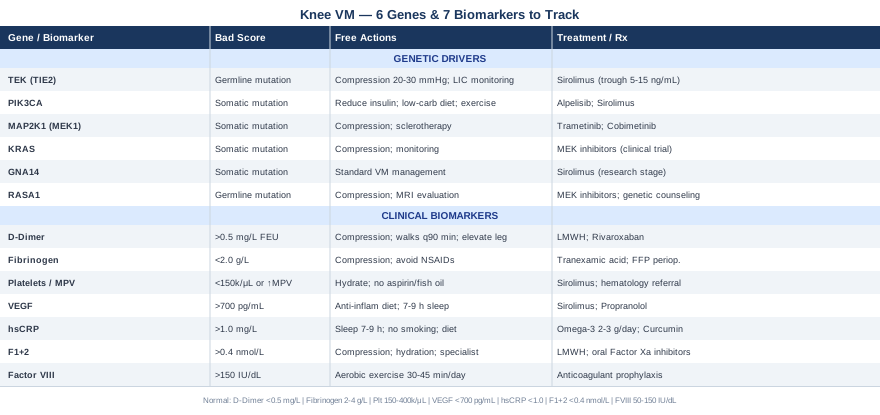
This article takes two evidence-grounded approaches. The first centers on the seven most clinically relevant biomarkers for knee venous malformation: what they reveal, how to measure them affordably, and what to do when they are out of range. The second covers the six most studied genetic factors behind venous malformations and what each finding may mean for treatment. Together, these strategies won't promise a cure — but they can help you stop guessing and start working with real information.
7 Biomarkers to Track When You Have a Knee Venous Malformation
Venous malformations are not static. The blood that pools in abnormal channels clots slowly and incompletely, generating a constant low-level coagulation activity that most standard panels miss. Tracking the right biomarkers regularly can reveal whether your malformation is stable, worsening, or responding to treatment — and it can flag dangerous trends before they become emergencies.
1. D-Dimer
Why it matters: D-dimer is a fibrin degradation product — a molecule released whenever a blood clot forms and begins to dissolve. In healthy individuals, baseline levels are very low. In patients with venous malformations, even those without symptoms of acute thrombosis, D-dimer is frequently elevated because the stagnant blood inside the malformation undergoes continuous low-level clotting. This state is called localized intravascular coagulopathy (LIC), and it is present in the majority of patients with large or symptomatic venous malformations.
A landmark study published in the Archives of Dermatology confirmed that elevated D-dimer directly correlates with the presence and severity of LIC in venous malformation patients, and that monitoring D-dimer before and after interventions predicts coagulation risk (Dompmartin et al., 2008). This makes it arguably the single most important biomarker in this context.
How to measure it: D-dimer is measured through a standard blood draw. Most clinical laboratories include it in coagulation panels. Cost is typically $20–$60 in the US depending on insurance; in Europe it is usually covered in hematology follow-up. A standard reference range is under 0.50 mg/L FEU, though labs vary slightly. Request a quantitative D-dimer, not just a qualitative "positive/negative" result — you need to track trends over time.
If the score is bad, the plan without supplements: If D-dimer is consistently elevated above 1.0 mg/L, the priority is reducing stagnation in the malformation. Daily use of a well-fitted graduated compression sleeve (15–20 mmHg at minimum, preferably 20–30 mmHg for larger lesions) reduces blood pooling mechanically and has been shown to lower D-dimer in some VM patients. Avoid prolonged immobility — short, frequent walks throughout the day (every 60–90 minutes) are more effective than one long walk. Elevating the limb when sitting also matters.
If the score is bad, the plan with supplements or equipment: Low-molecular-weight heparin (LMWH) such as enoxaparin is often used short-term before and after interventional procedures to reduce LIC risk. This requires a prescription and monitoring. Some practitioners use it in low prophylactic doses for extended periods in high-risk patients. Frequency: typically once daily subcutaneous injection, with dose adjusted by body weight and renal function. Side effects include bruising, bleeding risk, and, rarely, heparin-induced thrombocytopenia — always under specialist supervision. Rivaroxaban is a newer oral option increasingly being studied for LIC in venous malformations, with emerging clinical use at specialized centers.
2. Fibrinogen
Why it matters: Fibrinogen is a clotting protein synthesized by the liver. In the context of venous malformations, sustained low-level intravascular clotting can consume fibrinogen faster than the liver replaces it, leading to hypofibrinogenemia — low fibrinogen levels. This creates a paradoxical risk: the malformation is actively clotting, yet the patient may have a higher bleeding tendency because the clotting reserves are depleted. Fibrinogen below 1.5 g/L in a VM patient is a clinical warning sign.
How to measure it: Included in most coagulation panels. Cost: $15–$50. Normal range: 2.0–4.0 g/L. Best measured in the morning in a fasted state for consistency. Measure at baseline and then every 3–6 months if stable, or more frequently around procedures.
If the score is bad, the plan without supplements: Low fibrinogen in VM patients usually reflects active LIC. Addressing the underlying clotting burden through compression and mobility (as described above) can gradually allow fibrinogen to normalize. Avoid NSAIDs like ibuprofen, which further impair clotting; switch to acetaminophen for pain management.
If the score is bad, the plan with supplements or equipment: Fibrinogen concentrate or fresh frozen plasma may be considered perioperatively by your vascular team. No supplement reliably raises fibrinogen in this context. Tranexamic acid, an antifibrinolytic, is sometimes used to preserve existing fibrinogen and prevent excessive breakdown — always under specialist guidance. Do not attempt to manage this independently.
3. Platelet Count and Mean Platelet Volume (MPV)
Why it matters: Platelets are the first responders in clot formation. Large venous malformations can trap and consume platelets, sometimes leading to thrombocytopenia — a low platelet count that increases bleeding risk significantly. Conversely, reactive thrombocytosis (high platelets) can indicate compensatory bone marrow activity. Mean platelet volume (MPV) adds nuance: elevated MPV indicates larger, more metabolically active platelets, often in the context of ongoing consumption.
How to measure it: Part of any complete blood count (CBC). Cost: $10–$30. Normal platelet range: 150,000–400,000/μL. MPV normal: 7.5–12.5 fL. Measure baseline and track quarterly if stable.
If the score is bad, the plan without supplements: Thrombocytopenia in a VM patient requires specialist evaluation. Avoid aspirin, fish oil in high doses, and other antiplatelet agents until platelet count is stabilized. Ensure adequate hydration and avoid high-impact activities that could cause trauma to the malformation.
If the score is bad, the plan with supplements or equipment: If platelet counts fall below 100,000/μL, a hematology referral is warranted before any intervention on the malformation. Sirolimus, an mTOR inhibitor increasingly used for complex venous malformations (discussed in the genetics section), has shown effects on platelet dynamics in some patients and may stabilize coagulation parameters over time.
4. Vascular Endothelial Growth Factor (VEGF)
Why it matters: VEGF is the primary signaling molecule driving abnormal vessel formation. In venous malformations, the dysfunctional endothelial cells lining the malformation often overexpress VEGF receptors, contributing to vessel wall weakness and continued abnormal channel growth. Serum VEGF can serve as a rough indicator of malformation activity and may help track responses to treatment, particularly with anti-angiogenic or mTOR-targeting therapies.
How to measure it: VEGF serum levels are available through specialty labs, but not standard at all facilities. Cost: $80–$200. Reference ranges vary by lab (typically under 700 pg/mL). Timing matters — exercise, recent inflammation, or concurrent illness can elevate VEGF transiently. Measure in a controlled, fasted, rested state.
If the score is bad, the plan without supplements: Reducing systemic inflammatory load through dietary adjustments (minimizing ultra-processed foods, reducing refined carbohydrates) and normalizing sleep duration (7–9 hours) have modest, indirect effects on VEGF levels over time. Anti-inflammatory diet approaches are worth implementing as a foundation.
If the score is bad, the plan with supplements or equipment: Sirolimus (rapamycin), which inhibits the mTOR pathway downstream of VEGF signaling, has demonstrated measurable effects on VEGF-driven vascular anomalies in multiple clinical trials. Propranolol, originally studied in infantile hemangioma, has also been noted to reduce VEGF. Both are prescription medications. Sirolimus dosing for vascular anomalies typically targets serum trough levels of 5–15 ng/mL, adjusted by weight and tolerance. Side effects include immunosuppression, oral ulcers, and metabolic changes — requires experienced prescribers and regular monitoring.
5. High-Sensitivity C-Reactive Protein (hsCRP)
Why it matters: Inflammation is not the primary cause of venous malformations, but it is a significant amplifier of symptoms, pain, and functional limitation. When the stagnant blood inside the malformation produces micro-thrombi, the surrounding tissue responds with an inflammatory cascade. Elevated hsCRP reflects this ongoing low-grade inflammation and can also serve as a general cardiovascular risk marker — relevant because VM patients on sclerotherapy or with LIC are at elevated baseline cardiovascular risk.
How to measure it: Standard lab test. Cost: $10–$40. Optimal range per Peter Attia and Thomas Dayspring: below 1.0 mg/L; above 3.0 mg/L suggests significant systemic inflammation. Measure in the morning, avoiding measurement during acute illness or within two weeks of injury.
If the score is bad, the plan without supplements: Reducing inflammatory inputs matters here: 7–9 hours of sleep (cortisol dysregulation from sleep deprivation is a strong CRP driver), stress management, elimination of cigarette smoke, and reducing omega-6 heavy processed food intake. Compression therapy, by reducing blood stagnation in the malformation, may also reduce local inflammatory signaling.
If the score is bad, the plan with supplements or equipment: Omega-3 fatty acids (EPA/DHA, 2–3 g/day from fish oil or algal sources) have robust evidence for lowering hsCRP. Cycle consideration: take daily for 12 weeks minimum before reassessing. Side effects are minimal at standard doses but watch for mild GI discomfort; higher doses warrant physician supervision in anticoagulated patients. Curcumin with piperine (500–1000 mg/day of turmeric extract) has modest evidence for CRP reduction, though evidence is less robust than omega-3s.
6. Prothrombin Fragment 1+2 (F1+2)
Why it matters: Prothrombin Fragment 1+2 is a direct marker of thrombin generation — meaning it measures how actively your coagulation cascade is running at any given moment. Where D-dimer shows that clotting has occurred (retrospective), F1+2 shows that clotting is actively happening right now (prospective). For VM patients, F1+2 can help clinicians distinguish stable, compensated LIC from escalating coagulation activity that may precede a symptomatic thrombotic episode.
How to measure it: Requires a specialty coagulation lab. Not available at all clinical facilities. Cost: $100–$250. Normal reference: under 0.4 nmol/L in most labs. Often used together with D-dimer to create a more complete picture of coagulation burden in VM management.
If the score is bad, the plan without supplements: Elevated F1+2 in a VM patient should prompt immediate specialist consultation. In the meantime, all the compression and mobility strategies remain valid and important. Avoid dehydration, which concentrates clotting factors.
If the score is bad, the plan with supplements or equipment: Therapeutic anticoagulation (LMWH, direct oral anticoagulants like rivaroxaban) is the evidence-based intervention when F1+2 is markedly elevated and clinical picture supports intervention. Some centers are now using oral factor Xa inhibitors in a low-dose prevention protocol for VM patients with chronic LIC. This is a rapidly evolving clinical area — seek a vascular anomaly specialist familiar with current protocols.
7. Factor VIII Activity
Why it matters: Factor VIII is an amplifier in the coagulation cascade. It is also an acute-phase reactant — its levels rise with inflammation, stress, and obesity. In VM patients, chronically elevated Factor VIII (above 150 IU/dL) independently increases the risk of deep vein thrombosis and pulmonary embolism. Because venous malformations already predispose to intravascular stasis, the combination of high Factor VIII and LIC creates a significantly elevated thrombotic risk profile.
How to measure it: Included in comprehensive thrombophilia panels and available individually. Cost: $40–$120. Optimal: 50–150 IU/dL. Measure fasting, not during acute illness. Repeat if initially elevated to confirm (transient elevations occur).
If the score is bad, the plan without supplements: Sustained aerobic activity (30–45 minutes of low-impact exercise, 4–5 days per week) modestly lowers Factor VIII over time. Weight normalization also helps, as adipose tissue is a significant source of inflammatory signals that drive Factor VIII elevation. Avoid prolonged sedentary periods.
If the score is bad, the plan with supplements or equipment: No supplement reliably lowers Factor VIII to clinically meaningful degrees. If persistently elevated above 200 IU/dL in the context of a VM with LIC, anticoagulant prophylaxis should be discussed with a hematologist or vascular anomaly center. This combination is a legitimate indication for preventive anticoagulation in high-risk periods (travel, surgery, illness).
Taken together, these seven biomarkers form a practical monitoring panel that most VM patients never receive by default. Requesting them — and tracking them over time — transforms you from a passive recipient of care into an active participant who can detect trouble before it escalates.
The Genetic Drivers Behind Knee Venous Malformations
Understanding the genetics of venous malformations has accelerated dramatically in the past decade. What was once considered a developmental accident is increasingly understood as a defined set of molecular errors — most of them in signaling pathways that control vascular cell growth and survival. This matters clinically because several of these pathways are now druggable.
Gene 1: TEK (TIE2) — The Most Common Inherited Driver
The TEK gene encodes the TIE2 receptor tyrosine kinase, which is critical for maintaining normal venous endothelial integrity. Gain-of-function mutations in TEK cause constitutive receptor activation, meaning the vascular signaling pathway runs continuously without appropriate off-switches. The result is abnormal dilation and structural weakness of venous walls.
TEK mutations are found in the majority of familial venous malformation cases and in a significant subset of sporadic cases. They are typically present in all cells of the body (germline mutations) in inherited forms.
Without supplements/interventions: Structural management remains essential: graduated compression (20–30 mmHg), avoidance of prolonged standing or limb dependency, and regular LIC monitoring. Sclerotherapy performed by an experienced interventional radiologist directly targets the abnormal channels regardless of genetic cause.
With supplements or targeted therapy: TIE2-pathway dysregulation feeds into the PI3K-AKT-mTOR signaling axis, meaning sirolimus (an mTOR inhibitor) has biological rationale for TEK-mutation-positive malformations. Clinical evidence supports this: a study in The New England Journal of Medicine documented significant improvement in vascular anomalies including those with TEK-related pathology with sirolimus therapy (Adams et al., 2016). Sirolimus dosing: typically 0.8 mg/m² twice daily in pediatric patients; adult dosing individualized to trough levels of 5–15 ng/mL. Side effects: immunosuppression, mouth sores, elevated triglycerides, risk of infections. Long-term cycling strategies are still being studied.
Gene 2: PIK3CA — The Somatic Mutation Behind PROS
PIK3CA encodes the catalytic subunit of PI3K, a master regulator of cell growth, survival, and metabolism. Somatic (non-inherited, arising after conception) gain-of-function mutations in PIK3CA are found in a broad spectrum of vascular malformations collectively termed PROS (PIK3CA-Related Overgrowth Spectrum). These mutations hyperactivate the AKT-mTOR pathway, driving abnormal vascular channel proliferation.
Because these are somatic mutations, they may be present in the malformation tissue but absent from blood-based genetic tests. Tissue biopsy or liquid biopsy with ultra-sensitive sequencing is sometimes required for detection.
Without supplements/interventions: Lifestyle factors that reduce insulin resistance (which independently activates PI3K-AKT signaling) are worth addressing: maintaining a healthy body weight, limiting refined carbohydrates, and avoiding sedentary behavior. These won't reverse the mutation but may reduce the background activation level of the pathway.
With supplements or targeted therapy: Alpelisib, a PI3Kα-specific inhibitor originally developed for breast cancer, has shown significant efficacy in PIK3CA-driven overgrowth and vascular anomaly syndromes in clinical trials. This is a prescription oncology drug with significant side effects (hyperglycemia, rash, diarrhea) and requires specialized prescribing. Sirolimus remains the more broadly accessible alternative targeting the same downstream pathway. Emerging research suggests that combining a PI3K inhibitor with lifestyle strategies that reduce insulin (the primary PI3K activator) may enhance treatment response.
Gene 3: MAP2K1 (MEK1) — The RAS Pathway Link
MAP2K1 mutations activate the RAS-MAPK signaling cascade, another growth-regulating pathway found to be mutated in venous and lymphatic malformations. These somatic mutations cause constitutive ERK phosphorylation in endothelial cells, driving abnormal vessel formation.
Without supplements: Same foundational strategies as above: compression, regular LIC monitoring, and mechanical intervention through sclerotherapy or surgical resection where appropriate.
With targeted therapy: MEK inhibitors such as trametinib and cobimetinib have early clinical evidence for MAP2K1-driven vascular anomalies. These are prescription oncology agents currently being studied in specialized centers. Evidence is emerging but not yet at the level of sirolimus.
Gene 4: KRAS — Overlap with Low-Flow Malformations
Somatic KRAS mutations, long known in cancer biology, have been identified in a subset of venous and capillary-venous malformations. KRAS is an upstream RAS-family GTPase that, when mutated, chronically activates both the MAPK and PI3K pathways simultaneously.
Without supplements: No specific lifestyle intervention targets mutant KRAS directly. Standard compression and LIC monitoring remain the practical foundation.
With targeted therapy: The MEK inhibitors mentioned above (trametinib) have theoretical rationale for KRAS-driven malformations. This is early-stage in the vascular anomaly context; enrollment in a clinical trial at a specialized center may be an option worth discussing.
Gene 5: GNA14 — A Newer Finding in Sporadic VMs
GNA14 encodes a G-protein alpha subunit. Somatic mutations in GNA14 have been identified in sporadic venous malformations and cause activation of intracellular signaling pathways including MAPK and calcium signaling. This is a relatively recently characterized genetic driver.
Without supplements: The same management principles apply. The clinical significance of identifying a GNA14 mutation currently lies more in guiding research enrollment than in changing day-to-day management.
With targeted therapy: No approved targeted therapy specifically for GNA14 mutations in venous malformations currently exists. Research is ongoing. Sirolimus, as a broad mTOR inhibitor downstream of multiple signaling pathways, may still be considered based on clinical presentation.
Gene 6: RASA1 — Capillary-Venous Malformation Overlap
RASA1 encodes RAS p21 protein activator 1, a negative regulator of RAS signaling. Loss-of-function mutations in RASA1 lead to unopposed RAS-MAPK activation and are classically associated with capillary malformation-arteriovenous malformation (CM-AVM) syndrome. However, venous malformations can also be part of this phenotype.
Without supplements: Given the arteriovenous overlap in some RASA1 cases, imaging evaluation with MRI/MR angiography is important to characterize the lesion fully before any intervention. Standard compression applies for venous components.
With targeted therapy: MEK inhibitor trials are exploring RASA1-driven vascular anomalies, since loss of RASA1 causes overactivation of RAS-MAPK, which MEK inhibitors counteract. Evidence in the vascular anomaly context is early. Genetic counseling is warranted as RASA1 mutations are germline and have implications for family members.
What "Outlive" by Peter Attia Teaches Us About Vascular and Coagulation Health
Outlive: The Science and Art of Longevity by Peter Attia is not written about venous malformations specifically, but it may be the most practically useful book available for understanding the coagulation biomarkers that VM patients need to track — and for understanding why conventional medicine is so slow to use them. The book challenges the standard reactive model of vascular care and advocates for aggressive, proactive biomarker monitoring. Here are the ten most relevant insights for venous malformation patients.
1. Standard Lipid Panels Miss Most of the Story
Attia argues compellingly that LDL-cholesterol is a poor proxy for vascular risk. For VM patients, this parallels a deeper point: standard coagulation panels (PT, PTT) also miss most of the LIC story. D-dimer, F1+2, and fibrinogen are the equivalent of Attia's "ApoB" for the coagulation world — more specific, more predictive, but rarely ordered by default.2. Tracking Biomarkers Over Time Beats Single Snapshots
Attia emphasizes longitudinal tracking — the trend matters more than any single value. D-dimer rising gradually from 0.4 to 0.9 over six months is more concerning than a one-time reading of 0.9, even if neither triggers a clinical flag. Build a personal spreadsheet.3. Insulin Resistance Amplifies Vascular Dysfunction
Attia dedicates significant content to insulin resistance as a foundational driver of vascular and systemic inflammation. Elevated fasting insulin activates the PI3K-AKT pathway — the very same pathway dysregulated in PIK3CA-driven venous malformations. This is not a coincidence: keeping fasting insulin below 6 μIU/mL through diet and exercise may have genuine relevance for VM patients, particularly those with PIK3CA-related lesions.4. Zone 2 Aerobic Exercise Is the Most Powerful Anti-Inflammatory Tool Available
Sustained low-intensity aerobic work (the kind where you can just barely hold a conversation) improves mitochondrial function, lowers systemic inflammation, and reduces Factor VIII over time. Attia recommends 3–4 hours per week of Zone 2 for general health. For knee VM patients, low-impact modalities like swimming, cycling, or aquatic walking protect the joint while achieving the vascular benefits.5. Sleep Is Not Optional for Coagulation Health
Attia cites robust data showing that sleep deprivation elevates fibrinogen and CRP and impairs endothelial function. Targeting 7.5–8.5 hours with consistent sleep and wake times is as important as any supplement in this context.6. Omega-3s Are the Most Evidence-Backed Anti-Inflammatory Supplement
Attia recommends 2–4 g of EPA+DHA daily as the foundational anti-inflammatory supplement, noting that the evidence base is substantially more robust than for most other marketed products. For VM patients managing elevated hsCRP or who need to reduce inflammatory cofactors, this is the most rational starting point.7. Muscle Mass Is a Metabolic Reservoir That Protects Against Inflammatory Escalation
Lean muscle mass buffers glucose, reduces inflammatory cytokine output from adipose tissue, and improves insulin sensitivity — all of which reduce the inflammatory burden on vascular lesions. For knee VM patients, upper-body and contralateral leg resistance training remains possible and beneficial even when the affected knee limits lower-body work.8. The Danger of Polypharmacy Without Biomarker Monitoring
Attia warns against managing conditions through medications without tracking the relevant biomarkers to see if they're working. This applies directly to VM care: sclerotherapy, anticoagulants, and sirolimus all have measurable effects on D-dimer, fibrinogen, and platelet counts. Tracking these confirms whether treatment is working — or whether complications are developing silently.9. Visceral Fat Is a Biological Amplifier of Vascular Risk
Attia frames visceral adiposity not as a cosmetic issue but as an endocrine organ producing inflammatory signals. For VM patients, excess visceral fat elevates TNF-alpha, IL-6, and VEGF — directly feeding the abnormal vascular signaling environment of the malformation. Reducing visceral fat through dietary and exercise intervention is worth prioritizing.10. Personalized Medicine Requires You to Advocate for Yourself
Perhaps the most important message: the current medical system is not designed to proactively track coagulation biomarkers in chronic vascular anomaly patients. Attia's core argument — that patients need to become educated advocates who request specific tests and understand their results — applies with full force to anyone navigating a venous malformation diagnosis.Complementary Approaches with Genuine Relevance for Knee Venous Malformation
The following modalities have clinical evidence that may offer meaningful support for symptom management and quality of life in venous malformation patients. None replaces specialist care or the biomarker monitoring described above, but each addresses a real dimension of living with a chronic vascular condition in the knee.
Low-Level Laser Therapy (LLLT) / Photobiomodulation
What it is and why it may be relevant: Low-level laser therapy (also called photobiomodulation or LLLT) uses specific wavelengths of light — typically 630–850 nm — to stimulate mitochondrial function in tissue, reduce local inflammation, and promote tissue repair. In the context of venous malformations, LLLT is most relevant for managing the chronic pain and soft-tissue inflammation surrounding the malformation, particularly after sclerotherapy procedures when local tissue reactions can be prolonged.
Evidence and protocol: A systematic review published in Photomedicine and Laser Surgery found that LLLT at doses of 1–4 J/cm² significantly reduced pain and inflammation in musculoskeletal conditions involving the knee, with effects mediated through reduced prostaglandin E2 and IL-1β signaling. While no large RCT exists specifically for venous malformation-related knee pain, the anti-inflammatory mechanisms are well-characterized and the safety profile is excellent. A typical protocol involves 3–5 sessions per week for 4 weeks using a 830 nm near-infrared device at a power density of 30–60 mW/cm². Sessions last 5–15 minutes per treatment site.
Realistic application: LLLT devices are available for home use, though clinical-grade devices deliver more consistent results. For knee VM patients, treatments should avoid direct application over a known large malformation without specialist guidance, as light-tissue interactions in large vascular channels are incompletely characterized. Use on the perilesional tissue and joint margins is more appropriate. Expect gradual improvement in pain and swelling over 4–6 weeks of consistent application.
Mindfulness-Based Stress Reduction (MBSR)
What it is and why it may be relevant: Mindfulness-based stress reduction is an 8-week structured program developed by Jon Kabat-Zinn that combines meditation, body scanning, and gentle yoga to build moment-to-moment awareness and reduce psychological reactivity to pain and stress. For chronic venous malformation patients — who often experience ongoing, unpredictable pain, functional limitation, and the psychological burden of a poorly understood condition — MBSR addresses the emotional and neurological dimensions of pain that biomarkers and compression stockings cannot touch.
Evidence and protocol: A meta-analysis in JAMA Internal Medicine (Goyal et al., 2014) covering 47 randomized trials found that mindfulness meditation programs produced moderate evidence of improvement in anxiety, depression, and pain in chronic health conditions. The standard MBSR protocol involves weekly 2.5-hour group sessions over 8 weeks, plus a single day-long retreat, with daily home practice of 45 minutes. Online adaptations with comparable outcomes are now widely available and substantially more accessible.
Realistic application: For knee VM patients, the body scan component of MBSR can be particularly useful for developing a non-reactive relationship with chronic pain — learning to observe sensation without catastrophizing, which is documented to reduce pain amplification in chronic conditions. Start with 10 minutes of guided body scan daily for two weeks before committing to the full program. The investment is modest and the risk is essentially zero, making it worth incorporating alongside medical management.
Yoga
What it is and why it may be relevant: Therapeutic yoga — particularly yin yoga and restorative yoga styles — offers structured, low-intensity movement that can improve venous circulation in the lower limb, reduce soft-tissue tension around the knee, and support the lymphatic drainage mechanisms that help reduce malformation-related swelling. The key is choosing a style appropriate for a patient who may have limited weight-bearing capacity and a fragile vascular lesion in the joint.
Evidence and protocol: A randomized controlled trial published in Annals of Internal Medicine found yoga superior to stretching alone for chronic low back pain, with effects mediated partly through improved parasympathetic tone and reduced cortisol — which also has downstream benefits for vascular inflammation. For knee-specific applications, a study in Journal of Rheumatology found that chair-based and supine yoga reduced knee pain and improved function in chronic knee conditions without adverse events. Key poses for circulation: legs-up-the-wall (Viparita Karani), supine ankle circles, and gentle supine hip openers. Avoid deep knee flexion, kneeling positions, or poses that compress the popliteal fossa.
Realistic application: Practice three to four sessions per week of 20–30 minutes, focusing on supine and seated postures that encourage venous return from the lower limb. A yoga therapist with experience in vascular or orthopedic conditions is the safest starting point. Clearly communicate your diagnosis so poses that increase intravenous pressure in the affected limb can be avoided. Compression should be worn during active (non-supine) yoga positions.
Biofeedback
What it is and why it may be relevant: Biofeedback is a technique in which physiological signals — skin temperature, muscle tension, heart rate variability — are measured in real time and fed back to the patient, enabling voluntary regulation of normally unconscious processes. For venous malformation patients, biofeedback is particularly relevant for managing the chronic pain-stress cycle: pain increases sympathetic nervous system activation, which increases cortisol and inflammatory cytokines, which worsen vascular dysfunction and pain perception. Breaking this cycle has measurable physiological effects.
Evidence and protocol: A meta-analysis in Applied Psychophysiology and Biofeedback found that heart rate variability (HRV) biofeedback significantly reduced pain scores and improved functioning in chronic pain conditions, with effects mediated through improved vagal tone and reduced sympathetic overdrive. A standard protocol involves 10–15 sessions with a trained biofeedback therapist, each lasting 45–60 minutes, using HRV biofeedback at a resonance breathing frequency (typically 5–6 breaths per minute). Home devices (such as HeartMath or Polar chest straps with biofeedback apps) allow between-session practice.
Realistic application: HRV biofeedback is among the most accessible biofeedback modalities, as home devices now cost $80–$200 and are paired with validated software. Commit to 15–20 minutes of daily resonance breathing practice for a minimum of 6 weeks before assessing benefit. Track your resting HRV morning baseline as an objective indicator of progress — rising HRV correlates with improved autonomic regulation. This modality complements MBSR well and can be layered alongside it.
Conclusion
Knee venous malformations are complex, underdiagnosed, and poorly served by generic advice — but they are increasingly understood at both the molecular and physiological level. The core message of this article is that you do not have to navigate this condition blindly. Tracking seven specific biomarkers — especially D-dimer, fibrinogen, and prothrombin fragment 1+2 — gives you and your care team real-time insight into your coagulation status and treatment response. Understanding the genetic drivers, particularly TEK and PIK3CA mutations, can open doors to targeted therapies that were not available a decade ago.
The practical next step is not to implement everything at once, but to take one clear action: request a coagulation panel that includes D-dimer and fibrinogen at your next appointment, and ask whether genetic testing through a vascular anomaly program is appropriate for your case. Small, systematic moves toward better data consistently outperform large, unguided interventions. If you are not yet connected to a center specializing in vascular anomalies, that referral is worth pursuing — it is where the genetics, the biomarkers, and the interventional expertise converge.
Musculoskeletal: Joint Conditions
Cardiovascular: Blood Vessel Conditions Vascular Conditions
Autoimmune: Inflammatory Conditions