This article contains AI generated content.
Lateral Femoral Epicondyle Fracture: 7 Biomarkers And 3 Genes To Track
A fracture near the lateral epicondyle of the femur is not something most people think about until it happens — usually after a fall, a twisting sports injury, or a direct blow to the knee. Once the cast or brace comes off, the questions that matter most rarely get answered in a five-minute follow-up visit: why did this bone break the way it did, why is healing slower than expected, and what can actually be done about it beyond "wait and see."
Generic advice after an orthopedic injury tends to be the same for everyone — rest, calcium, physical therapy, patience. That advice isn't wrong, but it's built for an average person, and averages hide a lot. Two people with the same fracture, the same age, and the same treatment plan can heal at very different speeds, because their underlying bone biology, nutrient status, and genetic wiring are not the same.
This article takes a more specific approach. Instead of repeating generic recovery tips, it looks at the measurable biology behind bone strength and healing capacity — the blood markers that reveal how your bones are actually functioning right now, and the genetic factors that quietly shape how resilient your skeleton is over a lifetime. Neither replaces medical care for an acute fracture, but both can inform decisions that generic advice simply can't.
None of this promises faster healing or a guaranteed outcome — bone repair is a biological process with its own timeline. But better information, gathered honestly and interpreted carefully, tends to lead to better decisions. The sections below walk through the biomarkers worth tracking first, the genetic factors worth understanding as a longer-term layer, a set of research-backed lessons on connective tissue recovery, and complementary approaches with real supporting evidence.
Summary
This article centers on seven biomarkers — vitamin D, the calcium/PTH axis, two bone turnover markers (CTX and P1NP), bone mineral density, CRP, and homocysteine — that together paint a picture of bone quality, healing capacity, and fracture risk far more precise than "eat more calcium." It also covers three genes (VDR, COL1A1, and LRP5) that influence how your body builds and maintains bone, with realistic plans for each, with and without supplements. Beyond that, you'll find ten evidence-based lessons on connective tissue recovery drawn from current research on loading, collagen, and nutrient timing, plus a review of complementary approaches — from photobiomodulation to mindfulness-based pain management — that have actual human evidence behind them for fracture recovery. If you've ever wondered whether your slow healing, recurring injuries, or family history of fractures point to something measurable, this is the deeper layer most standard advice skips.
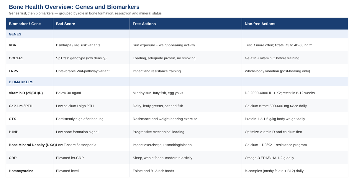
The 7 Biomarkers That Matter Most for Bone and Fracture Healing
A lateral femoral epicondyle fracture is a structural event, but what happens around it — inflammation, mineral balance, collagen turnover — is chemical, and chemistry can be measured. Below are the seven biomarkers with the most consistent human evidence for bone strength, healing speed, and future fracture risk. Peter Attia and Thomas Dayspring, both known for pushing beyond standard reference ranges in metabolic and bone health, are useful reference points here: they consistently argue that a "normal" lab value is not the same as an optimal one for a person actively trying to heal or protect their skeleton.
1. Serum 25-Hydroxyvitamin D (25(OH)D)
Vitamin D governs calcium absorption in the gut and calcium regulation in bone, which makes it the single most studied nutrient in fracture care. In orthopedic trauma populations, deficiency below 30 ng/mL is extremely common — some studies report it in 40 to 90 percent of fracture patients — and patients who remain deficient after injury show meaningfully higher rates of delayed union than those who correct their levels, as documented in a review of vitamin D supplementation in orthopedic trauma. It's worth noting plainly: the same research is clear that evidence directly proving supplementation speeds up acute healing is still limited, even though deficiency correction is strongly associated with fewer complications.
How to measure it: a standard 25(OH)D blood test, available through a physician or direct-to-consumer lab, typically costs between $40 and $120 in the US, though it's often bundled into broader panels for less.
If the score is bad, the plan without supplements: prioritize 15 to 20 minutes of midday sun exposure on bare skin several times a week (adjusted for skin tone and latitude), and increase dietary sources like fatty fish, egg yolks, and fortified dairy.
If the score is bad, the plan with supplements or equipment: vitamin D3, typically 2,000 to 4,000 IU daily for someone with confirmed deficiency, paired with vitamin K2 (MK-7, 100 to 200 mcg) to support proper calcium utilization. Retest in 8 to 12 weeks rather than guessing. Frequency: daily, not cyclical — vitamin D doesn't respond well to on-off dosing. Side effects: excessive dosing (typically above 10,000 IU/day sustained) can raise blood calcium; anyone with a history of kidney stones or hypercalcemia should test before supplementing at higher doses.
2. Calcium and Parathyroid Hormone (PTH)
Calcium and PTH move together: when calcium is low, PTH rises to pull it from bone, which is exactly the tradeoff you don't want during fracture repair. Checking both at once distinguishes a simple dietary shortfall from a parathyroid or kidney issue that needs separate management.
How to measure it: a basic metabolic panel with calcium is inexpensive, often $20 to $50; intact PTH adds roughly $50 to $100.
If the score is bad, the plan without supplements: increase dietary calcium through dairy, leafy greens, and canned fish with bones, spaced across the day since the body absorbs calcium more efficiently in smaller doses.
If the score is bad, the plan with supplements or equipment: calcium citrate (500 to 600 mg per dose, twice daily) if dietary intake is genuinely insufficient — food sources are preferred because high-dose calcium supplementation has been associated with cardiovascular concerns in some studies. Side effects: constipation is common with calcium carbonate; citrate forms are gentler. Anyone with elevated PTH should be evaluated for a parathyroid cause before supplementing further.
3. CTX (C-Terminal Telopeptide)
CTX reflects bone resorption — how fast old bone is being broken down. The International Osteoporosis Foundation and International Federation of Clinical Chemistry both recommend it as a preferred marker for tracking bone turnover and predicting fracture risk. During active healing some elevation is expected, but a persistently high CTX afterward can flag ongoing bone loss rather than a healing-related blip.
How to measure it: a specialty lab blood draw, typically $60 to $120, ideally drawn fasting in the morning since CTX has meaningful daily variation.
If the score is bad, the plan without supplements: resistance and weight-bearing exercise, resumed as soon as your surgeon or physiotherapist clears it, is the single strongest non-drug lever for shifting the resorption/formation balance favorably.
If the score is bad, the plan with supplements or equipment: adequate protein intake (1.2 to 1.6 g/kg body weight daily) supports the formation side of the equation; in postmenopausal women or older adults with confirmed high bone loss, this is also the marker doctors use to monitor antiresorptive medication — that decision belongs with a physician, not a supplement plan.
4. P1NP (Procollagen Type 1 N-Terminal Propeptide)
P1NP is the mirror image of CTX — a marker of bone formation. Tracking both gives a formation-versus-resorption picture instead of a single number. Interestingly, research shows that even in people already on bone-protective medication, higher P1NP is still associated with elevated fracture risk, underscoring that this marker reflects real biological turnover, not just a treatment response, per the same bone turnover marker research.
How to measure it: typically ordered alongside CTX, $70 to $150, same fasting-morning collection principle applies.
If the score is bad, the plan without supplements: consistent, progressive mechanical loading (walking, then resistance training as cleared) is the primary driver of healthy bone formation markers.
If the score is bad, the plan with supplements or equipment: ensure vitamin D and calcium are optimized first (see above), since formation markers respond poorly if raw materials are missing; a bone-density-focused physiotherapy program using graduated loading equipment (resistance bands progressing to weighted resistance) is more useful here than any supplement alone.
5. Bone Mineral Density via DXA Scan
A DXA scan doesn't just diagnose osteoporosis — it's the most direct window into whether a fracture happened because of an unusual mechanism of injury alone, or because underlying bone density was already compromised. This matters for anyone whose fracture seemed disproportionate to the trauma involved.
How to measure it: a DXA scan costs $100 to $400 out of pocket in the US, and is often covered by insurance for those over 65 or with fracture history, per general guidance summarized in NCBI's overview of osteoporosis biomarkers.
If the score is bad, the plan without supplements: impact and resistance exercise remain the best-documented non-pharmaceutical way to improve bone density over time, alongside smoking cessation and limiting excess alcohol, both of which independently impair bone remodeling.
If the score is bad, the plan with supplements or equipment: a structured program combining calcium, vitamin D3/K2, and progressive resistance training (2 to 3 sessions weekly) is the realistic baseline; whole-body vibration platforms have some supporting evidence for bone density in older adults but should only be used after full fracture healing and with physician sign-off, since acute or recently healed fractures are a contraindication.
6. C-Reactive Protein (CRP)
CRP is a general inflammation marker, not a bone-specific one, but it matters here because unresolved systemic inflammation is associated with delayed healing across orthopedic literature, and it's also one of the cheapest, most accessible markers to track over time.
How to measure it: a high-sensitivity CRP test runs $20 to $50 at most labs.
If the score is bad, the plan without supplements: address the basics that drive chronic low-grade inflammation — sleep consistency, reducing ultra-processed food intake, and moderate regular activity as cleared during recovery.
If the score is bad, the plan with supplements or equipment: omega-3 fatty acids (1 to 2 g EPA/DHA daily) have modest evidence for lowering CRP; cycling isn't necessary, but retesting every 8 to 12 weeks helps confirm whether the trend is improving. Side effects: high-dose fish oil can increase bleeding risk, which matters specifically around any planned orthopedic procedure — flag this with your surgeon.
7. Homocysteine
Homocysteine is one of the more overlooked fracture-risk markers. A meta-analysis of over 11,000 people found each 1 µmol/L increase in homocysteine raised fracture risk by roughly 4 percent, and elderly patients with elevated levels had about 70 percent higher hip fracture risk, independent of folate and B6 status, according to research summarized on homocysteine and fracture risk. Interestingly, the same body of research found that simply lowering homocysteine with B-vitamin supplementation hasn't been shown to reduce fracture risk in trials — a useful reminder that a marker being predictive doesn't automatically mean correcting it changes outcomes.
How to measure it: $40 to $90 at most labs, fasting recommended.
If the score is bad, the plan without supplements: increase dietary folate (leafy greens, legumes) and B12 (eggs, fish, meat), which are the natural cofactors in homocysteine metabolism.
If the score is bad, the plan with supplements or equipment: B-complex supplementation (methylfolate and B12 forms are often preferred for people with MTHFR variants) can reliably lower the number itself, even though — as noted above — it hasn't been proven to lower fracture risk directly. Frequency: daily, reassess at 3 months. Side effects: B12 supplementation is very low-risk; extremely high folic acid intake has been debated in relation to masking B12 deficiency, so use combined B-complex rather than folic acid alone.
Together, these seven markers shift the conversation from "am I healing normally" to "which specific system needs attention" — which is a far more actionable question. From here, it's worth looking at the genetic layer underneath these numbers, since genes influence how efficiently your body uses vitamin D, builds collagen, and responds to mechanical loading in the first place.
The Genetic Side: 3 Genes That Influence Bone Strength and Fracture Risk
Genetic research on this exact fracture location is essentially nonexistent — it's too specific and too rare a diagnosis to have its own genetic literature. What does exist, and is well studied, is the genetics of bone density, collagen quality, and skeletal loading response, all of which shape how resilient a femur is and how well it repairs itself after any fracture. Ali Torkamani's work on genomic risk prediction and Gary Brecka's approach to actionable genetic testing both make the same core point: a "bad" variant is rarely a life sentence, it's a signal for where to apply more deliberate lifestyle or nutrient support.
VDR (Vitamin D Receptor)
The VDR gene doesn't control how much vitamin D you have — it controls how effectively your cells respond to it. Certain polymorphisms (BsmI, ApaI, TaqI) have been linked to lower bone mineral density and higher fracture risk in a large meta-analysis of VDR variants and osteoporotic fracture, though the evidence across individual studies is mixed enough that this should be read as a risk modifier, not a diagnosis.
If the gene is unfavorable, the plan without supplements: consistent sun exposure and weight-bearing activity become more important, since you may need a higher effective vitamin D signal to get the same downstream effect as someone without the variant.
If the gene is unfavorable, the plan with supplements or equipment: many practitioners in this space suggest testing 25(OH)D more frequently (every 3 to 4 months instead of annually) and titrating D3 dose to an actual blood target of 40 to 60 ng/mL rather than the lab's minimum "sufficient" cutoff. Frequency: daily D3 dosing, not cyclical. Side effects: same hypercalcemia caution as above — this is a reason for closer monitoring, not higher blind dosing.
COL1A1 (Collagen Type I Alpha 1)
COL1A1 codes for the major structural protein in bone. The well-studied Sp1 binding site polymorphism is associated with reduced bone density and a meaningfully increased fracture risk — in one of the original studies, "ss" carriers showed nearly double the fracture odds of the more common genotype, per meta-analysis data on COL1A1 and osteoporotic fracture. Later, larger consortium studies found the individual effect size to be modest, so it's best treated as one contributing factor among several, not a dominant one.
If the gene is unfavorable, the plan without supplements: progressive mechanical loading, adequate total protein intake, and avoiding smoking (which independently impairs collagen cross-linking) are the foundation.
If the gene is unfavorable, the plan with supplements or equipment: 15 grams of gelatin or hydrolyzed collagen with roughly 50 mg of vitamin C, taken about an hour before loading exercise, has been shown to meaningfully increase collagen synthesis markers in a controlled human trial, per research on vitamin C-enriched gelatin and collagen synthesis. Frequency: on training days only — there's no established benefit to using it on rest days. Side effects: generally well tolerated; mild GI discomfort is possible, and those with fish or bovine allergies should check the collagen source.
LRP5 (Low-Density Lipoprotein Receptor-Related Protein 5)
LRP5 is a key gate in the Wnt signaling pathway that governs how bone responds to mechanical stress. Certain polymorphisms are linked to lower bone density, higher bone turnover markers, and increased fracture rates in elderly osteoporosis patients, as shown in research on LRP5 gene variants and fracture risk.
If the gene is unfavorable, the plan without supplements: since LRP5 governs the loading-response pathway itself, mechanical loading is arguably the single most direct lever available — impact activity and resistance training send the exact signal this pathway is built to detect.
If the gene is unfavorable, the plan with supplements or equipment: whole-body vibration training has some supporting evidence for bone density improvement in at-risk populations, but it is contraindicated during acute fracture healing and should only be introduced once a physician confirms the bone is fully healed. Frequency: 2 to 3 sessions weekly if used, short duration (10 to 15 minutes). Side effects: not appropriate for anyone with a recent fracture, pacemaker, or acute joint injury — this is equipment that requires medical clearance, not a home experiment.
Genes set the baseline; loading, nutrients, and time determine what actually gets built on top of it. That connects naturally to a body of research focused specifically on how connective tissue — bone included — responds to the way it's loaded and fed during recovery.
10 Lessons From the Research on Bone, Tendon, and Ligament Recovery
A recurring theme across recent sports-science and connective-tissue research, popularized by physiologists like Dr. Keith Baar and discussed at length on the Huberman Lab podcast, is that bone and connective tissue respond to specific, timed inputs — not just rest. These ten points challenge the older, purely rest-based model of recovery still common in general medical advice.
1. Tissue Adapts to Load, Not Rest Alone
Bone, tendon, and ligament are living tissues that remodel in response to mechanical signals. Complete rest beyond what's medically necessary can leave surrounding tissue weaker than a well-timed, gradually reloaded recovery process.
2. Collagen Synthesis Has a Narrow Window
Collagen-building activity in connective tissue rises sharply for roughly an hour after a targeted mechanical stimulus, then falls off — timing nutrient intake around activity, not randomly through the day, appears to matter.
3. Vitamin C Timing Before Loading May Help
Human trial data shows vitamin C taken shortly before intermittent loading activity measurably increases markers of collagen synthesis compared to loading alone, per the gelatin and vitamin C study referenced above.
4. Gelatin Supplies the Specific Amino Acids Collagen Needs
Glycine, proline, and hydroxyproline — the building blocks of collagen — rise in circulation after gelatin intake, giving the body raw material precisely when the post-loading synthesis window is open.
5. Anti-Inflammatories Immediately Post-Injury Are Debated
Some research suggests that heavy NSAID use in the earliest healing phase may blunt tissue remodeling signals, though the evidence is mixed and this is a conversation to have directly with your prescribing physician, not a reason to self-manage pain without guidance.
6. Sleep Is a Repair Window, Not Just Downtime
Growth hormone release and much of the cellular repair machinery relevant to bone and connective tissue are concentrated during deep sleep phases, making sleep consistency a genuinely practical recovery lever.
7. Prolonged Immobilization Weakens Tissue Faster Than Expected
Surrounding muscle, tendon, and bone density can decline surprisingly quickly during extended immobilization, which is part of why modern orthopedic protocols favor earlier, carefully controlled mobilization when the fracture pattern allows it.
8. Controlled Reloading Speeds Remodeling
Reintroducing graded mechanical load as soon as medically cleared appears to accelerate the tissue's transition from fragile repair scaffold to organized, load-bearing structure.
9. Collagen Turnover Slows With Age
Older adults naturally synthesize new collagen more slowly, which raises the relative value of deliberate loading and nutrient timing compared to relying on the body's baseline repair rate alone.
10. Small, Frequent Stimulus Beats Occasional Intensity
For connective tissue specifically, consistent, moderate loading sessions appear to build more durable adaptation than infrequent, high-intensity ones — a pattern that mirrors advice on tendon rehabilitation more broadly.
These lessons pair naturally with a set of complementary approaches that have direct clinical evidence in fracture and orthopedic recovery, rather than general wellness claims.
Complementary Approaches Worth Considering During Recovery
Low-Level Laser Therapy (Photobiomodulation)
Photobiomodulation uses specific wavelengths of light to stimulate cellular activity in healing tissue, and it's one of the few complementary approaches with direct, fracture-specific clinical evidence rather than borrowed evidence from other conditions.
In a randomized trial of wrist and hand fractures, patients receiving 830 nm laser treatment five times weekly for two weeks showed significantly better outcomes than the sham-treated group, and separate research has shown laser-treated fractures developing meaningfully denser callus formation on imaging, as described in a review of photobiomodulation and bone regeneration.
Realistically, this is something to pursue through a physiotherapy clinic that owns clinical-grade equipment rather than a consumer device, since study protocols use specific wavelengths and dosing; ask your treating physician or physiotherapist whether it's appropriate for your fracture stage and hardware (metal implants can affect candidacy).
Massage Therapy
Once a fracture is stable and immobilization ends, surrounding muscle and soft tissue are often stiff, shortened, and starting to form adhesions — exactly the pattern massage therapy is designed to address.
A published case study on humeral shaft fracture rehabilitation found that combining massage with strengthening exercise produced better functional outcomes than strengthening alone, attributed to improved tissue pliability and reduced scar adhesion, per the massage therapy case study.
Apply this only after your surgeon confirms the fracture is stable enough for hands-on work near the site, starting with gentle, broader-area massage before progressing to more localized scar or soft-tissue work.
Biofeedback
After a lower-limb fracture, relearning correct weight-bearing and gait mechanics is often harder than it sounds — patients tend to either overprotect or misjudge how much load they're actually placing on the healing leg. Biofeedback closes that gap by turning invisible load data into something you can see or hear in real time.
The COMFORT trial, a multicenter randomized study in elderly proximal femur fracture patients, used real-time visual biofeedback on weight-bearing to improve rehabilitation compliance, and related studies on musical and wearable biofeedback devices have shown measurable improvements in gait symmetry and force distribution during partial weight-bearing recovery, as outlined in the COMFORT trial protocol.
This is best accessed through a physiotherapy clinic with pressure-sensitive gait equipment or a wearable load sensor recommended by your rehab team, rather than attempted informally, since correct calibration is part of what makes it effective.
Mindfulness Meditation / MBSR
A traumatic fracture is frequently accompanied by acute stress and anxiety that generic pain management doesn't address, and unmanaged stress has its own documented effects on pain perception and recovery experience.
A study specifically on traumatic bone fracture patients found mindfulness-based stress reduction meaningfully improved outcomes for acute stress disorder following the injury, according to research on mindfulness decompression therapy in traumatic fracture patients, and separate trials in injured athletes have shown increased pain tolerance and reduced anxiety with MBSR practice during recovery.
A realistic starting point is 10 minutes of guided body-scan or breath-focused practice daily during the early recovery period, using a free app or a physiotherapy-referred program — this is a genuine complement to pain management, not a substitute for prescribed treatment.
Conclusion
A lateral femoral epicondyle fracture heals through bone biology that's measurable and, to a meaningful degree, influenceable — even if it can't be rushed or guaranteed. The seven biomarkers covered here turn a vague sense of "healing slowly" into specific, addressable numbers, while the underlying genetics explain why two people on the same recovery plan can respond differently. The research on connective tissue timing and the complementary approaches with real clinical support add practical tools that generic recovery advice usually leaves out.
None of this replaces your surgeon or physiotherapist — it's meant to make those conversations sharper. A reasonable next step is simple: ask for a basic vitamin D, calcium, and CRP panel at your next visit if you haven't had one, track how your recovery responds to graded loading once cleared, and bring any of these markers or approaches to your care team as questions rather than assumptions.
Musculoskeletal Endocrine & Metabolic
Musculoskeletal: Bone Conditions