This article contains AI generated content.
Lateral Meniscal Extrusion: 3 Genes and 7 Biomarkers to Track
Introduction
If you have been told your lateral meniscus is "extruded," you have probably already noticed that the advice that follows is thin. Lose weight if you carry extra weight. Strengthen the quadriceps. Avoid deep squats and pivoting sports. Wait and see. None of this is wrong, but none of it explains why your knee specifically behaves this way, why extrusion sometimes progresses quickly and sometimes stalls for years, or why two people with an identical MRI finding end up on very different paths.
That gap exists because meniscal extrusion is not one thing. It is the visible endpoint of several separate processes: cartilage matrix turnover, low-grade joint inflammation, connective tissue quality, mechanical loading history, and in some cases the anatomy you were born with, such as a discoid lateral meniscus. Generic advice treats all of these as interchangeable. They are not, and the blood tests, imaging measurements, and genetic markers that track each of them are far more informative than a single MRI report.
This article works through the evidence in that order. It looks at the biomarkers worth tracking to understand what is actually driving extrusion in your knee, then at the genetic and epigenetic research that helps explain individual susceptibility, then at a specific, evidence-backed protocol for supporting connective tissue that has gained attention outside standard orthopedic advice, and finally at complementary approaches with real, condition-relevant clinical data behind them.
None of this amounts to a cure, and anyone promising to "reverse" meniscal extrusion outright is overselling what the science supports. But better information changes decisions: which biomarkers to ask your doctor to order, which risk factors are modifiable, and which are simply part of your biology to manage rather than fight. That is a realistic and useful kind of hope.
Summary
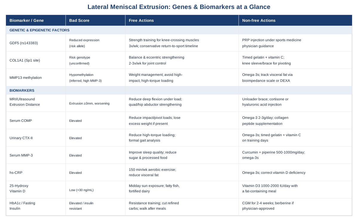
Lateral meniscal extrusion sits at the intersection of mechanics, biology, and sometimes anatomy you were born with. Below, you will find seven measurable biomarkers, ranging from a simple vitamin D test to specialized cartilage-turnover assays, that can tell you whether your knee's environment is pushing toward more extrusion or holding steady, plus realistic plans for improving each one, with and without supplements. You will also find three genetic and epigenetic factors, including a growth-factor gene and a collagen gene, that help explain why extrusion and meniscal injury do not affect everyone the same way. A closer look at a widely discussed Huberman Lab deep dive on connective tissue reveals a surprisingly specific, low-cost protocol for supporting collagen synthesis that most people have never heard of. And a review of complementary approaches, tai chi, yoga, massage therapy, and low-level laser therapy, shows which of them actually have knee-specific trial data behind them.
Seven Biomarkers That Reveal What's Happening Inside Your Knee
Meniscal extrusion is formally defined as displacement of the meniscus 3 millimeters or more beyond the edge of the tibial plateau, and it is now understood as a consequence of joint tissue degeneration interacting with mechanical stress rather than a purely mechanical event on its own, as described in research on meniscal extrusion and cartilage volume loss in a largely non-osteoarthritic cohort. Most of the research base focuses on the medial meniscus, since medial extrusion is more common and easier to study, but the underlying biology of cartilage breakdown, inflammation, and metabolic stress applies to the lateral compartment as well. Lateral extrusion has its own specific wrinkles too, particularly its association with a discoid lateral meniscus and with extrusion following partial meniscectomy, as reviewed in a narrative review of discoid lateral meniscus pathology in children. The seven markers below combine direct imaging of the extrusion itself with blood and urine tests that reveal the biological processes feeding it.
1. MRI or ultrasound-measured extrusion distance
This is the most direct biomarker there is: an actual measurement, in millimeters, of how far your lateral meniscus sits beyond the tibial plateau margin. It matters because extrusion distance correlates with faster cartilage loss and greater symptom severity, and tracking it over time tells you whether your knee is stable or deteriorating, independent of how it feels day to day, as shown in a 100-patient ultrasound and MRI analysis linking meniscal extrusion to symptom severity in knee osteoarthritis.
How to measure it: MRI remains the reference standard, typically performed with the knee unloaded, and costs roughly 400 to 1,200 dollars out of pocket in the United States depending on the imaging center and whether contrast is used. Ultrasound is a validated, far cheaper alternative for follow-up measurements once a baseline MRI exists, running around 100 to 300 dollars, and has been shown to track closely with MRI-based measurements, including under physiologic loading conditions, per research on ultrasound-detected medial meniscal extrusion with loading and flexion. For the lateral meniscus specifically, meniscal shape itself is a risk factor worth discussing with your radiologist, since certain morphologies predict a higher extrusion risk, as described in an MRI-based assessment of lateral meniscus shape and extrusion risk.
If the measurement is bad, the plan without supplements or equipment centers on load management: reducing deep flexion under load (deep squats, kneeling with rotation), addressing quadriceps and hip abductor weakness through supervised strengthening two to three times per week, and correcting any varus or valgus alignment issue with a physical therapist's guidance. If the measurement is bad, the plan with supplements or equipment includes an unloader-style knee brace for high-load activities, which redistributes force away from the affected compartment, and in select cases a cortisone or hyaluronic acid injection to manage symptoms while the mechanical plan takes effect; neither of these reverses existing extrusion, and both should be used as adjuncts, not replacements, for the loading changes above. Re-image no more than once every 6 to 12 months unless symptoms change abruptly, since extrusion changes slowly and frequent MRIs mostly add cost and radiation-free but unnecessary anxiety.
2. Serum COMP (cartilage oligomeric matrix protein)
COMP is a structural protein released into the bloodstream when cartilage matrix breaks down. Elevated serum COMP correlates with radiographic severity, pain, and disability in knee osteoarthritis, and can flag cartilage degeneration even before it shows up clearly on X-ray, according to a review of COMP as a biomarker for knee osteoarthritis, building on earlier population-level findings from the Johnston County Osteoarthritis Project showing COMP tracks with both presence and severity of disease.
How to measure it: this is not a standard primary-care blood draw. It requires a specialty or research-oriented lab (functional medicine clinics and some university-affiliated labs offer it), typically costing 80 to 150 dollars, and turnaround can take one to two weeks.
If the score is bad, the plan without supplements or equipment is the same anti-catabolic approach that helps most cartilage biomarkers: reduce high-impact and pivoting loads temporarily, lose excess weight if present (even 5 to 10 percent body weight reduces joint reaction forces meaningfully), and prioritize low-impact strength and aerobic conditioning. If the score is bad, the plan with supplements or equipment includes omega-3 fatty acids (roughly 2 to 3 grams combined EPA/DHA daily, cycling isn't necessary but reassess bloodwork every 3 to 4 months) and collagen peptide supplementation discussed further in the genetics section below; side effects are generally mild gastrointestinal upset with fish oil at higher doses, and fish oil should be used cautiously alongside anticoagulant medication. Retest COMP every 4 to 6 months; it moves slowly and monthly testing wastes money.
3. Urinary CTX-II
CTX-II is a breakdown fragment of type II collagen, the collagen that makes up both articular cartilage and the meniscus itself. Elevated urinary CTX-II independently predicts radiographic progression of knee osteoarthritis and has been shown to predict cartilage loss on MRI over 21 months, per a study on urinary CTX-II and cartilage loss prediction, though a more recent appraisal notes conclusive evidence for its use as an early-diagnosis tool is still lacking, as summarized in a review asking whether urinary CTX-II can serve as a knee osteoarthritis biomarker.
How to measure it: this requires a first- or second-morning urine sample sent to a specialty or research lab; expect 100 to 200 dollars and limited availability outside academic or sports-medicine-affiliated clinics.
If the score is bad, the plan without supplements or equipment is to reduce repetitive high-torque loading (deceleration sports, twisting under load) while symptoms and biomarkers are elevated, and to address any biomechanical driver such as quadriceps weakness or gait asymmetry with formal gait analysis. If the score is bad, the plan with supplements or equipment mirrors the COMP approach: omega-3s, adequate protein intake (1.2 to 1.6 grams per kilogram of body weight daily supports overall collagen substrate availability), and the timed gelatin-plus-vitamin-C protocol detailed in the podcast section below, taken on strength-training days only, not daily, to avoid unnecessary caloric and gastrointestinal load. Recheck every 4 to 6 months alongside COMP.
4. Serum MMP-3
MMP-3 (stromelysin-1) is an enzyme that actively degrades cartilage matrix components and activates other matrix-degrading enzymes. Serum MMP-3 has been shown to serve as a prognostic marker, with higher levels associated with greater cartilage volume loss over two years, according to research on serum MMP-3 as a prognostic marker for cartilage injury progression. Unlike COMP and CTX-II, which reflect matrix breakdown that has already happened, MMP-3 gives a window into ongoing catabolic enzyme activity.
How to measure it: specialty lab, typically 50 to 100 dollars, more commonly available through rheumatology-oriented labs than general practice, since it is also used to monitor rheumatoid arthritis activity.
If the score is bad, the plan without supplements or equipment focuses on reducing systemic and local inflammatory drivers: improving sleep quality (poor sleep raises inflammatory cytokines), managing stress, and reducing ultra-processed food and added sugar intake, which are linked to higher inflammatory enzyme activity. If the score is bad, the plan with supplements or equipment includes curcumin with piperine (500 to 1,000 mg daily, cycling 8 weeks on, 2 weeks off is a reasonable, though not strictly evidence-mandated, precaution for gastrointestinal tolerance) and the same omega-3 approach above; curcumin can interact with blood thinners and should be discussed with a physician if you take anticoagulants.
5. High-sensitivity CRP (hs-CRP)
Hs-CRP is a general marker of systemic low-grade inflammation, not specific to the knee, but it is one of the most consistently associated markers with osteoarthritis symptoms. It is significantly associated with knee osteoarthritis progression in community-based cohorts, as shown in the Yakumo study on hs-CRP and knee osteoarthritis progression, and a meta-analysis found circulating CRP is modestly but reliably elevated in osteoarthritis patients and correlates with pain and physical function, per a systematic review and meta-analysis of circulating CRP in osteoarthritis, even though its link to radiographic severity specifically is weaker than its link to symptoms.
How to measure it: this is a standard, widely available test, typically 15 to 40 dollars, and is included in many routine cardiovascular risk panels already.
If the score is bad, the plan without supplements or equipment is straightforward and well supported across conditions: improve sleep duration and consistency, reduce visceral fat through a modest caloric deficit if overweight, and add 150 minutes per week of moderate aerobic activity, which reliably lowers hs-CRP over 2 to 3 months. If the score is bad, the plan with supplements or equipment includes omega-3 fatty acids again (this is one of the better-supported anti-inflammatory supplements across multiple biomarkers) and, for those with confirmed vitamin D insufficiency (see below), correcting that deficiency, since low vitamin D and elevated CRP frequently travel together. Recheck hs-CRP every 3 months when actively intervening.
6. 25-hydroxyvitamin D
Vitamin D status matters here because low levels have been associated with worsening knee osteoarthritis in longitudinal cohorts, as shown in a study linking low vitamin D levels to knee osteoarthritis worsening. It is worth being candid, though: randomized trials of supplementation have been disappointing for structural outcomes. A well-conducted trial found vitamin D supplementation did not significantly change MRI-measured tibial cartilage volume or pain scores over two years, per a randomized trial of vitamin D supplementation and tibial cartilage volume. In other words, low vitamin D correlates with worse outcomes, but fixing a mild deficiency is unlikely to reverse existing extrusion. It is still worth correcting, since vitamin D has broader roles in bone and muscle health that matter for joint loading.
How to measure it: standard test, 40 to 80 dollars, widely available.
If the score is bad, the plan without supplements or equipment is 15 to 20 minutes of midday sun exposure on exposed skin several times a week, where climate and skin tone allow, plus dietary sources like fatty fish and fortified dairy. If the score is bad, the plan with supplements or equipment is vitamin D3, typically 1,000 to 2,000 IU daily for mild insufficiency or higher, physician-guided doses for more significant deficiency, taken with a fat-containing meal for absorption; no cycling is needed, but blood levels should be rechecked after 8 to 12 weeks to avoid over-supplementation, since excessive vitamin D can cause hypercalcemia over time.
7. HbA1c and fasting insulin (metabolic markers)
This pairing captures insulin resistance, which is increasingly recognized as a joint-relevant metabolic state independent of body weight. Insulin resistance, high waist circumference, and low HDL cholesterol are associated with tibial cartilage defects, and metabolic syndrome is linked to a meaningfully higher incidence of knee osteoarthritis in prospective cohorts, according to a meta-analysis of metabolic syndrome and knee osteoarthritis incidence and a pilot study correlating metabolic biomarkers with knee cartilage composition on MRI. This matters for meniscal extrusion because a metabolically unhealthy joint environment accelerates the cartilage and meniscal degeneration that drive extrusion, even in people who are not significantly overweight.
How to measure it: both are standard tests, together roughly 20 to 60 dollars, and often included in annual metabolic panels.
If the score is bad, the plan without supplements or equipment is the highest-leverage option on this entire list: resistance training two to three times weekly (muscle is the body's primary site of glucose disposal), reducing refined carbohydrate and added sugar intake, and walking after meals, which measurably blunts post-meal glucose and insulin spikes. If the score is bad, the plan with supplements or equipment includes a continuous glucose monitor for two to four weeks to identify personal food triggers (roughly 50 to 100 dollars for a short-term sensor), and, if physician-approved, berberine (500 mg two to three times daily, generally not recommended long-term without monitoring due to gastrointestinal side effects and interaction with diabetes medications) as an insulin-sensitizing option with meaningful clinical evidence in metabolic contexts. Recheck HbA1c every 3 months, since it reflects roughly a 3-month average.
Taken together, these seven markers separate meniscal extrusion into what can be measured directly (the extrusion itself), what reflects active tissue breakdown (COMP, CTX-II, MMP-3), and what reflects the systemic environment either accelerating or slowing that breakdown (CRP, vitamin D, metabolic markers). That distinction also sets up the next question naturally: why do two people with identical biomarker profiles and identical loading histories still end up with different outcomes? Part of the answer is genetic.
The Genetic Blueprint Behind Meniscal Extrusion
Genetic research on meniscal extrusion specifically is limited; most of the human evidence comes from broader osteoarthritis and ligament injury genetics, which overlaps meaningfully with meniscal biology since both involve collagen structure and joint tissue repair signaling. This is an area where researchers like Ali Torkamani, whose work has focused on translating genomic risk data into actionable individual insight, and Gary Brecka, known for popularizing accessible genetic panel interpretation, have pushed genetic literacy into mainstream health conversations, even though the underlying studies referenced below predate and stand independently of that popularization.
GDF5 (growth differentiation factor 5)
GDF5 is a signaling protein involved in joint formation and cartilage maintenance. A specific regulatory variant, rs143383, in the gene's 5' untranslated region reduces GDF5 expression and is associated with osteoarthritis susceptibility at genome-wide significance, as established in the original functional polymorphism study linking GDF5 to osteoarthritis susceptibility. More directly relevant here, this same SNP has been associated with meniscus injury and slower functional recovery in a cohort of Chinese soldiers, per a study on the GDF5 SNP and meniscus injury recovery. The practical implication is that carriers of the risk allele may have a joint environment with somewhat lower baseline regenerative signaling, which could mean slower recovery after meniscal stress even when mechanics are corrected.
If the gene is bad, the plan without supplements is built around compensating with controlled mechanical stimulus rather than trying to alter gene expression directly: consistent, moderate-load strength training for the muscles crossing the knee (quadriceps, hamstrings, hip abductors) 3 times weekly, since joint stability reduces the mechanical stress that would otherwise demand more repair capacity than the tissue can supply, and longer, more conservative return-to-sport timelines after any knee injury, since recovery may genuinely take longer for this genotype.
If the score is bad, the plan with supplements or equipment includes ensuring adequate dietary protein and vitamin D status (both support tissue repair broadly, as covered above), and considering platelet-rich plasma (PRP) injection under a sports medicine physician's guidance after significant meniscal injury, since PRP delivers a concentrated dose of growth factors, including members of the same signaling family GDF5 belongs to, directly to the joint; evidence for PRP in meniscal pathology specifically is still early and mixed, so this should be framed as a reasonable adjunct under specialist care rather than a proven fix, typically limited to 1 to 3 injections spaced several weeks apart, with mild post-injection swelling and soreness as the main side effect.
COL1A1 (collagen type I alpha 1, Sp1 binding site variant)
COL1A1 encodes a chain of type I collagen, a structural protein in ligaments, tendons, and the outer meniscus. The Sp1 binding site polymorphism (rs1800012) has been studied across multiple populations for its association with cruciate ligament rupture risk, with some studies suggesting the rare TT genotype is protective and more common genotypes carrying somewhat higher risk, as seen in a case-control study of COL1A1 polymorphism in Polish skiers with ACL injury. It is important to be direct about the evidence quality here: results are inconsistent across populations, and a study in Middle Eastern elite athletes found no significant association between this variant and ACL injury or outcomes, per research finding no association between COL1A1 rs1107946 and ACL injury outcomes. This gene is best treated as a plausible but unconfirmed contributor to individual connective tissue quality rather than a settled risk factor.
If the gene is bad, or simply unknown, the plan without supplements emphasizes proprioceptive and neuromuscular training, since ligament and capsule quality differences are best compensated for through better joint control: balance work, single-leg stability drills, and eccentric strengthening 2 to 3 times weekly, which has the strongest independent evidence for reducing knee injury risk regardless of underlying collagen genotype.
If the score is bad, the plan with supplements or equipment centers on the collagen-loading protocol detailed in the next section (timed gelatin or hydrolyzed collagen plus vitamin C around training), and a knee sleeve or brace during high-risk pivoting activity for proprioceptive feedback rather than structural support; braces do not strengthen tissue, but the added sensory input can reduce re-injury risk during higher-load activity.
MMP13 promoter methylation (an epigenetic factor)
This is not a gene variant you inherit but an epigenetic mark, a chemical tag on DNA that controls whether a gene is switched on, that can change over your lifetime in response to mechanical stress, obesity, and inflammation. In healthy cartilage, the MMP13 gene (which encodes a powerful cartilage-degrading enzyme) is kept silenced by DNA methylation. In osteoarthritic cartilage, that region becomes hypomethylated, and MMP13 gets switched on, driving matrix breakdown, as described in research on DNA methylation of the RUNX2 promoter and its role in mediating MMP13 transcription, with the broader epigenetic landscape of osteoarthritis reviewed in a review of epigenetics as a therapeutic target in osteoarthritis. Unlike GDF5 or COL1A1, this factor is genuinely modifiable, since methylation patterns respond to the same mechanical and metabolic inputs already discussed above.
If the epigenetic pattern is likely unfavorable (inferred from elevated MMP-3, obesity, or high inflammatory markers rather than direct testing, since clinical methylation testing for this specific site is not yet available outside research settings), the plan without supplements is weight management and reducing the specific loading pattern associated with this pathway, meaning high-impact, high-torque activity in an already extruded or inflamed knee, in favor of controlled, progressive loading instead.
If the score is bad, the plan with supplements or equipment includes omega-3 fatty acids, which have some evidence for favorably influencing inflammatory gene expression patterns, and body composition tracking (a simple bioelectrical impedance scale, 30 to 60 dollars, or periodic DEXA scan, 50 to 150 dollars per scan, every 6 months) to monitor visceral fat trends, since visceral adiposity is one of the more consistent drivers of the inflammatory signaling that promotes this kind of catabolic gene activation.
None of these three factors can currently be changed at the DNA level, and none should be used to justify fatalism about outcomes; the epigenetic example above shows the biology itself is responsive to the same lifestyle levers covered in the biomarker section. That responsiveness is exactly what the next section explores in more depth, through a specific, well-studied protocol for supporting connective tissue that has recently gained attention outside standard orthopedic circles.
What a Deep Dive on Connective Tissue Reveals
The Huberman Lab podcast's guest series with Dr. Andy Galpin, a kinesiology researcher specializing in exercise physiology, spent significant time on a subject most people never hear discussed: how tendons, ligaments, and fibrocartilage like the meniscus actually adapt, and why that process is fundamentally different from how muscle adapts. Below are ten of the most useful, evidence-grounded points from that body of work and the research it draws on, particularly the collagen synthesis research from Dr. Keith Baar's lab at UC Davis, each with a specific takeaway for someone managing meniscal extrusion.
1. Connective tissue remodels far slower than muscle
Muscle protein turnover happens on a scale of days; tendon, ligament, and cartilage-adjacent tissue turnover happens on a scale of months to years. This is a central reason meniscal extrusion does not resolve quickly even when every mechanical factor is corrected, and it argues for patience over the kind of 6-week fixes marketed for muscle-based injuries.
2. Loading is the primary signal for adaptation, but it has to be specific
Connective tissue adapts to the type of load it experiences, not just total activity volume. Generic cardio does little for meniscal or ligament resilience; controlled, progressive loading through the knee's actual range of motion is what drives adaptation, which is why physical therapy protocols emphasize specific movement patterns rather than general activity advice.
3. There is a real, testable window for boosting collagen synthesis around exercise
The most directly actionable finding from this research area comes from a controlled study showing that consuming vitamin C-enriched gelatin roughly one hour before a bout of connective-tissue-loading activity measurably increases markers of collagen synthesis, as demonstrated in a randomized crossover trial of vitamin C-enriched gelatin supplementation before intermittent activity.
4. Dose matters more than people assume
In that same trial, 15 grams of gelatin produced roughly double the collagen synthesis marker response compared to a 5-gram dose, meaning the commonly recommended "scoop" of collagen powder many people take may be a meaningfully sub-therapeutic dose for this specific purpose.
5. Timing is not optional, it is the mechanism
The supplement only works this way because it floods the bloodstream with collagen-building amino acids (glycine, proline, hydroxyproline) right before the loading stimulus tells the tissue to use them. Taking the same gelatin dose at a random time of day, disconnected from exercise, is not supported by this mechanism.
6. Longer-term collagen peptide use, combined with resistance training, changes real tissue structure
Beyond the acute synthesis marker studies, a 14-week randomized controlled trial found that daily specific collagen peptide supplementation combined with high-load resistance training produced significantly greater increases in Achilles tendon cross-sectional area compared to placebo, and other work has shown specific collagen peptides directly stimulate biosynthesis of tendon and ligament matrix molecules, per research on specific collagen peptides and matrix biosynthesis in tendons and ligaments, and a related trial found collagen peptides combined with calf-strengthening reduced pain and improved function in a study of specific collagen peptides in Achilles tendinopathy patients.
7. Isometric loading is an underused tool for irritated joints
For joints too irritable for dynamic loading, sustained isometric holds (for example, a wall-sit style quad contraction) still provide a meaningful mechanical signal to connective tissue without the shear stress of dynamic movement, making them a reasonable bridge strategy during flare-ups.
8. Recovery time between loading sessions needs to be longer than intuition suggests
Because connective tissue turnover is slow, loading the same tissue too frequently (daily high-intensity knee-loading work, for instance) does not allow enough time for the synthesis response to translate into structural change, and may instead tip the balance toward net breakdown; 48 to 72 hours between higher-load sessions targeting the same tissue is a more physiologically reasonable cadence.
9. Metabolic health blunts or amplifies this entire process
This directly connects back to the biomarker section: poor metabolic health and chronic systemic inflammation interfere with the body's ability to translate a good loading and nutrition protocol into actual tissue adaptation, which is one more reason the CRP and insulin markers above are not tangential to a "joint" problem.
10. None of this replaces fixing the mechanical driver
The consistent caveat throughout this body of work is that nutrition and supplementation support the tissue's capacity to adapt; they do not correct malalignment, weak stabilizing muscles, or a torn meniscal root. Skipping the mechanical correction and relying on collagen supplementation alone is a common and largely wasted effort.
Supporting connective tissue biology this way pairs naturally with approaches that reduce pain and improve movement quality without adding further joint stress, which is where several complementary modalities with real condition-specific trial data come in.
Complementary Approaches Worth Considering
The options below were chosen because they have meaningful clinical trial data specifically in knee osteoarthritis populations, the condition most closely tied to meniscal extrusion, rather than generic wellness claims.
Tai Chi
Tai chi combines slow, controlled weight-shifting movements with balance and proprioceptive training, which makes it relevant to meniscal extrusion because it loads the knee through a controlled range without the impact or twisting forces that aggravate an extruded meniscus. It also directly trains the neuromuscular control that compensates for reduced meniscal hoop-tension function.
In a 52-week single-blind randomized trial, tai chi performed twice weekly for 12 weeks produced improvements in pain and physical function equivalent to a standard course of physical therapy, according to a comparative effectiveness trial of tai chi versus physical therapy for knee osteoarthritis, a finding significant enough that the National Center for Complementary and Integrative Health highlighted it as a notable result.
Realistically, this means two 30 to 60 minute sessions weekly, ideally starting with an instructor familiar with modifying stances for people with knee pain, keeping range of motion within a pain-free zone and avoiding deep-knee stances until the extrusion and associated symptoms are stable.
Yoga
Yoga is relevant here for similar reasons to tai chi: controlled static and dynamic loading, improved hip and ankle mobility (which reduces compensatory stress on the knee), and better neuromuscular control, all without high-impact loading.
A 2024 systematic review and meta-analysis of randomized controlled trials found yoga produced significant improvements in pain, stiffness, and physical function in knee osteoarthritis patients, per a meta-analysis of yoga's impact on knee osteoarthritis patients, and a biomechanically-focused yoga program specifically reduced knee adduction moment, a key mechanical driver of medial compartment stress, in a randomized controlled trial of a biomechanically-based yoga program for knee osteoarthritis.
Apply this cautiously: avoid poses requiring deep knee flexion under load (deep lunges, hero pose) until symptoms and extrusion are stable, and favor a therapeutic or gentle style class over a vigorous vinyasa practice, ideally two to three sessions weekly.
Massage Therapy
Massage therapy is relevant less for structural change and more for reducing the muscle guarding and compensatory tension patterns that develop around a symptomatic knee, which can otherwise alter gait and increase asymmetric loading on the joint.
A randomized dose-finding trial found that 60-minute weekly Swedish massage sessions over 8 weeks produced significantly greater improvement in WOMAC scores (a validated pain and function measure) than usual care, per a randomized dose-finding trial of massage therapy for osteoarthritis of the knee, with shorter or less frequent sessions producing smaller benefits.
Realistically, weekly 60-minute sessions for 8 weeks is the best-supported starting protocol; this is a symptom and movement-quality tool, not a way to directly influence extrusion distance, and should be paired with the strengthening work covered earlier rather than used alone.
Low-Level Laser Therapy (Photobiomodulation)
Low-level laser therapy applies specific wavelengths of light to reduce local inflammation and pain, and it is relevant here as a low-risk adjunct for managing the pain and inflammation that often accompanies an extruded, symptomatic meniscus, without adding mechanical load.
A systematic review and meta-analysis of 14 randomized controlled trials involving over 800 patients found meaningful pain reductions immediately after therapy and at follow-up, particularly when combined with exercise therapy, per a systematic review and meta-analysis of low-level laser therapy for knee osteoarthritis pain and disability, though a more recent network meta-analysis found it outperforms sham treatment for pain but not clearly for function or stiffness, according to a network meta-analysis on optimal wavelength for low-level light therapy in knee osteoarthritis.
In practice, this means a course of sessions (commonly 2 to 3 times weekly for 4 to 6 weeks) delivered by a physical therapist or clinic using a device in the recommended 780 to 904 nanometer range, used alongside, not instead of, an active strengthening and loading program.
Conclusion
Lateral meniscal extrusion is not a single problem with a single fix; it is a mechanical finding shaped by cartilage turnover, systemic inflammation, metabolic health, and, for some people, an underlying genetic or anatomic predisposition. The seven biomarkers here give you a way to see which of those processes is actually active in your case instead of guessing, the genetic factors explain part of why identical mechanics produce different outcomes in different people, and the connective tissue research and complementary approaches offer realistic, evidence-grounded ways to support the tissue while the mechanical drivers are addressed. None of it replaces a proper orthopedic evaluation, and none of it promises to erase an existing extrusion, but all of it turns a vague diagnosis into a set of concrete, trackable numbers.
The next practical step is a conversation, not a purchase: bring this list of biomarkers to your next appointment, ask which ones are worth ordering given your specific history, and use the results to decide which of the plans above actually apply to you.
Musculoskeletal: Joint Conditions Tendon & Ligament Conditions
Endocrine & Metabolic: Diabetes & Blood Sugar Metabolic Syndrome
Autoimmune: Inflammatory Conditions Connective Tissue Conditions