This article contains AI generated content.
Lateral Meniscocapsular Separation: 6 Genes and 7 Biomarkers to Track
If you've been told you have a lateral meniscocapsular separation, you've probably already noticed how thin the information gets once you move past the MRI report. Surgeons will describe the mechanics well: the peripheral rim of the lateral meniscus has pulled away from its attachment to the joint capsule, usually because of a twisting injury, a tibial plateau fracture, or a combined ligament injury. What's harder to find is guidance on why some knees heal this kind of tear cleanly with conservative management or repair, while others stay unstable, re-tear, or drift toward early cartilage wear years later.
Generic advice tends to stop at "rest, physical therapy, maybe surgery." That's not wrong, but it treats every knee as identical, when the real variability comes from things that don't show up on an MRI: how efficiently your body builds and cross-links collagen, how much background inflammation is circulating through the joint, how your connective tissue genetics loaded the dice before the injury ever happened. Two people with the same tear pattern can have very different biological starting points for repair.
This article takes a more granular approach. Instead of repeating standard post-injury instructions, it looks at the specific biomarkers that reflect how your body is handling collagen turnover, inflammation, and tissue repair right now, and at the genetic variants that research has linked to meniscus and ligament injury risk in the first place. Neither replaces a surgeon's exam or an MRI, but both can sharpen the decisions you make around rehab pacing, nutrition, and long-term joint protection.
None of this promises a faster healing timeline or a guaranteed outcome, and anyone suggesting otherwise about a joint injury this specific should be treated with skepticism. What better information does is narrow the guesswork. Knowing your inflammatory load, your collagen-support nutrient status, and your genetic tendencies toward looser or stiffer connective tissue gives you and your care team a more precise map, and a more grounded reason to expect a plan tailored to your biology rather than a generic protocol.
Summary
A lateral meniscocapsular separation isn't just a local mechanical problem, it's a window into how well your whole system builds, repairs, and remodels collagen under stress. Below, you'll find seven measurable biomarkers, ranging from inexpensive blood panel staples like HbA1c to specialty cartilage-turnover markers, that reveal whether your body's chemistry is helping or hindering that repair, along with realistic plans (with and without supplements) for shifting the ones that are working against you. Beyond that, you'll find the six genes most consistently linked in human studies to meniscus and ligament injury risk, a research-backed breakdown of the collagen-loading science popularized on the Huberman Lab podcast, and a short list of complementary therapies with genuine evidence behind them for knee soft-tissue recovery. Read on to see which numbers are worth asking your doctor to order next.
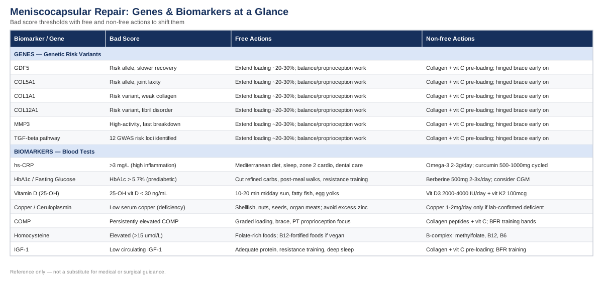
7 Biomarkers Worth Tracking When Your Lateral Meniscus Capsule Is Healing
The philosophy here borrows from how clinicians like Peter Attia, Thomas Dayspring, and Allan Sniderman have pushed cardiometabolic medicine toward earlier, more precise biomarker tracking instead of waiting for a problem to become obvious. The same logic applies to connective tissue: rather than waiting to see whether a meniscocapsular repair holds up, you can measure the biological conditions that make holding up more or less likely. The seven biomarkers below cover inflammation, glycemic control, collagen-support micronutrients, and direct markers of cartilage and connective tissue turnover. A few are cheap and available through any standard lab. A couple are specialty tests that are harder to access but worth knowing about if you want the fuller picture.
High-Sensitivity C-Reactive Protein (hs-CRP)
Chronic low-grade inflammation is one of the more overlooked variables in soft tissue healing. Elevated hs-CRP reflects systemic inflammatory load, and research on osteoarthritis phenotypes has shown that a subset of joint disease is meaningfully more inflammatory than others, with hs-CRP tracking that pattern reasonably well (George et al., Osteoarthritis and Cartilage). A meniscocapsular junction healing inside a chronically inflamed joint environment is working against a biochemical headwind: persistent inflammation upregulates the same matrix metalloproteinases that break down collagen, which is the opposite of what a healing capsular attachment needs.
How to measure it
hs-CRP is a standard blood draw, widely available through primary care or direct-to-consumer lab services, typically costing between $15 and $40 when ordered outside insurance coverage. It's frequently bundled into general wellness panels already.The plan without supplements or equipment
Prioritize a Mediterranean-style eating pattern (vegetables, olive oil, fatty fish, minimal ultra-processed food), get 7 to 9 hours of sleep consistently, and add 150 minutes per week of easy zone 2 cardio once your surgeon or physical therapist clears low-impact activity. Address dental and gum health, since chronic gum inflammation is an underrated contributor to systemic CRP. Retest after 8 to 12 weeks.The plan with supplements or equipment
Omega-3s (EPA/DHA, roughly 2 to 3 grams daily combined) have the best evidence for lowering CRP modestly over 8 to 12 weeks; expect mild fish-oil burps or, less commonly, loose stools, and use caution if you're on blood thinners. Curcumin with piperine (500 to 1,000 mg daily) is a reasonable add-on cycled in 8 to 12 week blocks with a few weeks off between cycles; it can cause GI upset and interacts with anticoagulants. Cold exposure tools (contrast baths, cold plunge) are popular but have weaker direct evidence for systemic CRP specifically and should be used cautiously, not immediately over a fresh surgical repair.HbA1c and Fasting Glucose
Collagen doesn't just need to be synthesized, it needs to stay flexible and organized. Chronically elevated blood glucose promotes advanced glycation end-products (AGEs) that stiffen and disorganize collagen fibers. In diabetic models, tendon-to-bone healing after soft tissue repair showed significantly more AGE accumulation and less organized collagen at the healing interface compared to normoglycemic controls (Bedi et al., Journal of Shoulder and Elbow Surgery). While that specific study involved an animal model of rotator cuff repair, the underlying glycation biology applies to any collagen-rich healing site, including a repaired meniscocapsular junction.How to measure it
Both HbA1c and fasting glucose are inexpensive, widely available blood tests, usually $10 to $30 each, and often already part of a standard metabolic panel.The plan without supplements or equipment
Reduce refined carbohydrate and added sugar intake, take a 10 to 15 minute walk after meals once mobility allows, and add resistance training 2 to 3 times weekly as your rehab progresses. Keep a consistent sleep and meal schedule, since irregular timing worsens glucose variability independent of diet quality.The plan with supplements or equipment
Berberine (500 mg, two to three times daily) has clinical evidence comparable to some oral glycemic agents for lowering fasting glucose and HbA1c; cycle it in 3-month-on, 1-month-off blocks, and expect GI side effects in some users. It interacts with several medications, so check with a prescriber first. A continuous glucose monitor is a useful, if optional, piece of equipment during the healing window to see how specific foods and inactivity (common after knee surgery) affect your glucose in real time.Vitamin D (25-Hydroxyvitamin D)
Vitamin D status is one of the more actionable levers in post-surgical knee recovery, largely through its role in muscle strength and neuromuscular function rather than a direct effect on the capsule itself. A registered clinical trial protocol is currently testing vitamin D supplementation specifically for improving quadriceps strength after ACL reconstruction (Ong et al., Trials), which reflects growing interest in this link even though the evidence base is still maturing. Quadriceps strength matters directly for a lateral meniscocapsular injury too, since a weak quad changes knee loading mechanics during the exact activities that stress a healing capsular attachment.How to measure it
A standard 25-OH vitamin D blood test costs roughly $40 to $80 direct-to-consumer, less if bundled into insurance-covered bloodwork.The plan without supplements or equipment
Aim for 10 to 20 minutes of midday sun exposure on bare skin several times a week, adjusted for your skin tone and latitude, and include fatty fish and egg yolks in your diet.The plan with supplements or equipment
If levels are below roughly 30 ng/mL, 2,000 to 4,000 IU of vitamin D3 daily, taken with a fat-containing meal and paired with 100 mcg of vitamin K2 (MK-7) to support proper calcium handling, is a standard and low-risk protocol. Retest in 8 to 12 weeks. Avoid self-dosing above 10,000 IU/day for extended periods without monitoring, since hypercalcemia is a real, if uncommon, risk at high sustained doses.Serum Copper and Ceruloplasmin
This is a less commonly checked pairing, but it matters directly for connective tissue. Copper is the essential cofactor for lysyl oxidase, the enzyme that cross-links collagen and elastin fibers into strong, stable structures (Rucker et al., American Journal of Clinical Nutrition). Without adequate copper, newly synthesized collagen at a healing meniscocapsular junction can be laid down but never properly cross-linked, leaving the repair mechanically weaker than it looks on imaging.How to measure it
Serum copper and ceruloplasmin together run about $50 to $90 and are available through most reference labs, though they're not part of a routine physical.The plan without supplements or equipment
Increase dietary copper through shellfish (especially oysters), nuts, seeds, and organ meats, and avoid high-dose zinc supplementation (above 40 mg/day long term) without a paired copper source, since excess zinc is a well-documented cause of copper deficiency.The plan with supplements or equipment
Only supplement copper if a confirmed deficiency shows up on testing. If so, 1 to 2 mg daily for 8 to 12 weeks, then retest, is a conservative approach. Copper toxicity is a genuine concern in people with Wilson's disease or unrecognized liver dysfunction, so this is one biomarker where self-supplementing without a lab-confirmed deficiency is not advisable.Cartilage Oligomeric Matrix Protein (COMP)
COMP is released into the bloodstream when cartilage and related joint matrix proteins are being broken down faster than they're rebuilt. A systematic review and meta-analysis found serum COMP, along with CTX-II and MMP-3, meaningfully elevated in knee and hip osteoarthritis compared to healthy joints (Hao et al., Osteoarthritis and Cartilage). For someone recovering from a meniscocapsular separation, a persistently elevated COMP can be an early signal that the joint is under more ongoing mechanical or inflammatory stress than the rehab plan is accounting for, well before that shows up as pain or on an X-ray.How to measure it
COMP is largely a specialty or research-lab test, typically $100 to $250, and not something you can order at a standard commercial lab counter. Ask a sports medicine physician or an academic orthopedic center if it's accessible in your area; availability varies a lot by region.The plan without supplements or equipment
Load management is the primary lever here: follow a graded return-to-activity plan rather than pushing through pain, use an unloading or hinged knee brace during higher-risk phases if your surgeon recommends one, and prioritize physical therapy focused on quadriceps, hamstring, and proprioceptive control rather than early high-torque pivoting drills.The plan with supplements or equipment
Collagen peptides (10 to 15 g daily) paired with roughly 50 mg of vitamin C taken about an hour before therapy or exercise sessions has direct human evidence for boosting collagen synthesis (detailed further below). Glucosamine and chondroitin have mixed evidence for cartilage-specific benefit; if used, cycle for 3 months and reassess rather than taking indefinitely on faith. Blood flow restriction (BFR) training bands are a useful piece of equipment for building strength around the joint with lower absolute mechanical load on the healing capsule.Homocysteine
Homocysteine is best known as a cardiovascular marker, but it has a direct connective tissue story too. In homocystinuria, the classic genetic condition of extreme homocysteine elevation, excess homocysteine physically interferes with collagen cross-linking through its sulfhydryl group, producing weaker, more disorganized connective tissue (Grieco, American Journal of the Medical Sciences). Most people will never approach homocystinuria-level elevations, but even moderately high homocysteine reflects a B-vitamin status that also matters for normal collagen and bone matrix maintenance (Fratoni & Brandi, Nutrients).How to measure it
A homocysteine blood test runs roughly $30 to $70 and is available through most standard labs.The plan without supplements or equipment
Increase folate-rich foods (leafy greens, legumes) and, if you eat a plant-based diet, pay close attention to B12-fortified foods, since B12 deficiency is a common and under-recognized driver of elevated homocysteine in that population.The plan with supplements or equipment
A B-complex containing methylfolate, methylcobalamin (B12), and B6 is a standard, low-cost approach; retest homocysteine after 8 to 12 weeks. Check B12 status specifically before starting high-dose folate, since folate can mask an underlying B12 deficiency. Avoid long-term B6 doses above 100 mg/day, which have been associated with peripheral neuropathy with prolonged use.Insulin-Like Growth Factor 1 (IGF-1)
IGF-1 is one of a handful of growth factors with a well-documented role in tendon and ligament healing, alongside PDGF, TGF-beta, and VEGF, orchestrating cell proliferation and matrix production at the injury site (Molloy et al., Sports Medicine). Low circulating IGF-1 doesn't doom a repair, but it's a reasonable proxy for whether your overall anabolic environment, sleep, protein intake, and training load, is supporting or undermining tissue repair at the systemic level.How to measure it
IGF-1 testing typically costs $60 to $120 and is available through most reference labs, sometimes requiring a physician order rather than direct-to-consumer access.The plan without supplements or equipment
Eat adequate protein (roughly 1.6 to 2.2 g per kilogram of body weight daily), maintain resistance training appropriate to your healing stage, and protect deep sleep, since growth hormone (and downstream IGF-1) release is concentrated in early-night slow-wave sleep. Avoid prolonged aggressive caloric deficits during active healing phases.The plan with supplements or equipment
There's no well-supported supplement that safely and directly raises IGF-1; be wary of anything marketed that way, and avoid unsupervised use of growth hormone or IGF-1 injectables, which carry real safety and legal concerns outside of diagnosed deficiency. Collagen peptides with vitamin C, timed before loading sessions, support the same downstream repair pathway more safely. BFR training equipment can stimulate a favorable anabolic signaling response using much lighter loads, which is useful when full-intensity resistance training isn't yet appropriate for the knee.What Your Genes May Reveal About Meniscus and Ligament Injury Risk
Biomarkers describe your current biology; genetics describes some of the tendencies you started with. Researchers like Ali Torkamani, who has spent years studying how genomic variation predicts real-world health risk, and communicators like Gary Brecka, who has popularized turning genetic and biomarker data into actionable protocols, have both pushed the same basic idea into the mainstream: a "risk" gene isn't a life sentence, it's information you can plan around. That framing applies well here, because several of the genes below have genuinely condition-relevant human evidence, not just general orthopedic speculation.
GDF5
Growth differentiation factor 5 helps regulate cartilage and joint tissue formation. A specific GDF5 variant was directly associated with meniscus injury susceptibility and slower postoperative functional recovery in a study of male soldiers (Ge et al., International Journal of Sports Medicine). This is one of the more condition-specific findings available, though it comes from a single population (Chinese male soldiers), so it shouldn't be treated as universally generalizable yet.COL5A1
COL5A1 helps regulate collagen fibril diameter in type I collagen-rich tissue, including ligaments and joint capsule. A particular COL5A1 variant has been associated with increased ACL rupture risk, notably in female athletes (Posthumus et al., American Journal of Sports Medicine), and the same collagen gene variants have separately been linked to greater generalized joint laxity (Bell et al., Sports Health). Looser structural collagen plausibly affects capsular stability at the meniscocapsular junction the same way it affects ligament tissue.COL1A1 and COL12A1
These collagen-family genes were examined alongside COL5A1 in a Chinese cohort and showed combined associations with ACL injury risk (Zhao et al., Journal of Athletic Training). COL1A1 affects type I collagen, the dominant structural protein in ligament and capsule tissue, while COL12A1 helps organize how collagen fibrils are arranged relative to each other. Variants here are associated with risk in combination rather than individually being decisive.MMP3
Matrix metalloproteinase 3 breaks down and remodels collagen matrix as part of normal tissue turnover. A specific MMP3 promoter polymorphism has been linked to altered soft tissue injury risk profiles across multiple studies (Gibbon et al., Journal of Sports Sciences). A high-activity MMP3 variant may mean your body breaks down existing collagen matrix faster than average under mechanical stress, which matters for how quickly a healing capsular repair should be allowed to bear load.TGF-Beta Signaling Pathway Variants
The newest and arguably most directly relevant finding comes from the first multiancestry genome-wide association study specifically on meniscus injury, which identified twelve independent genetic variants across the TGF-beta signaling pathway associated with meniscus injury risk (Umesh et al., Clinical Orthopaedics and Related Research). This is early-stage evidence, a first GWAS rather than a replicated finding, so treat it as an important signal that meniscus injury has a real heritable component, not yet as something individually actionable at the level of a single gene test.A General Compensation Plan for High-Risk Collagen Genetics
You can't edit these variants, but if you know (or suspect, based on a history of joint laxity or repeated soft tissue injuries in your family) that you're carrying several of these, the practical response is the same regardless of which specific gene is involved: assume your connective tissue needs more deliberate, gradual loading and more nutritional support than average, and plan rehab timelines accordingly rather than rushing back based on how the knee feels.Without supplements or equipment
Extend the loading progression phase of rehab by roughly 20 to 30% beyond a generic timeline, prioritize controlled eccentric and isometric strength work over ballistic or pivoting drills early on, and build in proprioceptive and balance training, which has genuine evidence for reducing repeat knee injury risk regardless of genetic background.With supplements or equipment
Combine the vitamin C plus collagen peptide protocol described in the next section with consistent, moderate resistance training rather than sporadic intense sessions, since genetically looser or slower-remodeling collagen appears to respond better to frequent, submaximal loading than to occasional heavy loading. A hinged knee brace during early return to cutting or pivoting sports is a reasonable precaution, cycled out once strength and proprioception benchmarks are met rather than worn indefinitely out of habit.The genetics and biomarker pictures connect naturally to a third body of research: how collagen-rich tissue actually responds to loading and nutrition day to day. That's where a lot of recent applied research, some of it popularized well outside traditional orthopedic circles, has started to change how clinicians think about tendon and ligament rehab timelines.
10 Lessons From the Research on Collagen, Loading, and Ligament Repair
Dr. Keith Baar, a physiologist who studies tendon and ligament biology, has discussed this research at length on the Huberman Lab podcast, and it's one of the more practically useful deep dives available for anyone recovering from a capsular or ligamentous knee injury. The core insight challenges a common assumption in general rehab advice: that healing connective tissue mainly needs rest and time. The research instead suggests it needs the right kind of mechanical signal and the right nutritional inputs at the right moments, and that generic timelines undersell how much you can influence the process.
1. Connective tissue heals far slower than muscle
Muscle has a rich blood supply and adapts within days to weeks. Ligaments and joint capsule tissue are far less vascularized, and their collagen turnover is measured in months, not weeks. This is part of why a meniscocapsular separation can look "healed" on imaging well before its mechanical strength has actually caught up.2. Collagen synthesis has a real, exploitable window after loading
Mechanical loading transiently increases collagen synthesis in tendon and ligament tissue for roughly 24 to 36 hours afterward, per the earlier growth factor research on tendon healing (Molloy et al.). This is the biological argument for consistent, frequent, submaximal loading rather than occasional heavy sessions.3. Vitamin C-enriched gelatin measurably increases collagen synthesis
A controlled human study found that subjects who consumed 15 g of vitamin C-enriched gelatin an hour before exercise showed roughly double the blood markers of new collagen synthesis compared to placebo, and connective tissue grown in a lab using their post-supplementation serum had improved mechanical properties (Shaw et al., American Journal of Clinical Nutrition). This is one of the more concrete, human, dose-specific findings in the entire connective tissue nutrition literature.4. Timing the vitamin C and collagen dose to the loading window matters
The benefit in that same study was specifically tied to consuming the gelatin and vitamin C roughly 60 minutes before the loading activity, not at a random time of day. Taking collagen peptides with breakfast while your rehab session happens at 5 p.m. likely captures less of this effect than timing intake before the session itself.5. Mechanical loading, not just rest, directs how new collagen is organized
Collagen fibers are laid down in response to the direction and magnitude of mechanical stress they experience during healing. Purely passive rest can lead to disorganized, weaker matrix, while controlled, progressive loading helps align new fibers along functional lines of stress, which is a large part of the rationale for early, carefully dosed physical therapy rather than prolonged immobilization.6. Isometric loading is a useful low-risk entry point
For irritated or healing connective tissue, sustained isometric holds provide mechanical stimulus with less shear and rotational stress than dynamic movement, making them a reasonable early-phase tool before progressing to eccentric and then more dynamic loading as the capsule tolerates it.7. Sex hormones influence collagen synthesis rates
Estrogen has been shown to reduce collagen synthesis in ligament tissue, which is part of the broader explanation researchers have proposed for why ACL and other knee soft tissue injuries occur more frequently in female athletes. This doesn't change the rehab approach directly, but it reinforces why generic, one-size-fits-all timelines can miss real biological variation between individuals.8. "Little and often" beats occasional intense loading for tendon-ligament tissue
Because the collagen synthesis window is time-limited and tissue remodels slowly, frequent, moderate mechanical stimulus appears to build stronger, better-organized tissue over time than infrequent maximal sessions, which mostly just risk re-aggravating a healing capsule without adding proportional benefit.9. Blood supply, not effort, is often the rate-limiting step
Because ligament and capsule tissue is poorly vascularized compared to muscle, willingness to "push through" rehab doesn't necessarily speed healing, and can backfire by causing repeated micro-trauma faster than the tissue can clear and rebuild. Patience with the loading progression is a biological requirement, not just caution.10. Nutrition and mechanical loading work together, neither is sufficient alone
Vitamin C-enriched collagen supplementation without any mechanical loading doesn't meaningfully build stronger ligament tissue, and loading without adequate collagen-building nutrients (protein, vitamin C, copper) underperforms too. The research consistently frames these as a combined input, which is a useful corrective to rehab plans that focus entirely on exercises and ignore the nutritional side, or supplement programs that ignore loading altogether.Nutrition and loading protocols address the biology of healing directly, but recovery from a knee injury isn't purely biomechanical, pain, fear of re-injury, and disrupted sleep or activity patterns all shape outcomes too. That's where a handful of complementary approaches, chosen carefully, have real supporting evidence.
Complementary Approaches With Real Supporting Evidence
None of the following modalities can substitute for surgical decision-making, structured physical therapy, or the biomarker and loading strategies above. Direct clinical trials on lateral meniscocapsular separation specifically don't exist, so the evidence below comes from closely related populations, knee osteoarthritis, ACL reconstruction rehab, and general knee pain, which is a reasonable but imperfect proxy.
Tai Chi
Tai chi combines slow, controlled weight-shifting movements with balance and proprioceptive demand, which makes it plausible as a low-impact way to rebuild neuromuscular control around a healing knee joint without the pivoting stress of most sports. It's gentle enough to introduce in mid-to-late rehab phases once basic strength and range of motion are restored, but still meaningfully loads the stabilizing muscles around the joint.A well-conducted randomized trial directly compared tai chi to standard physical therapy for knee osteoarthritis and found tai chi produced comparable improvements in pain and function (Wang et al., Annals of Internal Medicine), suggesting it's a legitimate alternative or complement to conventional PT rather than a lesser substitute.
For a healing meniscocapsular repair, this translates practically into introducing tai chi-style balance and weight-shifting work once your surgeon or physical therapist clears low-impact, non-pivoting activity, typically as a bridge between basic rehab exercises and a return to sport-specific movement, done two to three times weekly.
Massage Therapy
Massage therapy is relevant here mainly for managing the muscular guarding, stiffness, and compensatory tension that develops around a knee injury, particularly in the quadriceps, IT band, and calf, which can otherwise alter joint mechanics during rehab. It doesn't act directly on the capsule or meniscus tissue itself.A randomized controlled trial on massage therapy for knee osteoarthritis found meaningful improvements in pain and function compared to usual care (Perlman et al., Archives of Internal Medicine), and later dose-finding work from the same research group refined how much and how often massage needs to be delivered for benefit.
Realistically, this means scheduling regular massage focused on the surrounding musculature, not directly over a fresh surgical site, once your surgeon clears soft tissue work, generally once or twice weekly during active rehab, tapering as strength and mobility normalize.
Biofeedback
Electromyographic (EMG) biofeedback gives real-time visual or auditory feedback on muscle activation, which is particularly useful after knee injury because quadriceps inhibition, a reflexive weakening of quad activation, is extremely common and hard to overcome through willpower alone.A study on EMG biofeedback specifically in ACL reconstruction rehabilitation found it improved quadriceps strength recovery compared to standard therapy alone (Christanell et al., Sports Medicine, Arthroscopy, Rehabilitation, Therapy & Technology), directly supporting its use in knee soft tissue rehab more broadly, including after meniscocapsular repair where quad function is equally important for joint protection.
In practice, this means asking your physical therapy clinic whether EMG biofeedback equipment is available for quadriceps activation drills, typically used during supervised sessions two to three times weekly in early-to-mid rehab, until voluntary quad activation normalizes.
Mindfulness Meditation
Pain catastrophizing and fear of re-injury are well-documented barriers to full functional recovery after knee injury, independent of the tissue healing itself. Mindfulness-based approaches target that psychological layer directly, which matters because guarded movement patterns driven by fear can alter knee mechanics in ways that theoretically stress a healing capsule unevenly.A broad evidence review of noninvasive, nonpharmacological treatments for chronic pain, commissioned to inform clinical guidelines, included mindfulness-based stress reduction among the interventions with supportive evidence for musculoskeletal pain conditions (Skelly et al., Agency for Healthcare Research and Quality), though it's worth being clear that this evidence is general to chronic musculoskeletal pain rather than specific to meniscus injuries.
A realistic starting point is a structured 8-week mindfulness-based stress reduction course, or a shorter daily practice (10 to 20 minutes) using a guided app, introduced whenever pain-related fear or guarded movement seems to be limiting rehab progress more than tissue healing itself is.
Conclusion
A lateral meniscocapsular separation isn't just a mechanical tear to wait out. How well it heals, and how well it holds up afterward, is shaped by measurable factors: your inflammatory load, your glycemic control, your collagen-support micronutrient status, and the genetic tendencies your connective tissue started with. None of the biomarkers or genes covered here override a surgeon's clinical judgment, but together they turn a generic rehab timeline into something closer to a plan built around your actual biology, one where loading progression, nutrition timing, and supplement choices are informed rather than guessed at.
The most useful next step isn't dramatic. It's practical: ask your physician which of these seven biomarkers are worth ordering given your specific injury and history, track them before and after your rehab milestones rather than once, and bring the results into the conversation with your surgeon or physical therapist so the loading and nutrition plan can actually reflect what your tissue needs, not just how many weeks have passed on a calendar.
Musculoskeletal: Joint Conditions Tendon & Ligament Conditions Sports Injuries