This article contains AI generated content.
· UpdatedLateral Meniscus Cyst: 6 Genes And 6 Biomarkers To Track
Introduction
If you've received a diagnosis of lateral meniscus cyst, you probably already know the standard recommendations: rest, physical therapy, possibly a corticosteroid injection, and surgery if nothing else works. That guidance isn't wrong — but for many people dealing with persistent symptoms, recurrent cysts, or slow recovery after treatment, it's not enough.
A lateral meniscus cyst doesn't appear in a vacuum. It develops in a knee where the fibrocartilage was already under a specific kind of stress — mechanical, biochemical, or both. The cyst itself, a fluid-filled sac forming at the outer edge of the lateral meniscus, is usually a secondary response to a horizontal tear or degenerative change in the meniscal tissue. The question worth asking isn't just "how do I drain or remove this cyst" but "why did this tissue fail, and what is preventing it from healing the way it should?"
Generic rehabilitation protocols don't account for the fact that some individuals carry genetic variants that make fibrocartilage inherently more fragile or more prone to enzymatic degradation. They don't check whether your baseline inflammatory markers are chronically elevated, silently raising your risk of re-injury. They don't measure whether your cartilage matrix is degrading faster than it's being rebuilt. These aren't fringe considerations — they are measurable, modifiable factors that go routinely unexamined in orthopedic care.
This article takes a more targeted approach. The primary section covers six of the most informative biomarkers you can track — markers that reflect your knee's inflammatory state, collagen breakdown rate, and tissue repair capacity — with concrete plans for addressing them, both with and without supplements. A second section explores six genetic variants that influence connective tissue resilience and cyst susceptibility, with practical compensation strategies for each. Neither replaces a qualified orthopedic evaluation, but both give you and your clinician a sharper picture of what is actually driving your condition — and what to do about it.
6 Biomarkers to Track for Lateral Meniscus Cyst
Most people with a lateral meniscus cyst never connect their blood work to their knee. That gap matters. The six markers below can tell you whether your body is running in a pro-inflammatory state that impedes healing, whether your cartilage matrix is under active enzymatic attack, and whether deficiencies in key nutrients are quietly undermining tissue repair. Together, they give a biochemical portrait of why recovery may be stalling — and where to intervene first.
1. High-Sensitivity C-Reactive Protein (hs-CRP)
Why it matters
hs-CRP is the most accessible measure of systemic low-grade inflammation. The liver produces CRP in response to inflammatory signals from multiple tissues, and the high-sensitivity assay picks up elevations that standard CRP panels miss. In the context of meniscal pathology, elevated hs-CRP indicates a systemic inflammatory environment that slows tissue repair, sensitizes nociceptors (making pain worse than it otherwise would be), and promotes continued cartilage catabolism. A chronically elevated baseline is not just a downstream marker — it is an active contributor to tissue destruction and delayed healing.
Research consistently shows that systemic inflammation accelerates the progression of cartilage damage and worsens outcomes after meniscal procedures. Keeping hs-CRP below 0.5 mg/L is a reasonable target for tissue healing contexts; values between 1–3 mg/L indicate moderate systemic inflammation; above 3 mg/L is high.
How to measure it
Standard blood draw, available through any general practitioner. Cost: $15–$40 as a standalone test, often included in cardiovascular risk panels. No fasting required, though avoiding intense exercise in the 24 hours prior improves baseline accuracy. Results are available in 24–48 hours.
If hs-CRP is elevated: the plan without supplements
The most impactful interventions are dietary. Eliminating ultra-processed foods, refined seed oils (soybean, corn, canola), and excess refined sugar consistently lowers hs-CRP within 6–8 weeks in clinical trials. Time-restricted eating — confining food intake to an 8–10 hour window without caloric restriction — has demonstrated meaningful reductions in inflammatory markers in randomized controlled trials. Moderate-intensity aerobic exercise (30–45 minutes, 3–4 times per week, targeting approximately 65% of maximum heart rate) is among the most powerful anti-inflammatory tools available and has a dose-response relationship with CRP. Finally, optimizing sleep to 7–9 hours per night with a consistent circadian schedule is non-negotiable: sleep fragmentation and short sleep duration are among the strongest predictors of elevated CRP independent of other factors.
If hs-CRP is elevated: the plan with supplements or equipment
Omega-3 fatty acids (EPA + DHA): 2–4g/day of combined EPA and DHA from high-quality fish oil or algae oil. Take with the largest meal of the day. No cycling required; clinical benefit becomes measurable after 8–12 weeks of sustained use. Mild gastrointestinal discomfort can occur at higher doses — splitting into two doses resolves this in most cases. Individuals on blood thinners should consult a physician before using doses above 2g/day.
Curcumin (as BCM-95 or CurcuWIN formulations): 500–1000mg/day of a high-bioavailability curcuminoid extract. Standard curcumin has very poor absorption without a phospholipid or piperine carrier. Multiple randomized trials have demonstrated hs-CRP reductions with these formulations. Take with food; no cycling required at standard doses.
Sauna (traditional Finnish or infrared): 3–4 sessions per week, 15–20 minutes per session at 80–90°C (traditional) or 50–60°C (infrared). Consistent sauna use has been linked to reduced circulating inflammatory markers, including CRP, in observational and prospective studies. Stay well hydrated and avoid immediately post-exercise if experiencing acute joint swelling.
2. CTX-II (C-Terminal Crosslinked Telopeptide of Type II Collagen)
Why it matters
CTX-II is the most studied biochemical marker of type II collagen breakdown — the primary structural collagen in articular cartilage and the fibrocartilage of the meniscus. When meniscal tissue is degenerating or under sustained mechanical stress, type II collagen fibers are fragmented and CTX-II fragments appear in urine. Studies published in rheumatology and sports medicine literature have consistently shown elevated urinary CTX-II in patients with meniscal pathology compared to healthy controls, and higher baseline levels are associated with faster progression of cartilage damage over time.
For lateral meniscus cysts specifically, elevated CTX-II signals that the tissue directly adjacent to — or within — the cyst is undergoing active matrix degradation, not just passive distension. This is a signal to act, not just monitor.
How to measure it
First-morning urine sample, corrected for urinary creatinine. Available through specialty labs including ZRT Laboratory and certain functional medicine panels. Cost: $60–$120. Not yet standard in routine orthopedic practice, but increasingly used in sports medicine, functional medicine, and longevity-oriented practices. Results are expressed as ng/mmol creatinine; lower is better. Significant elevation warrants an urgent look at both mechanical load reduction and matrix-protective interventions.
If CTX-II is elevated: the plan without supplements
Reducing the mechanical source of type II collagen fragmentation is the first priority. This means a gait analysis to identify compensatory movement patterns — often the actual driver of lateral compartment overload — and adjusting activity accordingly. Aquatic exercise and cycling maintain joint mobility and muscular activation without the compressive and shear forces that drive CTX-II elevation. Targeted strengthening of the hip abductors, gluteus medius, and VMO corrects the valgus tracking patterns that disproportionately stress the lateral knee compartment. Avoid prolonged immobilization: controlled loading is essential for tissue remodeling, but the dose and type must be appropriate to the healing stage.
If CTX-II is elevated: the plan with supplements or equipment
Hydrolyzed collagen peptides + vitamin C: 10–15g of hydrolyzed type I/III or type II collagen, consumed with 200–500mg of vitamin C, ideally 30–60 minutes before a rehabilitation session or any structured mechanical loading. Research from Keith Baar's lab (Shaw et al., 2017, American Journal of Clinical Nutrition) demonstrated that this timing significantly increases collagen synthesis in loaded connective tissues. Vitamin C is essential for hydroxylation of proline and lysine residues in collagen structure — without adequate vitamin C, new collagen is structurally compromised.
Boswellia serrata (AKBA-standardized): 100–250mg of AKBA (acetyl-11-keto-β-boswellic acid) twice daily. Boswellia inhibits 5-LOX (leukotriene synthesis) and MMP activity — both of which drive type II collagen fragmentation. Trial minimum: 8 weeks. Take with food; well-tolerated in most individuals.
Low-level laser therapy (LLLT) / photobiomodulation: Covered in detail in the complementary approaches section. There is preliminary human evidence that LLLT reduces cartilage catabolism markers including CTX-II by stimulating mitochondrial activity in chondrocytes and tenocytes.
3. COMP (Cartilage Oligomeric Matrix Protein)
Why it matters
COMP is a non-collagenous pentameric protein that provides structural stability to the cartilage extracellular matrix by bridging collagen fibrils. When cartilage or meniscal fibrocartilage is mechanically stressed or undergoing degradation, COMP is released into the bloodstream. In healthy joints, serum COMP rises transiently after vigorous physical activity and returns to baseline within hours — a sign of normal mechanobiology. In damaged joints, the resting baseline is chronically elevated, indicating cumulative structural disruption.
For lateral meniscus cysts, elevated resting COMP is a signal that the surrounding cartilage is contributing to a destructive cycle: structural disruption → matrix protein leakage → ongoing enzymatic activity → further degradation. Peter Attia has highlighted COMP alongside CTX-II as two of the most practically informative markers for people trying to understand their musculoskeletal longevity trajectory.
How to measure it
Standard blood draw. Available through ARUP Laboratories, Mayo Medical Laboratories, and select functional medicine panels. Cost: $50–$100. Ideally measured fasting and at rest (not within 24 hours of intense exercise), as post-exercise COMP elevation is normal and would confound interpretation.
If COMP is elevated: the plan without supplements
Identify and correct the biomechanical source of abnormal joint stress. For lateral compartment issues, common contributors include: IT band tightness (daily foam rolling of the lateral thigh, 5–7 minutes, plus targeted IT band stretching), hip abductor weakness driving excessive tibial internal rotation during weight-bearing, and inappropriate loading volume (training frequency, duration, or surface hardness). Replace high-impact activity (running on asphalt, plyometrics) with low-impact alternatives during the healing phase. A physical therapist with gait analysis capability is the most efficient path to identifying individual mechanical drivers.
If COMP is elevated: the plan with supplements or equipment
Glucosamine sulfate + chondroitin sulfate: 1500mg/day glucosamine sulfate and 800–1200mg/day chondroitin sulfate, ideally split into two or three doses with meals. The GAIT trial (Clegg et al., New England Journal of Medicine, 2006) showed mixed overall results but significant benefit in the moderate-to-severe OA subgroup. Effect on COMP specifically requires a minimum 12-week trial; response rates vary and are not universal.
Unloader knee brace: Lateral or medial unloader braces redistribute tibiofemoral contact stress and reduce mechanical input to the damaged compartment. Prescription custom orthotics: $300–$800. OTC functional braces: $50–$200. This is a mechanical intervention that directly reduces the daily COMP release driven by abnormal joint loading.
Hyaluronic acid injections: Intra-articular HA injections (a medical procedure, typically $200–$500 per injection series) improve synovial fluid viscosity and have shown some capacity to normalize COMP levels over a treatment course. Evidence quality is moderate; discuss with an orthopedic specialist.
4. Interleukin-6 (IL-6)
Why it matters
IL-6 is a cytokine with a complex profile: acutely and transiently, it has anti-inflammatory roles (notably during exercise); chronically elevated, it promotes synovial inflammation, inhibits chondrocyte and fibroblast repair activity, and increases vascular permeability — contributing directly to the kind of fluid accumulation seen in meniscal cysts. Chronically elevated IL-6 is also a powerful driver of central sensitization, meaning that elevated baseline IL-6 amplifies perceived pain independent of tissue damage severity.
Tracking IL-6 alongside hs-CRP provides a more nuanced inflammatory picture. hs-CRP reflects liver-mediated systemic inflammation; IL-6 reflects cytokine-level activity that is closer to the joint tissue itself.
How to measure it
Blood draw, available as a standalone cytokine test or as part of an inflammatory cytokine panel. Cost: $40–$80 standalone. Optimal fasting resting levels: below 1.8 pg/mL in most reference ranges. Values consistently above 3 pg/mL in a musculoskeletal context warrant attention, particularly when combined with elevated hs-CRP.
If IL-6 is elevated: the plan without supplements
Visceral adipose tissue is among the most potent drivers of chronic IL-6 overproduction. Reducing waist circumference through consistent resistance training (3x/week) and sustained dietary moderation produces measurable IL-6 reductions over 3–6 months. Cold exposure (cold showers or brief cold water immersion at 10–15°C, 3–5 minutes, 3–4 times per week) has demonstrated acute modulation of IL-6 signaling in multiple human studies. Psychological stress is an underappreciated driver of cytokine elevation: evidence-based stress reduction practices — covered in the complementary approaches section — have documented effects on both IL-6 and hs-CRP in randomized trials.
If IL-6 is elevated: the plan with supplements or equipment
Magnesium glycinate or malate: 300–400mg elemental magnesium at night. Magnesium deficiency — estimated to affect over 50% of Western adults — is independently associated with elevated inflammatory cytokines including IL-6. Glycinate and malate forms are preferred for bioavailability and tolerability over oxide forms. No cycling needed; long-term use is safe at this dose.
Resveratrol: 150–500mg/day with food. Multiple human clinical trials have documented modest but consistent IL-6 reductions with resveratrol supplementation, particularly in the context of metabolic dysregulation. Avoid high doses (above 1g/day) if taking anticoagulants. No cycling required at standard doses.
Infrared sauna: 3 sessions per week, 20–30 minutes per session. Consistent infrared sauna use has demonstrated reductions in circulating IL-6 and other inflammatory cytokines in several prospective human studies. Portable infrared saunas are available at $200–$600 if clinic access is limited.
5. 25-OH Vitamin D
Why it matters
Vitamin D is not merely a bone metabolism nutrient. The vitamin D receptor (VDR) is expressed in virtually every tissue including synovium, chondrocytes, tenocytes, and immune cells, and active vitamin D (1,25-OH₂ D₃) directly regulates the expression of hundreds of genes involved in collagen synthesis, anti-inflammatory signaling, and musculoskeletal repair. Deficiency is widespread — affecting roughly 40% of adults in Western nations — and has been associated with increased musculoskeletal injury rates, slower healing post-orthopedic procedure, and greater pain intensity in joint conditions. For anyone recovering from a meniscal cyst or trying to prevent recurrence, vitamin D optimization is one of the highest-leverage single interventions available.
How to measure it
Standard 25-hydroxyvitamin D (25-OH D) blood test. Cost: $25–$60, available through any GP. Optimal range for musculoskeletal recovery: 40–70 ng/mL (100–175 nmol/L). Values below 30 ng/mL represent clinical deficiency; values between 30–40 ng/mL are suboptimal for tissue healing purposes.
If Vitamin D is low: the plan without supplements
Midday full-body sun exposure remains the most efficient natural source. Depending on skin tone and latitude, 10–30 minutes of direct sun exposure to the arms, legs, and torso produces 10,000–20,000 IU of vitamin D3 — but this is seasonally unavailable at northern latitudes above 35°N between October and April. Dietary sources contribute modestly: fatty fish (wild-caught salmon, mackerel, sardines) 3–4 times weekly provides meaningful but insufficient amounts for deficient individuals to reach optimal levels through diet alone.
If Vitamin D is low: the plan with supplements or equipment
Vitamin D3 + MK-7 (vitamin K2): 2000–4000 IU/day of D3 paired with 100–200mcg of MK-7 form K2. K2 ensures calcium mobilized by vitamin D is directed to bone rather than soft tissue. Take with the largest fat-containing meal of the day for optimal absorption. Retest at 8–12 weeks and adjust dose to maintain the 50–70 ng/mL range. For severe deficiency (below 20 ng/mL), supervised loading protocols of 5000 IU/day may be appropriate. Toxicity is only a real concern at sustained doses exceeding 40,000 IU/day without monitoring. Side effects at standard supplementation doses are rare.
6. Serum MMP-3 (Matrix Metalloproteinase-3)
Why it matters
MMP-3 (stromelysin-1) is one of the primary enzymes responsible for degrading the structural components of the meniscal extracellular matrix: aggrecan, fibronectin, and collagen types II, III, and IV. In joints with meniscal tears or cysts, MMP-3 activity is significantly upregulated compared to healthy controls, and serum MMP-3 serves as a systemic proxy for this local enzymatic dismantling of matrix tissue. Elevated MMP-3 signals that the balance between matrix degradation and matrix synthesis is currently tilted toward destruction — a condition that must be addressed for healing to occur.
How to measure it
Blood draw. Available through specialty and rheumatology panels; not universally included in standard blood work. Cost: $60–$120. In healthy adults, serum MMP-3 typically ranges from 2–8 ng/mL. Elevation within a joint pathology context warrants both anti-inflammatory and matrix-protective intervention.
If MMP-3 is elevated: the plan without supplements
Controlled progressive mechanical loading — timed appropriately to the healing stage — stimulates mechanosensitive signaling pathways (mechanotransduction via integrin receptors) that suppress excess MMP activity. A progressive rehabilitation protocol under a qualified physiotherapist's supervision is the most effective non-supplemental intervention for normalizing the MMP-3/TIMP balance. Prolonged immobilization paradoxically increases matrix degradation by removing the mechanical signals that maintain matrix homeostasis.
Dietary polyphenols exert meaningful MMP-inhibitory effects: green tea, dark berries, pomegranate, extra-virgin olive oil, and dark chocolate (≥70% cacao) should be part of the daily dietary pattern. Human intervention studies have demonstrated measurable MMP-3 reductions with consistent polyphenol-rich dietary patterns over 6–12 weeks.
If MMP-3 is elevated: the plan with supplements or equipment
EGCG (epigallocatechin gallate — green tea extract): 400–600mg/day of standardized EGCG. Human clinical trials have demonstrated meaningful MMP-3 suppression at this dose in joint pathology contexts. Cycle 8 weeks on, 2–4 weeks off. Take with food to avoid mild gastric irritation; avoid on an empty stomach.
Boswellia serrata (AKBA-standardized): 100–250mg of AKBA-standardized Boswellia, twice daily with food. Boswellia inhibits both MMP-3 and 5-lipoxygenase (5-LOX), making it one of the more broadly targeted natural MMP inhibitors with genuine human trial support for joint conditions. Minimum trial: 8 weeks.
Platelet-rich plasma (PRP) injection: A medical procedure in which a concentration of the patient's own platelets is injected into the affected joint. PRP introduces a mixture of growth factors (TGF-β, PDGF, IGF-1) that downregulate MMP activity and stimulate matrix synthesis. Evidence for knee and meniscal applications is growing and now moderate in quality. Cost: $500–$1500 per injection, generally not covered by insurance. Discuss with an orthopedic or sports medicine physician.
Genetics and Epigenetics: What Your DNA May Reveal
While biomarkers reflect your body's current state, genetic variants set the baseline conditions under which your connective tissue operates. Not everyone with a lateral meniscus cyst carries these variants — and carrying them doesn't guarantee pathology. But if you have had difficulty healing, a recurring cyst, or early-onset meniscal degeneration, the following six genes are among the most relevant to investigate through a consumer genetic test (23andMe or AncestryDNA raw data) or a dedicated panel from a company such as StrateGene or Genomic Life.
COL2A1 — The Core Collagen Gene
COL2A1 encodes type II collagen alpha-1 chain, the primary structural protein of articular cartilage and the inner fibrocartilaginous zone of the meniscus. Variants in COL2A1 are associated with reduced collagen fibril organization, earlier-onset fibrocartilage degradation, and increased susceptibility to meniscal tears — the precursor lesion to most lateral meniscus cysts.
If the gene is bad, the plan without supplements: Emphasize controlled progressive loading in rehabilitation to stimulate collagen remodeling without exceeding the reduced structural threshold. Avoid high-impact activities long-term; favor cycling, swimming, and elliptical over running and jumping. Gait analysis is particularly important to identify loading asymmetries that place the lateral meniscus under cumulative stress.
If the score is bad, the plan with supplements or equipment: Daily hydrolyzed type II collagen (10g/day) + vitamin C (200–500mg) timed before loading — see the CTX-II section for protocol specifics. Long-term glucosamine sulfate (1500mg/day) to support proteoglycan synthesis. COL2A1 variants represent a structural predisposition, not a death sentence; the goal is to provide the building blocks and mechanical environment for optimal collagen remodeling within whatever structural capacity the variant allows.
MMP3 — The Degradation Enzyme Gene
The MMP3 gene (stromelysin-1) has a well-characterized promoter polymorphism: the 5A/6A variant. The 5A allele is associated with approximately threefold higher MMP-3 gene expression compared to the 6A allele. Individuals homozygous for 5A have significantly elevated MMP-3 activity, translating to accelerated extracellular matrix turnover and faster degradation of meniscal proteoglycans and collagen. This variant is one of the best-studied genetic contributors to cartilage and connective tissue pathology in knee conditions.
If the gene is bad, the plan without supplements: Emphasize polyphenol-rich diet consistently (not just during flares): green tea (3–4 cups/day), berries daily, olive oil as primary fat. Avoid dietary AGEs (advanced glycation end-products) from high-heat cooking of animal proteins, which independently stimulate MMP activity. Cold-water therapy may modulate MMP expression acutely.
If the score is bad, the plan with supplements or equipment: EGCG (400–600mg/day, cycle 8 weeks on / 3 weeks off), Boswellia AKBA (100–250mg twice daily). These two directly inhibit MMP-3 activity. For 5A/5A individuals specifically, this is a high-priority intervention worth maintaining long-term. Monitor serum MMP-3 to confirm response.
GDF5 — The Joint Shape Gene
GDF5 (Growth Differentiation Factor 5) is a key regulator of joint morphogenesis and the synthesis of cartilage and periarticular structures. The rs143384 SNP in GDF5 is one of the most replicated genetic associations with osteoarthritis risk in genome-wide association studies, affecting both knee and hip joint geometry and the quality of fibrocartilaginous structures. Individuals carrying the risk allele tend to have slightly altered joint geometry and a reduced capacity for fibrocartilage repair after injury.
If the gene is bad, the plan without supplements: Joint geometry cannot be changed, but its consequences can be managed. A biomechanical assessment focusing on reducing asymmetric load distribution across the knee (particularly the lateral compartment) is the most impactful intervention. Hip and glute strengthening programs that normalize femoral rotation and reduce knee valgus under load are especially important for GDF5 risk allele carriers.
If the score is bad, the plan with supplements or equipment: GDF5 signaling involves the BMP pathway. Early human and in vitro research suggests that vitamin D optimization supports BMP-pathway cartilage signaling; maintaining serum 25-OH D at 50–70 ng/mL is a reasonable low-cost intervention. Custom orthotics and lateral wedge insoles may reduce lateral compartment load and mitigate the functional consequences of altered joint geometry. Cost: $50–$300.
ACAN — The Proteoglycan Blueprint Gene
The ACAN gene encodes aggrecan — the primary large proteoglycan of cartilage and fibrocartilage, responsible for the compressive resistance and hydration of meniscal tissue. Aggrecan holds water within the matrix through its densely sulfated chondroitin and keratan sulfate side chains. Variants affecting ACAN expression or aggrecan protein structure reduce the meniscus's ability to absorb compressive loads, making it more susceptible to tearing and degenerative change. ACAN variants have been associated with early-onset cartilage and intervertebral disc degeneration in human studies.
If the gene is bad, the plan without supplements: Hydration management takes on additional importance for ACAN risk variants — aggrecan function is exquisitely sensitive to tissue hydration state. Maintaining adequate fluid intake (approximately 35ml/kg body weight/day) is a free, evidence-based intervention for proteoglycan function. Graduated loading through rehabilitation stimulates chondrocyte aggrecan synthesis via mechanotransduction.
If the score is bad, the plan with supplements or equipment: Chondroitin sulfate (800–1200mg/day) provides sulfated glycosaminoglycan building blocks directly relevant to aggrecan structure. Combined with glucosamine, these are the most directly ACAN-relevant supplements available. MSM (methylsulfonylmethane, 1–3g/day) provides sulfur for proteoglycan sulfation. Minimum trial: 12 weeks.
VEGF (VEGFA) — The Cyst Fluid Dynamics Gene
VEGFA (Vascular Endothelial Growth Factor A) regulates angiogenesis and vascular permeability. Its role in lateral meniscus cysts is specific and underappreciated: higher VEGF expression increases vascular permeability in periarticular tissue, promoting fluid accumulation and cyst enlargement. Several VEGFA SNPs are associated with elevated baseline VEGF levels. Additionally, VEGF drives neovascularization into normally avascular meniscal tissue — a process that initially represents a healing attempt but can chronically increase inflammatory signaling and fluid production within the meniscal cyst.
If the gene is bad, the plan without supplements: Consistent aerobic exercise — paradoxically — normalizes baseline VEGF expression over time, despite acutely raising it. Anti-inflammatory dietary patterns (Mediterranean-style) reduce pro-VEGF signaling by lowering the IL-6/TNF-α milieu that stimulates VEGF production. Avoiding prolonged immobilization is important: stagnant venous and lymphatic flow raises local VEGF signaling.
If the score is bad, the plan with supplements or equipment: Quercetin (500–1000mg/day with food) is a well-studied natural VEGF modulator; it reduces excessive VEGF expression in inflammatory contexts. Cycle: 8 weeks on, 2 weeks off. Mild anticoagulant effect at high doses — avoid if on warfarin. Compression knee sleeves (20–30mmHg) reduce local vascular permeability and fluid accumulation and are inexpensive ($20–$50) with no side effects.
TGFB1 — The Tissue Repair Regulator Gene
TGF-β1 (Transforming Growth Factor Beta 1) is a central regulator of tissue repair, fibrosis, and immune modulation in musculoskeletal tissue. Its role in meniscal healing is dual: adequate TGF-β1 signaling is essential for initiating fibrocartilage repair; excessive or dysregulated TGF-β1 promotes fibrosis and scar formation that impairs functional recovery. The rs1800469 and rs1982073 variants in TGFB1 affect both expression level and receptor sensitivity. Variants associated with low TGF-β1 output impair the initial repair response; those associated with chronically elevated TGF-β1 signal can contribute to excessive scar formation in and around the cyst site.
If the gene is bad, the plan without supplements: For low-TGF-β1 variants: PRP and progressive mechanical loading are the most practical ways to introduce the growth factor signaling that the gene is underproducing. For high-TGF-β1 variants: scar management through manual soft tissue work, progressive eccentric loading, and avoiding prolonged rest (which amplifies fibrotic signaling) is the more relevant strategy.
If the score is bad, the plan with supplements or equipment: Vitamin D3 (at optimal serum levels) modulates TGF-β1 signaling and helps prevent fibrotic dysregulation; this is another reason to maintain 50–70 ng/mL. PRP injections deliver concentrated TGF-β1 and PDGF locally, directly compensating for low-output TGFB1 variants. For high-output variants, anti-fibrotic strategies including resveratrol and quercetin have demonstrated TGF-β1 pathway modulation in human cell studies, though clinical evidence for meniscal fibrosis specifically remains early-stage.
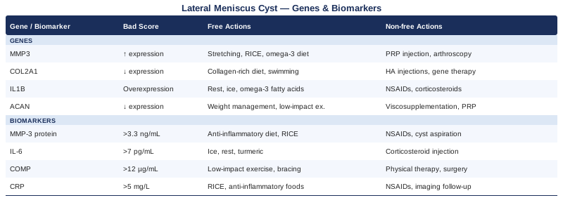
Summary Table: Genes and Biomarkers at a Glance
Rebuilding Knee Health from the Ground Up: The Knees Over Toes Approach
Few approaches in the past decade have challenged conventional physical therapy thinking as directly — or with as much practical traction — as the methodology developed by Ben Patrick (the "Knees Over Toes Guy"). Drawing on a body of biomechanics research showing that most conventional knee rehabilitation programs prioritize pain avoidance over true tissue rebuilding, Patrick's approach focuses on progressively and systematically loading the knee through its full range of motion — including knee-over-toe positions that standard PT often prohibits.
The core insight behind the method is that the tissues of the knee — tendons, cartilage, meniscus — adapt to load. Avoiding load doesn't build resilience; it atrophies it. The research foundation includes work on tendon mechanobiology by Keith Baar and Jill Cook, studies on the dose-response relationship between progressive loading and collagen synthesis, and tibial bone density research showing that full-range knee loading produces protective structural adaptations not seen with partial-range protocols.
The 10 Most Impactful Principles for Lateral Meniscus Health
1. Load is not the enemy — uncontrolled load is. The meniscus degenerates faster with complete unloading than with progressive, well-dosed loading. Most lateral meniscus cysts are associated with years of either overloading (repetitive sport) or underloading (sedentary individuals). The goal is the appropriate dose.
2. The tibialis anterior is the forgotten muscle for knee health. Strengthening the shin (tibialis anterior, tibialis posterior) via reverse Nordic-type exercises and calf raises with full dorsiflexion dramatically improves knee tracking and reduces lateral compartment shear. This is almost never addressed in standard knee rehab.
3. Sled work is the foundation, not the finale. Backward sled pulling loads the knee through knee-over-toe ranges at minimal joint compression and essentially zero joint shear — making it one of the safest progressive overloads for compromised knees. Start at bodyweight equivalent; progress over weeks.
4. Hip flexor length directly affects knee load distribution. Tight hip flexors tilt the pelvis anteriorly, shift loading posteriorly, and force the knee to compensate with altered tracking. Daily hip flexor work (not just stretching — active loaded stretching like the couch stretch with posterior pelvic tilt) is essential.
5. Terminal knee extensions (TKEs) should be the first exercise reintroduced after acute pain subsides. They selectively activate the VMO, reduce lateral patellar tracking, and generate joint-stabilizing signals without meniscal compression.
6. ATG (ass-to-grass) squats are a long-term goal, not a contraindication. For individuals with healthy meniscus tissue, full-depth squatting distributes load more evenly than partial squatting and maintains cartilage hydration through the full arc. For cyst patients, it is a rehabilitation destination — to be worked toward progressively, not imposed immediately.
7. Hamstring and calf tendon strength is the most reliable predictor of knee resilience in athletes. Nordic hamstring curls, single-leg calf raises on a raised surface through full range, and GHD (glute-ham developer) work are prioritized over quad isolation in the methodology.
8. Consistency over intensity — always. The connective tissue adaptation cycle (collagen synthesis and cross-linking) operates on a 48–72 hour lag from mechanical stimulus. Daily light work outperforms three-weekly heavy sessions for tissue remodeling.
9. Proprioception training is often what's missing. The lateral meniscus plays a significant role in proprioception. After cyst aspiration or resolution, single-leg balance drills on unstable surfaces (balance boards, BOSU) re-train the joint's mechanoreceptor density before returning to sport.
10. Chronic pain in the knee after a cyst is often a loading deficit, not persistent structural damage. Once structural integrity is confirmed (MRI, orthopedic review), persistent pain is frequently a sign of de-conditioned tissue signaling — addressed through progressive loading, not avoidance.
Complementary Approaches Worth Considering
Conventional orthopedic care and the biomarker/genetic framework above address the condition structurally and biochemically. Several evidence-informed complementary modalities can add meaningful support — particularly for pain management, tissue recovery, and inflammation modulation. Three stand out as having the most relevant evidence for lateral meniscus cyst specifically.
Low-Level Laser Therapy (Photobiomodulation)
Low-level laser therapy (LLLT), also called photobiomodulation (PBM), uses specific wavelengths of red and near-infrared light (typically 630–1000nm) to penetrate tissue and stimulate mitochondrial activity in cells. In the musculoskeletal context, this translates to enhanced ATP production in chondrocytes and fibroblasts, reduced oxidative stress, and modulation of pro-inflammatory cytokine expression — including MMP-3 and IL-6.
A 2015 systematic review and meta-analysis by Stausholm et al., published in BMJ Open (PMID 31771993), found that LLLT produced clinically meaningful reductions in knee pain and improvements in function in patients with knee osteoarthritis, with an acceptable safety profile. For lateral meniscus cysts specifically, the evidence is extrapolated from adjacent knee pathology studies rather than cyst-specific trials — a limitation worth noting.
Practically, LLLT is delivered via clinical devices (physiotherapy clinics, sports medicine practices) or class II home devices. Clinic protocols typically involve 8–12 sessions over 3–4 weeks, 5–10 minutes per session, with the probe placed directly over the lateral joint line. At-home devices (808nm laser pens, Joovv or Mito Red panels for broader near-infrared application) provide a more accessible option for ongoing maintenance at $200–$600. Avoid direct application over active hemorrhagic areas or immediately post-surgical sites.
Mindfulness-Based Stress Reduction (MBSR)
MBSR is an 8-week structured program combining mindfulness meditation, body scan practices, and yoga-based movement. Its relevance to lateral meniscus cyst is primarily through two mechanisms: chronic stress is a well-established driver of elevated IL-6, CRP, and MMP-3 — all biomarkers directly relevant to meniscal pathology — and central sensitization (amplified pain perception) is a common complication in individuals with persistent knee pain. MBSR directly addresses both.
A landmark randomized controlled trial by Cherkin et al. (JAMA Internal Medicine, 2016, PMID 27002445) demonstrated that MBSR produced significant reductions in pain and functional limitations in chronic musculoskeletal conditions, with effects comparable to cognitive behavioral therapy. Reductions in circulating inflammatory markers have been documented as secondary outcomes in MBSR trials across multiple chronic inflammatory conditions.
Practically, MBSR programs are available through hospitals, mindfulness centers, and online platforms (Palouse Mindfulness offers a free evidence-based online version). The time commitment is approximately 45 minutes per day during the 8-week program. For individuals with a lateral meniscus cyst accompanied by persistent or disproportionate pain, MBSR represents a low-cost, low-risk adjunct with genuine physiological mechanisms behind it — not simply a relaxation tool.
Massage Therapy
Massage therapy for lateral meniscus cyst is not a direct tissue intervention — it does not resolve the cyst itself — but it addresses the functional consequences that typically surround it: IT band and lateral quadriceps tightness that redistributes load to the lateral knee, inhibited glute and hip abductor function from compensatory guarding, and restricted knee range of motion from pericapsular tissue tightness.
A 2015 systematic review by Best et al. in the Journal of Athletic Training found that soft tissue massage combined with exercise rehabilitation consistently outperformed exercise alone for functional outcomes in knee conditions including meniscal pathology, with improvements in pain scores and functional mobility. Specific techniques relevant to lateral meniscus cyst include deep transverse friction to the lateral joint line, myofascial release to the IT band and lateral retinaculum, and trigger point therapy to the tensor fasciae latae and gluteus medius.
Practically, a clinical sports massage therapist with musculoskeletal specialization is the most effective route. Weekly sessions of 45–60 minutes during the active treatment phase (6–8 weeks), transitioning to bi-weekly maintenance, is a reasonable protocol. Foam rolling at home (lateral thigh, 5–7 minutes daily) serves as a self-applied adjunct between sessions. Massage to the acutely inflamed cyst area itself should be avoided; the surrounding musculature and soft tissue are the primary targets.
Conclusion
A lateral meniscus cyst is not a single problem with a single solution. It sits at the intersection of mechanical load, biochemical tissue quality, inflammatory state, and — for some individuals — genetic predisposition. The biomarkers covered here (hs-CRP, CTX-II, COMP, IL-6, vitamin D, and MMP-3) give you a measurable, actionable picture of the biochemical environment driving your knee's failure to heal. The genetic variants reviewed (COL2A1, MMP3, GDF5, ACAN, VEGF, TGFB1) provide a structural context that explains why some individuals are more susceptible and what compensatory strategies are most relevant for them.
None of this replaces a proper orthopedic evaluation or a well-supervised rehabilitation program. But it adds a layer of precision that generic advice cannot provide. The smartest next step for most readers is straightforward: get the accessible bloodwork (hs-CRP, 25-OH vitamin D, IL-6) ordered, review the results in context, and use this information to have a more informed conversation with your physiotherapist or orthopedic specialist about what your recovery actually needs.
Musculoskeletal: Joint Conditions Sports Injuries
Autoimmune: Inflammatory Conditions Connective Tissue Conditions