This article contains AI generated content.
· UpdatedLateral Patellar Compression Syndrome — 5 Genes and 6 Biomarkers to Track
Introduction
If you have lateral patellar compression syndrome, you have almost certainly been told to strengthen your VMO, stretch your IT band, and tape your kneecap. That advice is not wrong — but for a significant portion of people, it is incomplete. Months of faithful rehabilitation sometimes produce minimal improvement, and nobody explains why your case is different from someone who recovered in six weeks.
The reason often lies in biology that standard physical therapy does not measure. How much your lateral retinaculum is structurally predisposed to tightness, how quickly your cartilage degrades under compressive load, how much systemic inflammation is feeding pain sensitization in your joint — these factors vary considerably between individuals, and increasingly, they are measurable. Without knowing where your specific biology stands, you are essentially applying average solutions to a problem that may not be average.
This is not a pitch for a dramatic cure. It is a case for precision. When you know that your systemic inflammation is elevated, your cartilage is breaking down faster than it is rebuilding, your vitamin D receptor is functionally impaired, or your muscle fiber composition makes VMO activation particularly difficult — you have something concrete to act on. The guesswork shrinks, and the interventions become more targeted.
This article takes two main approaches. The first examines 6 trackable biomarkers — measurable signals in blood and urine that reflect the state of your joint tissue, your inflammatory burden, and your nutritional status. The second explores 5 genetic variants that influence how your connective tissue is built and maintained, and what to do if you carry an unfavorable version. Additional sections cover 10 key insights from the Huberman Lab on connective tissue and recovery, and five complementary modalities with meaningful clinical evidence for patellar conditions.
Summary
Most people with lateral patellar compression syndrome track their pain. This article argues you should also track six biological markers: hsCRP (inflammation), COMP and CTX-II (cartilage degradation), 25-OH Vitamin D (muscle and bone signaling), RBC Magnesium (muscle relaxation and nerve coupling), and IL-6 (inflammatory cytokine). Each comes with a cost range, optimal values, and a concrete action plan — both with and without supplements, including dosing, cycling, and side effects. The genetics section reveals five variants — COL1A1, MMP3, ACAN, VDR, and ACTN3 — that may explain why your lateral retinaculum tears down faster, your cartilage cushion degrades sooner, or your VMO simply refuses to respond to conventional training. A Huberman Lab summary follows, including one counterintuitive insight about cold therapy timing that most rehabilitation programs get wrong. Finally, five complementary modalities — biofeedback, photobiomodulation, massage, yoga, and tai chi — are reviewed with specific protocols and supporting clinical trials.
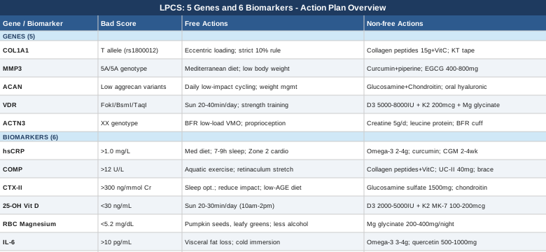
6 Biomarkers to Track for Lateral Patellar Compression Syndrome
Most people with LPCS are assessed structurally — through imaging, biomechanical observation, and movement screening. Blood and urine biomarkers are rarely part of the evaluation, yet they reveal critical information about whether the joint's internal chemistry is working for or against recovery. The six markers below represent the most actionable combination: affordable enough to access, specific enough to matter, and actionable enough to change what you do.
1. High-Sensitivity C-Reactive Protein (hsCRP)
Why it matters for LPCS. hsCRP is a liver-produced acute-phase protein that rises in response to cytokine signaling from inflamed tissues. In lateral patellar compression syndrome, repetitive mechanical irritation of the lateral retinaculum, the subchondral bone at the lateral facet, and Hoffa's infrapatellar fat pad generates persistent local inflammation. When this is sustained — because the underlying biomechanical issue is never fully corrected — it produces measurable systemic inflammatory activity. Elevated hsCRP (above 1.0 mg/L, and particularly above 3.0 mg/L) is independently associated with accelerated cartilage breakdown and impaired soft tissue healing. It also amplifies pain sensitization via cytokine-driven neuroinflammation, which is why some patients report that pain feels disproportionate to their structural damage. Thomas Dayspring and Peter Attia both place hsCRP among their first-tier biomarkers for any chronic musculoskeletal condition.
How to measure it
Request "high-sensitivity CRP" specifically — standard CRP is less precise at low levels. Available at any major lab (Quest Diagnostics, LabCorp) or through a GP. Cost: $10–$30 out of pocket. Most insurance plans cover it with a physician order. Optimal range: below 0.5 mg/L. Borderline: 0.5–1.0 mg/L. Elevated: above 1.0 mg/L. Above 3.0 mg/L is high-risk territory for both musculoskeletal deterioration and cardiovascular disease. Retest every 3 months when implementing interventions.
If hsCRP is elevated: the plan without supplements
The most powerful dietary intervention is a Mediterranean-style eating pattern — rich in vegetables, extra-virgin olive oil, legumes, fatty fish, and limited in refined carbohydrates and processed oils. A randomized trial by Esposito et al. (2004, JAMA) showed participants following this pattern reduced hsCRP by nearly 40% over two years. Sleep is equally critical: fewer than 6 hours per night significantly elevates hsCRP, and this relationship is dose-dependent. Aim for 7–9 hours with a consistent schedule. Zone 2 aerobic exercise (150–180 minutes per week at conversational pace, using low-impact modalities that do not aggravate the knee — cycling, swimming, incline walking) reduces systemic inflammation reliably without adding load to the lateral patella. Avoiding prolonged sitting is also important: static knee flexion increases compressive stress at the lateral patellar facet and sustains local inflammatory signaling.
If hsCRP is elevated: the plan with supplements or equipment
Omega-3 fatty acids (EPA+DHA): 2–4g combined daily. Take with food, preferably with the largest fat-containing meal (improves absorption). Triglyceride form (fish oil) absorbs approximately 70% better than ethyl ester form. Retest hsCRP at 3 months. Cycling: continuous use is appropriate; some practitioners retest every 6 months to calibrate dose. Side effects: fishy aftertaste (use enteric-coated capsules), mild anticoagulation at doses above 4g/day — caution if on blood thinners. Curcumin with piperine: 500–1000mg standardized extract (95% curcuminoids) plus 5–10mg piperine to enhance bioavailability. Take with food. Cycle: 8–12 weeks on, reassess. Side effects: GI discomfort in some individuals; reduces iron absorption if taken with iron-rich meals; potential interaction with blood-thinning medications. For equipment, a continuous glucose monitor (CGM) worn for 2–4 weeks can identify postprandial glucose spikes that are chronically driving hsCRP elevation — particularly useful if metabolic health is suspected as a root cause.
2. Cartilage Oligomeric Matrix Protein (COMP)
Why it matters for LPCS. COMP is a pentameric glycoprotein found in cartilage, tendons, and ligaments. When these structures are mechanically stressed or degenerating, COMP fragments are released into synovial fluid and subsequently into the bloodstream. In LPCS, the lateral facet of the patella undergoes abnormally high compressive loading with every knee flexion cycle — over years, this damages articular cartilage. Elevated serum COMP is one of the earliest measurable signals that cartilage is being damaged faster than it is being repaired. Research published in Osteoarthritis and Cartilage has demonstrated that serum COMP elevates meaningfully before structural changes appear on MRI, making it a genuinely early warning marker rather than a confirmation of already-advanced damage.
How to measure it
COMP is available through specialty lab panels (LabCorp, Quest, or direct-to-consumer services such as Ulta Lab Tests with a physician order). Cost: approximately $80–$150. Normal serum COMP is generally below 12 U/L, though reference ranges vary by lab and age. Measure consistently — always collect the sample before or always after your typical daily activity window, as COMP fluctuates with physical loading. A morning fasting sample before exercise provides the most stable baseline. Retest every 3–6 months when implementing cartilage-targeted interventions.
If COMP is elevated: the plan without supplements
The primary goal is to reduce compressive loading on the lateral patellar facet without deconditioning. Aquatic exercise (pool walking, water aerobics, swimming) removes ground reaction forces while maintaining cardiovascular fitness and muscle activation — the optimal combination during this phase. Cycling with proper seat height (full knee extension at the bottom of the pedal stroke, foot in neutral) is well tolerated. Lateral retinaculum mobilization through self-stretching (cross-legged kneeling stretch, medial patellar glide mobilization) reduces the compressive contact force at the lateral facet by improving patellar mobility. Sleep quality becomes especially important: articular cartilage has no direct blood supply and receives nutrition primarily through synovial fluid exchange during non-weight-bearing periods. Poor sleep disrupts this repair window. Maintaining healthy body weight is also fundamental — each kilogram of excess weight adds approximately 3–5kg of patellofemoral compressive force.
If COMP is elevated: the plan with supplements or equipment
Hydrolyzed collagen peptides: 10–15g per day, taken with 500mg vitamin C approximately 30–60 minutes before low-impact exercise. Shaw et al. (2017, American Journal of Clinical Nutrition) demonstrated that this protocol significantly increased collagen synthesis markers in connective tissue compared to placebo. Cycle: 12 weeks minimum; reassess COMP at 3 months. Side effects: generally minimal — occasional mild GI discomfort. Undenatured type II collagen (UC-II): 40mg/day (taken on an empty stomach). This works via an immune tolerance mechanism rather than as a raw building block. A 2016 randomized trial by Lugo et al. in Journal of the International Society of Sports Nutrition showed significant reductions in joint discomfort markers. Cycle: 3–6 months. Side effects: very well tolerated; rare GI sensitivity. For equipment, McConnell patellar taping or a lateral-relief patellar tracking brace (DonJoy, Bauerfeind) during all weight-bearing activities immediately reduces lateral facet compression and should accompany any supplement strategy targeting cartilage.
3. CTX-II (C-Terminal Telopeptide of Type II Collagen)
Why it matters for LPCS. CTX-II is a urinary degradation marker of type II collagen — the primary structural protein of articular cartilage. Unlike COMP, which reflects general cartilage matrix stress, CTX-II specifically quantifies the rate of type II collagen breakdown. In LPCS, the compressed lateral patellar cartilage undergoes accelerated collagen turnover. High CTX-II predicts cartilage loss on longitudinal imaging studies and is associated with faster progression toward patellofemoral osteoarthritis if the underlying load issue is not corrected. European research programs on knee osteoarthritis have used CTX-II as a primary endpoint in cartilage trials precisely because it detects active damage before structural changes are visible — making it one of the most clinically meaningful early markers in this article.
How to measure it
CTX-II is measured from a second morning urine sample (discard the first void; collect the second) and normalized to urinary creatinine to control for concentration variation. Available through specialty labs (in Europe, Nordic Bioscience's Artus-II assay is the gold standard; US alternatives through specialty lab panels). Cost: approximately $100–$200. Elevated values are generally above 300 ng/mmol creatinine, though reference ranges vary by age (values are higher in younger individuals with active growth plates). Retest every 3–6 months when targeting cartilage health.
If CTX-II is elevated: the plan without supplements
Sleep optimization is the single highest-leverage non-supplement intervention for CTX-II. Cartilage type II collagen breakdown peaks nocturnally, and the repair cycle depends on adequate growth hormone release during slow-wave sleep. Anything that disrupts slow-wave sleep — alcohol within 3 hours of bedtime, irregular sleep schedules, screen light exposure before sleep, room temperatures above 68°F — directly impairs this repair window. A consistent sleep schedule with a cool, dark room and no alcohol on weeknights is a high-impact, zero-cost intervention. Second, reduce high-impact loading volume: running, stair descent with load, jumping, and deep squats under load should be temporarily replaced with aquatic exercise and cycling. Third, eliminate dietary advanced glycation end products (AGEs): sugar and refined carbohydrate consumption generates AGEs that crosslink and stiffen collagen, making it substantially more vulnerable to enzymatic degradation by matrix metalloproteinases.
If CTX-II is elevated: the plan with supplements or equipment
Glucosamine sulfate: 1500mg per day (sulfate form specifically — not glucosamine hydrochloride, which has significantly weaker evidence). Several clinical trials have shown measurable reduction in CTX-II with glucosamine sulfate in knee OA populations. Take in a single daily dose with food. Cycle: 3-month trial minimum; if no symptomatic or biomarker benefit by 3 months, discontinue. Side effects: generally very well tolerated; nausea is rare; contraindicated in shellfish allergy. Chondroitin sulfate: 1200mg/day, frequently paired with glucosamine. Multiple meta-analyses confirm its benefit for cartilage degradation markers. Side effects: minimal; occasional mild GI symptoms. Vitamin C: 500–1000mg/day — vitamin C is an obligate cofactor in prolyl and lysyl hydroxylation, the enzymatic steps that crosslink collagen and make it resistant to degradation. Deficiency at the subclinical level is not uncommon and directly accelerates type II collagen breakdown. Side effects at this dose: minimal (GI tolerance is individual; buffered forms are gentler). For equipment: an offloading or patellar tracking brace worn during all weight-bearing activity reduces compressive stress at the lateral facet and allows supplementation to work alongside reduced mechanical damage, not against continued loading.
4. 25-Hydroxyvitamin D
Why it matters for LPCS. Vitamin D deficiency is among the most prevalent nutritional deficiencies in adults worldwide, yet its consequences extend far beyond bone density. For LPCS specifically, vitamin D signaling influences three critical areas: type II fast-twitch muscle fiber development (directly relevant to VMO power and patellar stabilization capacity), anti-inflammatory gene expression via NF-κB pathway modulation (reducing the cytokine burden in chronically irritated joint tissue), and subchondral bone metabolism (deficient subchondral bone fails to absorb shock adequately, increasing cartilage compressive stress). In LPCS, VMO weakness is the proximate neuromuscular cause of lateral patellar maltracking in most cases — and vitamin D deficiency impairs the neuromuscular capacity to build and maintain that muscle, creating a vicious cycle where rehabilitation yields slower-than-expected results. Peter Attia has consistently placed 25-OH Vitamin D among his highest-priority musculoskeletal and metabolic biomarkers.
How to measure it
A standard 25-hydroxyvitamin D blood test costs $30–$80 and is covered by most insurance plans once per year. Request "25-OH Vitamin D total" (combined D2 + D3). Optimal range: 40–60 ng/mL. Below 30 ng/mL is considered deficient. Below 20 ng/mL is severely deficient and will meaningfully impair muscle function, immune regulation, and healing. Retest every 3 months when supplementing to avoid over-supplementation and to calibrate dose.
If 25-OH Vitamin D is low: the plan without supplements
Sun exposure is the most effective natural source. Full skin exposure of arms, legs, and face for 20–30 minutes between 10am and 2pm can generate 10,000–20,000 IU of vitamin D3, depending on skin tone, latitude, and season. Darker skin tones require 30–60 minutes for the same output. Beyond September in northern latitudes (above 40°N), UVB radiation is insufficient for vitamin D synthesis regardless of exposure duration — supplementation becomes mandatory in winter for these individuals. Dietary sources contribute modestly: fatty fish (salmon, mackerel, sardines), egg yolks, and fortified foods provide meaningful amounts, but dietary intake alone is rarely sufficient to correct a clinical deficiency.
If 25-OH Vitamin D is low: the plan with supplements or equipment
Vitamin D3 (cholecalciferol): 2000–5000 IU/day for maintenance; 5000–10,000 IU/day to correct deficiency (with medical guidance at higher doses). Always combine with Vitamin K2 (MK-7 form): 100–200mcg/day. K2 directs calcium into bones and teeth rather than arterial walls and soft tissues — an essential safety co-supplement at higher D3 doses. Take D3 and K2 together with a fat-containing meal for optimal absorption. Magnesium (see biomarker 5 below) is also required for vitamin D conversion and should accompany supplementation. Cycle: supplement continuously; retest at 3 months and titrate dose to maintain 40–60 ng/mL. Side effects: hypercalcemia is possible with sustained doses above 10,000 IU/day without monitoring — regular retesting prevents this. At 5000 IU/day with K2, risk is minimal in most otherwise healthy adults.
5. Red Blood Cell (RBC) Magnesium
Why it matters for LPCS. An estimated 50–60% of adults fail to meet magnesium intake requirements, yet the standard serum magnesium test is a poor indicator of actual cellular magnesium status — it can appear normal even when intracellular stores are substantially depleted. RBC magnesium, measured from the interior of red blood cells, reflects true intracellular magnesium concentration far more accurately. Magnesium is a cofactor in over 300 enzymatic reactions, including those governing muscle relaxation, nerve-muscle coupling, collagen synthesis, and ATP production. For LPCS, this is directly relevant: chronic tightness of the lateral retinaculum, the iliotibial band, and the vastus lateralis is a central driver of lateral patellar compression. Magnesium deficiency impairs smooth muscle relaxation and interferes with neuromuscular signaling — making it harder to release chronically tight lateral structures and harder to selectively activate the VMO. It also impairs collagen hydroxylation (overlapping with vitamin C and vitamin D pathways).
How to measure it
Request "RBC magnesium" specifically — not standard serum magnesium. It requires a specific tube and processing protocol. Available through integrative medicine providers, functional medicine panels, or direct-to-consumer lab services. Cost: $40–$80. Optimal RBC magnesium: 5.2–6.5 mg/dL. Below 5.2 mg/dL suggests intracellular depletion, regardless of whether serum magnesium appears normal. Retest every 3 months when supplementing.
If RBC Magnesium is low: the plan without supplements
Dietary improvement is the essential first step. The richest food sources of magnesium include pumpkin seeds (the single richest source per gram), dark leafy greens (spinach, Swiss chard), almonds, avocado, dark chocolate (70%+), black beans, and whole grains. Practical changes: add pumpkin seeds to daily meals, include a large leafy green salad with olive oil (fat enhances magnesium absorption), and reduce processed foods (processing strips magnesium from grains). Reduce alcohol and caffeine — both increase urinary magnesium excretion significantly. Chronic psychological stress promotes cortisol-driven renal magnesium wasting, so any intervention that reduces the stress response also improves magnesium retention.
If RBC Magnesium is low: the plan with supplements or equipment
Magnesium glycinate: 200–400mg elemental magnesium per day, taken at night 30–60 minutes before sleep. Glycinate is amino acid-chelated, highly bioavailable, and has minimal laxative effect — the key advantage over magnesium oxide, which is cheap and poorly absorbed. Magnesium malate is an equally good alternative if muscle pain or fatigue is prominent, as malate supports mitochondrial function. Cycle: continuous use is appropriate and safe long-term. Retest RBC magnesium at 3 months to confirm correction and titrate dose. Side effects: loose stools if dose is too high (reduce by 50mg increments until tolerated; this is the main limiting symptom). Transdermal magnesium chloride spray or Epsom salt baths (magnesium sulfate) may provide supplementary absorption and offer a useful local muscle-relaxing effect for tight lateral knee structures, though the evidence for systemic correction via transdermal routes is weaker than oral supplementation.
6. Interleukin-6 (IL-6)
Why it matters for LPCS. IL-6 is a pleiotropic cytokine produced by adipose tissue, immune cells, and chronically stressed musculoskeletal tissues. In LPCS, the persistently irritated lateral retinaculum and inflamed infrapatellar fat pad (Hoffa's fat pad) can generate sustained IL-6 secretion at the joint level. Systemically elevated IL-6 perpetuates the inflammatory cycle, directly sensitizes joint nociceptors (making pain feel worse than structural damage alone would predict), inhibits muscle protein synthesis — impairing VMO rehabilitation progress — and drives hsCRP elevation. Tracking IL-6 alongside hsCRP allows you to determine whether your inflammatory burden is primarily metabolic and systemic in origin or whether it is locally driven by joint tissue, which changes the most appropriate intervention strategy.
How to measure it
IL-6 is available through specialty labs and some academic medical centers, and through direct-to-consumer services (e.g., Ulta Lab Tests). Cost: $50–$150. Standard insurance reimbursement varies. Normal levels: typically below 7 pg/mL. Values consistently above 10–15 pg/mL indicate chronic low-grade inflammatory activity. The sample must be processed promptly (plasma separated and frozen within 30 minutes of collection) — follow lab-specific instructions precisely to avoid false elevation from poor sample handling.
If IL-6 is elevated: the plan without supplements
Body composition improvement — specifically visceral fat reduction — is the most powerful non-supplement intervention for chronically elevated IL-6. Visceral adipose tissue is the primary non-joint source of excess IL-6 in most adults. Even a 5–7% reduction in total body weight via a calorie-controlled anti-inflammatory diet produces measurable IL-6 reductions. Time-restricted eating (limiting food intake to an 8–10 hour window) has been shown in recent randomized trials to lower fasting IL-6 independently of caloric restriction, likely through circadian rhythm-mediated insulin and cortisol optimization. Cold water immersion (15–20 minutes at 60–65°F water, 3–4 times per week) produces anti-inflammatory effects through catecholamine modulation. Reduce or eliminate alcohol: even moderate consumption of 1–2 drinks daily is associated with chronically elevated IL-6 in long-term studies.
If IL-6 is elevated: the plan with supplements or equipment
Omega-3 fatty acids (as detailed in biomarker 1): 3–4g EPA+DHA daily. EPA specifically inhibits the arachidonic acid pathway that feeds IL-6 production. Quercetin: 500–1000mg/day, taken with a fat-containing meal for absorption (quercetin is fat-soluble and poorly absorbed without dietary fat). Multiple randomized trials have shown meaningful reductions in circulating IL-6 with quercetin supplementation over 8–12 weeks. Cycle: 8–12 weeks on, 2 weeks off, then reassess. Side effects: generally well tolerated; at doses above 1g/day, quercetin can inhibit CYP3A4 (an enzyme involved in metabolizing many drugs) — caution if taking medications processed by this enzyme. Vitamin C: 1000mg/day. Multiple clinical studies confirm vitamin C as an inhibitor of IL-6 signaling at the transcription level. Side effects: GI discomfort above 2g/day; use buffered form if gastric sensitivity is a concern.
What Recent Genetics Research Reveals About Lateral Patellar Compression Syndrome
Genetic testing for musculoskeletal health is no longer limited to academic research settings. Consumer-accessible platforms and clinical DNA panels now make several relevant variants detectable in any individual. These variants do not predetermine outcomes — but they do explain why some people are more prone to lateral retinaculum tightness, cartilage degradation, and VMO dysfunction, and they point toward which interventions are most likely to produce meaningful results for that specific biology.
Gary Brecka, a human biologist and health researcher, has argued that genetic methylation variants — particularly those in collagen, inflammatory, and receptor pathways — are among the most commonly overlooked explanations for chronic musculoskeletal conditions that fail to resolve with standard care. Ali Torkamani, genomics researcher at Scripps Research Institute, has similarly proposed that polygenic risk scores for connective tissue resilience may become routine in musculoskeletal medicine as evidence accumulates. The five genes below represent the most clinically actionable variants for someone navigating LPCS. The evidence base is mostly drawn from connective tissue injury, osteoarthritis, and muscle physiology research rather than LPCS specifically — but the mechanistic relevance is direct.
Gene 1: COL1A1 — The Collagen Structural Blueprint
What it affects. COL1A1 encodes the alpha-1 chain of type I collagen — the dominant structural protein in tendons, ligaments, joint capsules, and the lateral retinaculum. A well-characterized polymorphism at the Sp1 binding site (rs1800012, G→T substitution) produces a weaker, less mechanically stiff collagen architecture. Carriers of the T allele show higher rates of ligament and tendon injury, slower structural recovery, and — critically for LPCS — greater susceptibility to lateral retinaculum laxity combined with paradoxical tightening after chronic irritation. A lax but chronically irritated lateral retinaculum allows the patella to drift laterally under load and then fails to release this compression as tissue becomes fibrotic.
If the gene variant is unfavorable: the plan without supplements
Progressive tendon loading is the cornerstone. Collagen responds to mechanical stimulation by upregulating synthesis — even in carriers of the T allele, though the response is slower. Eccentric exercises (slow controlled lowering phase) are particularly effective: terminal knee extensions with a 4-second lowering phase, slow mini-squats (3–4 seconds eccentric), and single-leg step-downs. Frequency: 3–4 sessions per week, 3–5 sets of 10–15 reps. For T-allele carriers, connective tissue adaptation lags muscle adaptation — follow the 10% rule strictly: increase weekly loading volume by no more than 10% per week to allow collagen remodeling to keep pace with training demands. Avoid sudden loading spikes, which are the primary trigger for acute retinaculum aggravation in this variant.
If the gene variant is unfavorable: the plan with supplements or equipment
Hydrolyzed collagen peptides (type I): 15g/day with 500mg vitamin C, taken 30–60 minutes before loading exercise. Shaw et al. (2017, American Journal of Clinical Nutrition) demonstrated that this specific protocol significantly increases collagen synthesis markers in tendons and ligaments compared to a placebo control. Frequency: daily, indefinitely — this is a food-based supplement with no meaningful cycling requirement. Side effects: minimal; occasional mild GI discomfort. Vitamin C is non-negotiable as a cofactor: prolyl and lysyl hydroxylase enzymes (which crosslink collagen chains, giving them structural strength) require vitamin C. A deficient intake undermines any dietary or supplemental collagen investment. Dose: 500–1000mg/day with the collagen supplement. Side effects: GI sensitivity at higher doses; use a buffered form if needed. For equipment, kinesiology tape (KT Tape) applied along the lateral retinaculum and patellar tracking line during activity reduces the mechanical demand on structurally weaker collagen and allows progressive strengthening without chronic tissue overloading.
Gene 2: MMP3 — The Cartilage Breakdown Enzyme
What it affects. MMP3 (matrix metalloproteinase-3, also called stromelysin-1) is an enzyme that degrades multiple extracellular matrix components: aggrecan, fibronectin, and collagen types II, III, IV, and V. The 5A/6A promoter polymorphism (rs3025058) directly regulates how much MMP3 your cells produce: the 5A/5A genotype drives approximately twice the promoter activity of the 6A/6A genotype, meaning homozygous 5A carriers produce substantially more cartilage-degrading enzyme at rest. In LPCS, where the lateral patellar cartilage is already under abnormal mechanical stress, an overactive MMP3 system can accelerate cartilage matrix degradation to a degree that structural damage outpaces repair. The evidence for this variant is primarily drawn from osteoarthritis research rather than LPCS specifically, but the mechanistic pathway is directly applicable: the compressed lateral facet in LPCS subjects the cartilage to the same MMP3-mediated damage observed in early-onset OA.
If the gene variant is unfavorable: the plan without supplements
The same Mediterranean dietary pattern that reduces hsCRP also downregulates MMP3 transcription through NF-κB inhibition — NF-κB is the master transcription factor that drives MMP3 gene expression in response to inflammatory signals. Maintaining healthy body weight is equally critical: adipose tissue produces adipokines (leptin, resistin) that upregulate MMP3 expression independent of mechanical loading. Each kilogram of excess body fat modestly but measurably increases articular MMP3 activity through these adipokine pathways. Reduce loading-induced MMP3 spikes by avoiding high-impact activity during symptomatic phases and using the low-impact modalities described in the biomarker section.
If the gene variant is unfavorable: the plan with supplements or equipment
Curcumin: 500–1000mg/day (standardized 95% curcuminoid extract with piperine). Curcumin is a well-characterized NF-κB inhibitor that directly suppresses MMP3 transcription. Both in vitro and clinical studies support this effect. Cycle: 8–12 weeks continuously, then reassess inflammatory biomarkers (hsCRP, COMP). Side effects: GI upset in some individuals; reduces iron absorption if taken with iron-rich meals; potential interaction with anticoagulant medications. EGCG (epigallocatechin gallate from green tea extract): 400–800mg/day standardized extract, preferably decaffeinated. EGCG inhibits MMP3 at both the transcriptional and enzymatic levels. Take with food. Cycle: 8 weeks on, 2 weeks off. Side effects: at doses above 800mg/day, potential hepatotoxicity has been reported in case studies — do not exceed this dose; take with food to reduce risk. The human evidence base is mostly drawn from in vitro and early OA studies — effect size in humans is promising but should be considered preliminary rather than definitively established.
Gene 3: ACAN — The Cartilage Cushion Gene
What it affects. ACAN encodes aggrecan, the largest proteoglycan in articular cartilage and the primary molecule responsible for its compressive resilience. Aggrecan's sulfated glycosaminoglycan chains attract and retain water within the cartilage matrix, creating the hydraulic cushioning that protects underlying bone during loading. Variants in ACAN that reduce aggrecan synthesis or alter its molecular structure produce cartilage that is less hydrated and less capable of distributing compressive load. In LPCS, where lateral patellar cartilage bears abnormally concentrated load with every knee flexion, a structurally compromised aggrecan-deficient matrix fails faster than average. This may explain why some individuals develop cartilage damage visible on MRI at a stage of life and activity level that would not typically predict such findings.
If the gene variant is unfavorable: the plan without supplements
Cartilage has no direct blood supply and depends on cyclical mechanical loading and unloading for nutrition — fluid is forced in and out of the matrix during compression and release cycles, delivering nutrients and removing waste products. Prolonged immobilization starves cartilage. Excessive impact loading crushes it. For ACAN variant carriers, the optimal protocol is consistent, low-impact cyclical loading: 30–60 minutes of daily cycling, walking, swimming, or elliptical training. Avoid prolonged sitting without movement breaks — get up and move for 5 minutes every 60–90 minutes. Aggressive weight management: the 3–5kg of patellofemoral force added per kilogram of excess body weight is the most direct variable controllable through lifestyle.
If the gene variant is unfavorable: the plan with supplements or equipment
Glucosamine sulfate + chondroitin sulfate: 1500mg glucosamine + 1200mg chondroitin per day (combination form). These are the precursor building blocks for proteoglycan synthesis, including aggrecan. The GAIT trial showed benefit specifically for moderate-to-severe knee symptoms; benefit in mild cases is more variable. Cycle: 3-month trial; continue if symptomatic improvement is clear. Side effects: shellfish allergy precaution (glucosamine is typically shellfish-derived); mild GI in some. Oral hyaluronic acid: 80–200mg/day of low-molecular-weight form (e.g., Hyabest). Randomized trials on knee OA patients have shown modest but statistically significant improvements in joint comfort and lubrication markers. Cycle: 3 months minimum; reassess. Side effects: very well tolerated. For equipment, a patellofemoral unloading brace with lateral cutout worn during activities substantially reduces compressive force on the lateral facet, directly protecting already-compromised aggrecan-deficient cartilage while rebuilding the biology underneath.
Gene 4: VDR — The Vitamin D Receptor Gatekeeper
What it affects. Even when vitamin D blood levels test as adequate, the VDR (vitamin D receptor) gene determines how effectively cells respond to that vitamin D. Three well-studied VDR polymorphisms — FokI (rs2228570), BsmI (rs1544410), and TaqI (rs731236) — alter receptor structure, binding affinity, or expression levels, effectively reducing the signal strength of vitamin D even when its concentration is normal. For LPCS, this matters acutely: vitamin D signaling is essential for type II fast-twitch muscle fiber development (critical for VMO power), for anti-inflammatory gene expression that reduces joint cytokine burden, and for calcium regulation in the subchondral bone. Individuals carrying unfavorable VDR variants may show entirely normal 25-OH Vitamin D on a standard test yet still have functionally impaired vitamin D signaling in musculoskeletal tissues. Gary Brecka has identified VDR variants as among the most clinically significant for unexplained chronic musculoskeletal dysfunction — noting that standard testing completely misses receptor-level impairment.
If the gene variant is unfavorable: the plan without supplements
Maximize all non-supplement vitamin D inputs simultaneously. Sun exposure (20–40 minutes of direct skin exposure at peak UV hours, depending on skin tone and season) provides the highest natural D3 production. Dietary sources (fatty fish, egg yolks) add modest incremental amounts. Crucially, adequate dietary magnesium is required for VDR expression — a point often overlooked: magnesium is a cofactor not only in vitamin D conversion but in the VDR gene itself. Resistance training that recruits fast-twitch fibers has been shown to upregulate VDR expression in skeletal muscle — particularly relevant for the VMO. Frequency: 3–4 strength sessions per week with VMO-focused exercises.
If the gene variant is unfavorable: the plan with supplements or equipment
VDR variant carriers typically need to target the upper end of the optimal vitamin D range (60–80 ng/mL rather than 40–60 ng/mL) to achieve the same effective cellular signaling. This may require 5000–8000 IU of D3 daily. Always combine with Vitamin K2 (MK-7): 200mcg/day and Magnesium glycinate: 300–400mg/day (VDR expression and vitamin D hydroxylation both depend on magnesium). These three form a functionally interdependent triad. Cycle: continuous; retest 25-OH Vitamin D at 3 months. Side effects: at 8000 IU/day sustained, monitor calcium, phosphorus, and PTH alongside vitamin D levels — hypercalcemia risk exists with prolonged high doses without laboratory monitoring. This is why the target range (60–80 ng/mL) is critical: once reached and confirmed, the supplemental dose can be reduced to a maintenance level.
Gene 5: ACTN3 — The Muscle Fiber Composition Gene
What it affects. ACTN3 encodes alpha-actinin-3, a structural protein found exclusively in type IIx fast-twitch muscle fibers. The R577X polymorphism (rs1815739) is one of the most widely studied athletic performance variants in human genetics: approximately 18% of Caucasian adults are homozygous for the X allele (XX genotype) and produce zero alpha-actinin-3, with a substantially reduced fast-twitch fiber capacity. For LPCS, this is directly relevant: the VMO functions as the primary active patellar medial stabilizer, and its fibers include a significant proportion of type II fast-twitch units responsible for rapid, powerful patellar stabilization during loading. XX genotype individuals have a structural disadvantage in generating the quick, powerful VMO contractions needed to counteract the lateral pull of the retinaculum — a potential explanation for why some people fail to respond to standard VMO strengthening programs despite months of consistent effort.
If the gene variant is unfavorable: the plan without supplements
For XX genotype individuals, the evidence points toward slow, sustained VMO activation protocols outperforming explosive power-based approaches. Rather than traditional leg press sets, shift focus to: terminal knee extensions with a 4-count eccentric phase, wall slides held at 30° knee flexion for 30–60 seconds, and — particularly valuable — blood flow restriction (BFR) training. BFR applies a restrictive cuff (60–80% of limb occlusion pressure) to the proximal thigh during light-resistance exercise, inducing the same muscle adaptation stimulus as high-load training at a fraction of the joint stress. Multiple peer-reviewed studies support BFR as a tool for inducing meaningful muscle hypertrophy in populations with reduced fast-twitch fiber capacity. Frequency: 4–5 short VMO sessions per week. Complement with proprioceptive training (single-leg balance on unstable surfaces, perturbation training) — XX individuals compensate effectively by improving neuromuscular coordination, which provides patellar stabilization through a different mechanism than raw fast-twitch power.
If the gene variant is unfavorable: the plan with supplements or equipment
Creatine monohydrate: 3–5g/day continuously. Creatine supplementation preferentially benefits individuals with lower baseline phosphocreatine stores in muscle — which includes XX genotype individuals with fewer fast-twitch fibers. It improves power output in type I-dominant muscle populations and supports the BFR training adaptation. No loading phase is required at 5g/day. Cycle: continuous use is established as safe in the literature for years; no physiological need to cycle. Side effects: intracellular water retention (not subcutaneous), mild GI discomfort if taken as a single large bolus (split into 2 doses if needed). Leucine-enriched protein: 2.5–3g of leucine per meal (achievable through whey protein or BCAA supplementation). Leucine directly triggers mTOR-mediated muscle protein synthesis even in slow-twitch fiber-dominant muscle populations — supporting the VMO hypertrophy that XX individuals need despite their genetic disadvantage. For equipment, a BFR training cuff (B Strong, Saga Fitness, or similar) is a direct investment for the XX genotype: it enables the precise low-load, high-stimulus training protocol that best matches type I-dominant muscle physiology. Used 3–4 times per week for lower limb work, integrated into a VMO-specific protocol.
With a detailed picture of your biological landscape — what is inflamed, what is degrading, what your genes predispose you toward — the question becomes: what broader recovery principles can accelerate the return of function and tissue health? The Huberman Lab has assembled and synthesized a body of research on connective tissue, inflammation, and neuromuscular recovery that challenges several standard clinical assumptions.
The Huberman Lab on Joint and Connective Tissue Recovery: 10 Key Insights
Dr. Andrew Huberman (Stanford neuroscience professor and host of the Huberman Lab podcast) has produced multiple episodes synthesizing research on connective tissue biology, inflammation regulation, and musculoskeletal recovery — referencing peer-reviewed studies throughout. The following 10 insights are the most directly actionable for someone with LPCS and are drawn from those episodes and their cited sources.
1. Collagen Synthesis Has a Timing Window You Can Exploit
Huberman extensively cites the work of Dr. Keith Baar (UC Davis) showing that collagen synthesis in tendons peaks approximately 6 hours after mechanical loading — and that consuming 15g of hydrolyzed collagen with 50mg vitamin C approximately 30–60 minutes before exercise significantly amplifies this synthesis window. The mechanism: vitamin C is required for prolyl hydroxylase activity, which crosslinks nascent procollagen into structurally strong fibers. This protocol costs less than one dollar per day and is one of the few evidence-based interventions that directly stimulates retinaculum and tendon remodeling.
2. High-Repetition Work Outperforms Heavy Loading for Connective Tissue
Contrary to common assumption, Huberman and Baar explain that high-repetition, lower-load work (20–30 reps per set) produces superior collagen synthesis in tendons compared to heavy low-rep work. For the lateral retinaculum and patellar tendon in LPCS, this means daily light-resistance exercises (resistance bands, bodyweight squats, terminal knee extensions) may accomplish more for tissue remodeling than twice-weekly heavy training.
3. Deep Sleep Drives Cartilage and Connective Tissue Repair
Huberman has detailed how the majority of growth hormone release occurs during slow-wave (deep) sleep, and growth hormone is the primary driver of cartilage matrix synthesis and connective tissue repair. Three specific behaviors suppress slow-wave sleep and must be avoided: alcohol within 3 hours of sleep, eating a large meal before sleep, and exposure to bright or blue-spectrum light after dark. A consistent sleep schedule (within 30 minutes of the same time daily) substantially increases slow-wave sleep proportion.
4. Zone 2 Cardio as a Primary Anti-Inflammatory Tool
Huberman consistently highlights 150–200 minutes per week of Zone 2 aerobic work (conversational pace, nasal breathing) as one of the most powerful inflammation-reduction interventions available. For LPCS, this is best delivered through low-impact modalities (cycling, swimming, incline treadmill walking) that do not add compressive load to the lateral patella while simultaneously reducing IL-6, hsCRP, and visceral fat.
5. Cold Exposure Timing Is Non-Obvious — and Matters
Huberman and Dr. Susanna Søberg's research highlight a key nuance: cold exposure within 4 hours of strength training blunts hypertrophic adaptation by suppressing the inflammatory signaling cascade that drives muscle protein synthesis. For LPCS, this means cold (ice, cold water immersion) should be reserved for days separate from VMO strength training, or applied more than 4 hours after a strength session. Cold for pain and inflammation management remains valid — it simply must be strategically timed.
6. Nasal Breathing and Sleep Quality Drive Nocturnal Inflammation
Huberman has highlighted research showing that chronic mouth breathing and subclinical sleep apnea produce nocturnal hypoxia, which activates NF-κB and drives systemic inflammatory cytokine production overnight. For LPCS patients with persistently elevated hsCRP or IL-6 that does not respond to dietary interventions, a sleep apnea screening is warranted. Mouth taping at night (a simple strip of surgical tape applied to closed lips) is a free, immediately applicable intervention for promoting nasal breathing during sleep.
7. Morning Sunlight Shapes the Cortisol-Inflammation Relationship
Huberman's foundational recommendation — 10–30 minutes of outdoor sunlight exposure within 60 minutes of waking — amplifies the natural morning cortisol spike. This is relevant to LPCS because cortisol has an anti-inflammatory function: its morning peak normally suppresses overnight cytokine accumulation. When circadian rhythms are disrupted (irregular sleep, artificial light at night, no morning light exposure), this anti-inflammatory cortisol peak flattens, leaving a chronically elevated late-day inflammatory state.
8. Omega-3 Dosing Should Be Bodyweight-Adjusted
Huberman has discussed that omega-3 requirements scale with body mass and inflammatory load. His referenced guidance: 1–2g EPA+DHA daily for maintenance; 2–4g for active inflammatory conditions. He emphasizes taking triglyceride form omega-3s (whole fish oil) with the largest fat-containing meal of the day, which improves absorption by approximately 70% compared to ethyl ester forms taken on an empty stomach.
9. Connective Tissue Adapts More Slowly Than Muscle — and Requires Daily Stimulation
While muscle requires 48–72 hours between loading sessions to recover optimally, tendons and ligaments benefit from more frequent, lower-intensity stimulation. Daily light loading (not maximal effort) may produce better connective tissue collagen turnover rates than infrequent heavy loading. This supports a daily light VMO activation protocol (mini-squats, terminal knee extensions with resistance band) in addition to 2–3 more intensive strength sessions per week.
10. Central Sensitization Is Treatable — but Requires Specific Inputs
Huberman has addressed how chronic musculoskeletal pain can become self-sustaining through neuroplastic changes in the spinal dorsal horn, even after the underlying structural issue is addressed. This central sensitization is reversible — but specific inputs are required: consistent adequate sleep (which is the most powerful documented reversal mechanism), graded progressive exposure to loading (rather than avoidance, which reinforces sensitization), and psychological stress reduction (chronic stress amplifies pain signaling through HPA axis → spinal sensitization pathways). This explains why LPCS pain sometimes persists long after biomechanical corrections are made.
Beyond biology-targeted interventions, several physical and movement-based modalities have accumulated meaningful clinical evidence for patellofemoral conditions. The five below are the most evidence-supported options from the approved complementary categories.
Complementary Approaches Worth Considering for LPCS
Biofeedback
Biofeedback for LPCS uses surface electromyography (EMG) sensors placed over the VMO to provide real-time visual or auditory feedback about its activation level during exercise. In patellofemoral pain syndrome — the broader category that includes LPCS — the VMO is consistently found to activate later and at lower amplitude than the vastus lateralis during functional movements. This imbalance allows the patella to drift and compress laterally. Standard exercise instruction alone cannot teach selective VMO activation by proprioception; biofeedback makes the muscle's invisible electrical signal visible in real time.
A randomized controlled trial by Ng et al. published in Clinical Rehabilitation (2008) demonstrated that EMG biofeedback combined with exercise produced significantly greater VMO:VL activation ratio improvements and superior pain reduction compared to exercise alone in patients with patellofemoral pain, with benefits maintained at 8-week follow-up. This is one of the more directly relevant trials for the LPCS population.
For practical application, clinical sessions with a physiotherapist trained in EMG biofeedback are the gold standard (3 sessions per week for 6–8 weeks). Consumer-grade EMG biofeedback units (Myon, Thought Technology) are available in the $200–$800 range for home use. Focus exercises: terminal knee extensions, mini-squats, stair ascent tasks. This modality is particularly valuable for ACTN3 XX genotype individuals who have structural difficulty generating selective fast-twitch VMO activation and benefit most from real-time neural feedback.
Low-Level Laser Therapy / Photobiomodulation
Photobiomodulation (PBM) delivers near-infrared light (typically 808–1064nm wavelength) to penetrate tissue and stimulate mitochondrial energy production via cytochrome c oxidase. In musculoskeletal applications, this drives localized anti-inflammatory effects, accelerates fibroblast collagen synthesis, and reduces pain through endorphin modulation and reduced nociceptor sensitization. For LPCS, PBM applied over the lateral retinaculum and the lateral patellar facet targets two of the primary sites of chronic irritation — tissue that is often inaccessible to surface treatments but within the penetration depth of near-infrared wavelengths.
A systematic review and meta-analysis by Bjordal et al. (2003, Physical Therapy Reviews) found that low-level laser therapy produced significant pain reduction and functional improvement in patellofemoral pain conditions, with the strongest evidence at the 830nm wavelength and doses of 4–8 joules per treatment point. This meta-analysis remains one of the more methodologically rigorous pieces of evidence for a complementary modality in this condition specifically.
For home application, class II near-infrared devices (Joovv, Mito Red, or handheld LLLT devices in the $200–$600 range) can be positioned directly over the lateral knee. Protocol: 5–10 minutes per session at manufacturer-specified distance, 3–5 sessions per week. The strongest tissue penetration and anti-inflammatory evidence is at 830–904nm wavelength; visible red light (630–680nm) provides superficial effects but less joint penetration. Caution: do not use directly over active infection, malignancy, or on the eyes.
Massage Therapy
Soft tissue massage targeting the lateral retinaculum and iliotibial band directly addresses the excessive lateral tension that compresses the patella against the lateral trochlear groove. Lateral retinaculum tightness is a primary structural driver in LPCS — and manual therapy is one of the few approaches that directly treats this tightness rather than compensating around it. COL1A1 variant carriers, whose collagen responds unusually to chronic mechanical stress, may particularly benefit from regular soft tissue work to prevent fibrotic adaptations in the lateral retinaculum.
A pragmatic randomized trial by Crossley et al. published in the American Journal of Sports Medicine (2002) showed that a 6-week physical therapy course including soft tissue mobilization of the lateral retinaculum produced clinically meaningful improvements in pain and function compared to sham ultrasound control, with lateral retinaculum mobilization identified as a key active component of the treatment protocol.
Practical protocol: 6–12 sessions with a sports physiotherapist or deep tissue massage therapist, focusing on transverse friction and deep tissue work along the lateral retinaculum (lateral femoral condyle to lateral patella), the ITB, and the vastus lateralis. Frequency: weekly initially, progressing to fortnightly as tissue releases improve. Between sessions, self-massage with a firm foam roller (lateral thigh) and a small lacrosse ball (medial retinaculum and patellar mobility work) maintains gains. Caution: avoid deep aggressive work during acute pain flares — wait until pain is below 4/10 on the visual analog scale.
Yoga
Yoga combines stretching of the lateral hip, iliotibial band, and quadriceps complex with controlled isometric strengthening of the medial stabilizers — simultaneously addressing both the structural cause (lateral tightness) and the compensation deficit (medial stabilizer weakness) in LPCS. Hip-opening postures (pigeon, reclined figure-four, lizard lunge) stretch the tensor fascia latae and lateral retinaculum through their anatomical connections. Slow, controlled transitions between poses develop VMO activation capacity through gradual, controlled knee tracking demands that can be safely progressed over time.
A randomized controlled trial by Ni et al. published in Journal of Alternative and Complementary Medicine (2011) found that an 8-week yoga intervention produced significant improvements in knee pain, stiffness, and physical function in patients with knee osteoarthritis — a condition sharing the cartilage degradation mechanisms at work in LPCS. Evidence for LPCS specifically is primarily extrapolated from patellofemoral pain studies and yoga's established biomechanical effects on lateral hip and IT band structures.
Practical application: begin with Yin or restorative yoga (slow, held postures of 2–5 minutes each) rather than dynamic Vinyasa, which can aggravate the patella through rapid, uncontrolled knee flexion sequences. Key postures for LPCS: pigeon, lizard lunge, reclined hero (with modifications if needed), and wall-supported chair pose (30° knee flexion held for VMO activation). Frequency: 3–5 sessions per week for 8 weeks; maintain 2–3 sessions ongoing. Caution: avoid deep unsupported squats and aggressive lunge variations in the first 4 weeks; progress gradually with pain as the guide.
Tai Chi
Tai chi's slow, deliberate weight-shifting movements challenge knee proprioception and neuromuscular control without high-impact loading. In LPCS, chronic pain reduces mechanoreceptor input from the lateral retinaculum — impairing the nervous system's real-time ability to modulate patellar tracking during movement. This proprioceptive deficit is a poorly recognized contributor to ongoing dysfunction that persists even after structural issues are addressed. Tai chi's specific demands on balance, weight transfer, and knee alignment train the precise neuromuscular pathways needed for dynamic patellar stabilization.
A well-designed randomized controlled trial by Wang et al. published in Arthritis and Rheumatism (2009) showed that 12 weeks of tai chi in patients with knee osteoarthritis produced significant reductions in pain, stiffness, and physical dysfunction, with improvements in muscle strength and balance exceeding those of the control group. A subsequent meta-analysis by Yan et al. (2013, PLOS ONE) confirmed these findings across multiple knee pain populations.
Practical application: a beginner 24-form tai chi program practiced 3 sessions per week for 12 weeks is the evidence-supported starting protocol. Online programs from established certified instructors provide accessible alternatives to in-person classes. The weight transfer sequences and slow knee-aligned stepping are the most proprioceptively relevant components for LPCS. Caution: verify that the instructor is trained in therapeutic applications; some faster-paced tai chi styles involve lateral knee shear patterns that can aggravate lateral patellar compression and should be avoided in the first 6 weeks.
Conclusion
Lateral patellar compression syndrome does not resolve uniformly with the same protocol for every person. The biology driving it — the inflammatory state of your joint tissue, the rate at which your cartilage is degrading, how efficiently your VMO stabilizes your patella, and how resilient your lateral retinaculum is under repeated loading — varies significantly between individuals, and that variability is increasingly measurable. The six biomarkers in this article give you a way to see that biology directly. The five genetic variants give you a framework for understanding why your body may be responding in ways that standard care does not anticipate.
None of this replaces a careful clinical evaluation, appropriate imaging, or work with a qualified sports medicine physician or physiotherapist. But it makes those conversations more informed and more productive. Knowing that your hsCRP is elevated, your vitamin D receptor function may be impaired, your cartilage collagen is degrading faster than average, and your muscle fiber composition makes conventional VMO training sub-optimal changes both what you should be asking and which interventions deserve priority. The next intelligent step is simple: begin with the two most accessible and affordable biomarkers — hsCRP and 25-OH Vitamin D. They are inexpensive, widely available, and will reveal whether systemic inflammation and vitamin D insufficiency are undermining your recovery. Build the picture progressively from there. Better information leads to better decisions — and in a condition where many people accept partial, slow recovery as normal, that clarity is genuinely worth pursuing.
Musculoskeletal: Joint Conditions Muscle Conditions Tendon & Ligament Conditions
Autoimmune: Inflammatory Conditions Connective Tissue Conditions